




Servicios Personalizados
Revista

Articulo
 Inglés (pdf)
 Articulo en XML
 Referencias del artículo

 Traducción automática
 Enviar articulo por email
Indicadores
Links relacionados
-
 Citado por Google
-
 Similares en Google
Compartir

 Permalink

South African Journal of Surgery
versión On-line ISSN 2078-5151versión impresa ISSN 0038-2361
S. Afr. j. surg. vol.63 no.3 Cape Town 2025
https://doi.org/10.36303/SAJS.01315
TRAUMA SURGERY
A comparison of presenting symptoms and signs to CT-angiogram and contrast swallow in penetrating neck injury
KA MisraI; TC HardcastleI, II
IDepartment of Surgical Sciences, Nelson R Mandela School of Clinical Medicine, University of KwaZulu-Natal, South Africa
IIInkosi Albert Luthuli Central Hospital, South Africa
ABSTRACT
BACKGROUND: Penetrating neck injuries account for a significant trauma burden on the provincial healthcare system. Penetrating neck trauma ranges from obvious aerodigestive and/or vascular injuries with unstable physiology, to stable patients with subtle injuries which may cause morbid complications in the future if overlooked. The majority of the hospitals in the province have major inadequacies in terms of radiology staff and equipment, leading to a significant burden on Inkosi Albert Luthuli Central Hospital (IALCH).
METHODS: A retrospective descriptive study was performed using data from the IALCH trauma unit databases, reviewing patient charts between 1 January 2018 and 31 December 2020. Data retrieved encompassed the age, demographics, referral hospitals, mechanism of injury, type of injuries, zones, imaging indications and results.
RESULTS: Patients were referred from surrounding hospitals, the majority of which were young males and the lead mechanism was stab wounds. The vast majority of injuries were found in zone 2 and the majority of patients were referred exclusively due to proximity of skin injury and positive imaging findings formed the minority.
CONCLUSION: For patients with soft signs of vascular injury there is room for clinical observation without routine imaging. Similarly, patients with soft signs of oesophageal injury, such as proximity, dysphagia, dysphonia, odynophagia, retropharyngeal air on CT and hematemesis may be admitted for observation rather than routinely imaged, provided they can access surgical care if they fail non-operative management.
Keywords: CT-angiogram, contrast swallow, penetrating neck, outcomes
Introduction
The majority of the hospitals in the KwaZulu-Natal province have major inadequacies in terms of radiology staffing and functional equipment, leading to a significant burden on the quaternary facility Inkosi Albert Luthuli Central Hospital (IALCH). There is also a huge trauma burden, much of which requires imaging to include or exclude potential organ damage.1 Many of the referrals to the institution for penetrating neck injuries reveal normal scans and swallows, thereby wasting resources and manpower.
The no-zone approach for penetrating neck trauma has revolutionised and superseded the previous gold standard of the neck zone approach.2 Patients presenting with soft signs of vascular or trachea-oesophageal injuries are currently being routinely imaged out of fear of omission of elusive injuries, which if overlooked, can have ominous potential consequences for the patient. This has resulted in imposing an unnecessary workload and pressure on an already overburdened provincial healthcare system.
This aim of the study was to review presenting symptoms and signs for penetrating neck injury referrals to IALCH and comparing these to CT-angiogram (CTA) and contrast swallow findings with the goal to reduce unnecessary imaging referrals to IALCH hence decreasing the workload.
Method
Retrospective descriptive quantitative data was collected from the trauma unit databases, including electronic patient records, of all patients treated for penetrating neck injuries with signs and symptoms of vascular or signs of digestive tract injuries which were referred to IALCH between 1 January 2018 and 31 December 2020.
Data was gathered from archived electronic patient records and captured onto a spreadsheet in Microsoft Excel® (Microsoft corp, Redmund WA). Age, demographic details, referring hospital, injury mechanisms, zones of the neck and imaging findings were included as part of the data gathering process. Statistical analysis was performed with SPSS 28 (IBM Corp, Armonk NY), reported as medians and interquartile ranges, or means and standard deviations, as appropriate. Statistical significance was set two-tailed with a /rvalue of < 0.05. Positive and negative predictive values were calculated for the sensitivity and specificity of the imaging studies.
Results
Two hundred and eleven referrals were identified from the review of the charts for imaging requests between January 2018 and December 2020. The total number of patients referred for both CTA and contrast swallow were 98, the total number of patients referred for CTA were 54 and the total number of patients referred for contrast swallow only were 59. It is unknown whether the patients sent for contrast swallow only had normal CT scans done at base.
Based on the analysis of demographics of the population, 9 patients were female (4%) and the remainder 202 (96%) were male. The median age of patients in the cohort was 30. The interquartile range (IQR) is equal to 30.72.
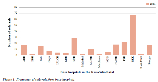
Figure 1 illustrates the distribution of patients referred from hospitals in and around KwaZulu-Natal. The majority of patients were referred from R K Khan (RKK) 67 (31.8%), King Edward (KEH) 29 (13.7%), Port Shepstone (PSH), 21 (9.95%), Prince Mshiyeni Memorial (PMMH) 17 (8.06%), Addington (ADH) 16 (7.58%), Stanger 16 (7.58%), GJ Crookes (GCJ) 14 (6.63%) hospitals and 31 (14.70%) collectively from the remaining hospitals.
The lead mechanism of injury was stab wound (SW) injuries with a total of 177 (84%) while gunshot wounds (GSW) comprised 34 (16%) (Table I). Patients were also evaluated according to location of neck zone of the skin injury; zone 2 injuries comprised 107 (51%), while zone 1 were 61 (29%) and zone 3 made up 11 (5%) respectively. In addition, the data analysis reflected that 32 patients had injuries in multiple zones (15%).
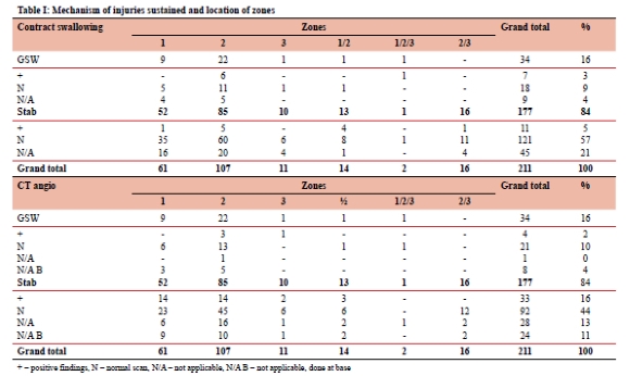
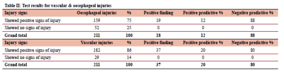
Table II illustrates that patients with clinical signs and symptoms of digestive tract injuries accounted for 159 (75%), while 52 (25%) had no apparent clinical injury present. Of the 159 patients who had a clinical suspicion of oesophageal injury only 19 (12%) were positive. Upon evaluation of the contrast swallow findings, 19 (12%) had a positive finding, 140 (88%) were normal. Of the 19 positive imaging findings, 7 were secondary to GSW and 12 were from SWs. Table II further highlights that the majority of patients presented with clinical signs and symptoms of vascular injuries (182, 86%), while 29 (14%) had no apparent clinical finding apart from the neck wound. Upon evaluation of the CTA findings, 37 (20%) had a positive finding, 113 (62%) were normal and 32 (18%) scans done at the base hospitals were normal (the patients were sent only for the contrast swallow). Of the 37 positive imaging findings, 4 were secondary to GSW and 33 were from SWs. With regards to CTA, zone 1 and zone 2 injuries were most commonly associated with positive imaging findings, followed by combined zone 1 and 2, and then isolated injury to zone 3. Unfortunately, in patients without signs and symptoms who were only referred for contrast swallow, it was not always clear if they had a normal CTA at the base hospital, thereby excluding any vascular injury.
The data from Table III illustrates that of the total patients (157) imaged with fluoroscopy, 138 were found to be normal. The data further breaks down the indication for requesting the imaging. The main indication for requesting the imaging was due to proximity of the skin injury, followed by odynophagia and surgical emphysema. Table III also depicts that of the contrast fluoroscopy, 19 (12%) were positive for oesophageal injuries. Zone 2 injuries were most associated with positive imaging finding followed by combined zone 1 and 2 and then zone 1 injury. Table III further analyses the signs/symptoms of patients referred for contrast swallow to exclude oesophageal injury. Of the 53 patients tested based on proximity, only 2 revealed positive imaging findings, thereby demonstrating a positive result of 4%, a negative result of 96% and a positive predictive value (PPV) of 3.77%. Of the 8 patients tested based on history of dysphagia, all revealed normal imaging, thereby demonstrating a negative result of 100% and a PPV of 0. Of the 47 patients tested based on odynophagia, 6 scans revealed positive findings, thereby demonstrating a positive result of 13%, a negative result of 87% and a PPV of 12.77%. Of the 3 patients tested based on the presence of dysphonia, none had positive imaging findings, thereby demonstrating a negative result of 100% and a PPV of 0. Of the 7 patients tested based on the finding of retropharyngeal air on CT scan, all 7 revealed normal scans, thereby demonstrating a negative result of 100% with a PPV of 0. Of the 10 patients tested based on the finding of prevertebral air on X-ray, 5 had positive imaging findings thereby demonstrating a positive result and negative result of 50% respectively with a PPV of 50%. Of the 25 patients tested based on the presence of surgical emphysema clinically, 4 revealed positive imaging findings, thereby demonstrating a positive result of 16% with a negative result of 86% and a PPV of 16%. Of the 2 patients tested based on the presence of food/saliva in the neck wound, both scans revealed injuries, thereby demonstrating a positive result and PPV of 100%. Of the 2 patients tested based on hematemesis, both revealed normal scans, thereby demonstrating a negative result of 100% and a PPV of 0. The above findings emphasise the importance of differentiating between hard and soft signs of oesophageal injury. What is apparent from the data is that not many patients with hard signs (airway compromise, massive hematemesis or bubbling through the wound) were imaged. Two patients with food and saliva from the wound were sent for imaging and inevitably revealed positive imaging findings. Soft signs of oesophageal injury include proximity, odynophagia, dysphonia, dysphagia, and subcutaneous or mediastinal air. The PPV calculation was again performed in accordance with the standard formula, and it was not possible to calculate negative predictive values (NPVs) due to data limitations. The majority of the soft signs revealed a PPV of less than 15%, with the exception of prevertebral air on X-ray, surgical emphysema and food/saliva in wound.
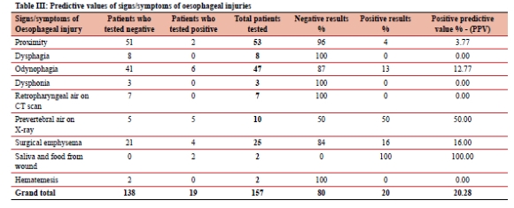
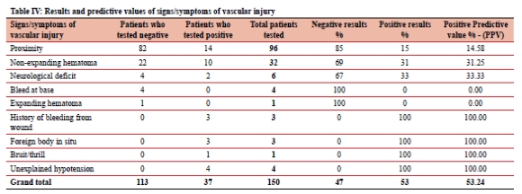
Table IV illustrates that of the total patients imaged with CTA (150), 113 were found to be normal. The indication for requesting the imaging is further broken down in Table IV The main indication for requesting the imaging was proximity of the skin injury, followed by non-expanding hematomas, with suspected neurological deficit or an alleged bleed at base, and a single expanding hematoma also included as indications for imaging. Of the 96 patients tested based on proximity, 14 revealed positive imaging findings, thereby demonstrating a positive result of 15%, a negative result of 85% and a PPV of 14.58%. Of the 32 patients tested based on a non-expanding hematoma, 10 revealed positive imaging findings, thereby demonstrating a positive result of 31%, a negative result of 69% and a PPV of 31.25%. Of the 6 patients tested based on a neurological deficit, only 2 revealed positive findings, thereby demonstrating a positive result of 33%, a negative result of 67% and a PPV of 33.33%. Of the 4 patients tested based on history of a bleed at base, none had positive imaging findings, thereby demonstrating a negative result of 100% and PPV of 0. The 1 patient tested based on an expanding hematoma, had negative imaging finding, thereby demonstrating a negative result of 100% and a PPV of 0. Of the 3 patients tested based on history of active bleeding, from wounds requiring bleeding-control, all 3 had positive imaging findings, thereby demonstrating a positive result and a PPV of 100%. Of the 3 patients tested based on having a foreign body impaled in the neck, all 3 revealed positive imaging findings, thereby demonstrating a positive result and PPV of 100%. The one patient that had a bruit and a thrill had a positive imaging finding, thereby demonstrating a positive result and PPV of 100%. Of the 4 patients tested on basis of unexplained hypotension, all 4 revealed positive imaging findings, demonstrating 100% positive result and a PPV of 100%. The above findings highlight the importance in differentiating between hard and soft signs, all the patients with hard signs (active bleeding from wound, shock/unexplained hypotension and bruit/ thrill) harboured an underlying vascular injury. The CTA for the patient with the sign of an expanding hematoma strangely demonstrated a normal study, it is therefore assumed to have resulted from an uncontrolled muscle bleeder, as usually an expanding, pulsatile hematoma is accompanied by a vascular injury. Soft vascular signs include proximity, non-expanding hematomas, history of a bleed at base and focal neurological deficit. The majority of the patients who presented with soft signs demonstrated negative imaging results. NVPs could not be calculated due to testing limitations as a retrospective study was conducted with pre-existing data and the fact that "TN" (true negatives) in the NPV formula could not be derived. In terms of predictive values, important to note, all signs/symptoms except proximity demonstrated a PPV of more than 15%.
Discussion
This study demonstrated that penetrating SWs to the young male population dominated the cohort, with a large number of the patients who were imaged revealing normal scan and swallow results. How this can translate to reducing the imaging burden is, however, less clear.
Two separate studies conducted in the United States of America and England, respectively, exhibited comparable results in terms of demographics and mechanism.3,4 The study from England included 214 patients mostly young males, with penetrating SWs forming the predominant mechanism of injury.3 The US study by Isaza-Restrepo et al.4 with 207 patients, revealed similar findings in terms of male predominance and penetrating stab injuries.4
Similarly, most of the patients referred for imaging to IALCH for penetrating neck injuries were male (96%) and the mean age of patients was 30, which highlights that trauma is rife among younger male patients in our setting. SWs dominated the mechanism of injury in this cohort. Numerous patients were referred from surrounding tertiary hospitals due to either non-functional or non-existing CT scans and non-existing fluoroscopy machines. Patients with signs and symptoms of either vascular and digestive tract injuries accounted for 86% and 75% respectively, highlighting the reason for referral. The remainder of the patients imaged, who had no signs and symptoms, were soft referrals due to proximity only. The CTA findings and the contrast swallow findings revealed a positive finding in 20% and 12% respectively. Evidently only 1/5 of patients with signs and symptoms of vascular injuries had positive image findings and 1/6 of those with signs and symptoms suggestive of digestive tract injuries had positive findings on imaging.
These figures highlight that the majority of patients referred for imaging potentially do not actually warrant imaging and even those that have signs and symptoms of injuries, mostly have no injuries. Their signs and symptoms could be attributed to other variables, such as soft tissue trauma, swelling, neuropraxia, anxiety, etc. In our review, the vast majority of the negative CTA findings were referred based on proximity and non-expanding hematomas, both soft signs, and with regards to the fluoroscopy, the majority were referred due to proximity, odynophagia and surgical emphysema. Upon determining the PPV for both signs and symptoms of vascular and oesophageal injury, it was shown that proximity had a PPV of less than 15% in both imaging groups, suggesting the need for more conservatism in resource-limited settings if this is the only additional clinical marker. Similarly, vascular injury proximity without other soft signs has a very low predictive rate for significant injury, even with the presence of a small or non-expanding haematoma. In addition, other symptoms such as dysphagia, dysphonia, odynophagia, retropharyngeal air on CT and hematemesis had a similar low PPV, suggesting again observation, or the use of other evaluation modalities available at the local hospital (e.g. endoscopy) to avoid unnecessary transfers and treatment delays. On the contrary prevertebral air on lateral neck X-ray, the presence of surgical emphysema and suspected salivary or food leak were highly predictive of injury, with the former suggesting the need for imaging while the last of the three should dictate the need for empiric surgical or other therapeutic intervention, without further imaging. This begs the question of whether these patients should rather be monitored and re-examined prior to being imaged urgently or at least be reviewed by a consultant surgeon. A similar study that was conducted by Isaza-Restrepo et al.4 mirrored the findings of our study. Their study also validated that it was safe to exclude vascular and digestive tract injuries in patients who had no signs or symptoms (only wound proximity) thus emphasising the importance of clinical examination in these patients.4
As mentioned earlier, the validation of the no-zone approach has assisted in triaging patients. Patients with hard signs of vascular or aerodigestive injuries warrant urgent operative intervention, either as an emergency or after image-guided approaches.5,6 However, controversy exists when assessing and managing patients without significant symptoms or soft signs. Appropriate and timely treatment of these patients with subtle signs can affect outcomes and quality of life. Delayed identification of vascular injuries such as pseudoaneurysms and arteriovenous fistulae may hold unfavourable long-term effects on patients, hence failing to identify them heralds potentially dire consequences and even death. In a recent literature review by Ntola et al.,6 the importance of thorough clinical examination and appropriate imaging options were highlighted for vascular trauma patients. Doppler ultrasound (DUS) may be beneficial in patients with zone 2 injuries, as it is not only is readily available, more cost-effective and easier to perform, but also does not expose patients to contrast or ionising radiation. However, DUS is operator dependent and is limited by bony structures, dressings and large hematomas, limiting its use in zone 1 and 3 injuries.6 Regarding penetrating neck trauma, their review is synchronous with international views, regarding CTA as a gold standard for injuries in all zones with soft signs as it has a high sensitivity, specificity and a high negative predictive value of 97-100%. They further advised that CTA be performed in neck injuries that have penetrated the neck fascia regardless of zone, in keeping with the no-zone concept.6
Digestive tract injuries or perforations carry a high mortality rate. In terms of penetrating injuries to the oesophagus, it has been shown that clinical findings are unreliable, identifying just 80% of injuries.7 In a study conducted by Nel et al.8 in 2007, it was found that odynophagia, concerning as it may be, was not a reliable clinical sign of oesophageal injury. Currently water-soluble fluoroscopy is the gold standard investigation used to exclude oesophageal injuries and has been shown to be safe and accurate as shown by Nel et al.8 However, considering that IALCH is one of only 2 hospitals in the province that offers this service, it is not surprising that the hospital is inundated with referrals, thus warranting a review of the referral criteria and suggesting proximity alone is insufficient reason to do a swallow-study.
This highlights the potential role of computed tomography in excluding oesophageal injuries. Studies show that CT oesophagography has high sensitivity and NPV when ruling out oesophageal perforations, especially in critically ill patients who are not able to swallow the contrast on demand. The fact that CT oesophagography can be readily performed with a CTA is reassuring in terms of reducing the number of patients being referred for fluoroscopy, however this requires a well-defined protocol and well-trained radiology staff.9,10,11 In the past, injuries to the hypopharynx and some cervical oesophageal injuries were managed operatively, especially if there is communication to the skin. However, local studies by Madiba et al.12 in 2003 and subsequently by Madsen et al.13 from Pietermartizburg showed that in selected cases non-operative management could be safe and effective.
In this review, the low yield of positive results leaves a few questions, that, if answered could lead to less unnecessary scans and reduced burden on our overburdened health system. A high index of suspicion with retropharyngeal air on postero-anterior and lateral neck x-rays in addition to symptoms warrants further imaging. Injury to the oesophagus has shown to be uncommon in penetrating neck trauma based on its anatomical location and protection by surrounding structures with its incidence being as low as 5%. Then, would it be safe to discharge patients who present with no signs or symptoms of oesophageal injuries? Patients presenting with signs and symptoms of digestive tract injuries who do not meet criteria for urgent exploration should be imaged with fluoroscopy using iso-oncotic water-soluble contrast if there is a high index of suspicion. This was shown to be accurate and safer than barium contrast in the large study by Nel et al.8 in 2008. In centres with no fluoroscopy, CT-oesophagography is a formidable alternative. Upper endoscopies could also be used to exclude larger perforations and patients with significant symptoms and no obvious injuries seen on endoscopy may then be referred for fluoroscopy if doubt still exists.
Physical examination has again shown to be unreliable in predicting vascular injuries. GSW were shown to more likely cause a vascular injury. However, it has been shown by Madsen et al.14 that patients asymptomatic for vascular injuries secondary to SWs to the neck do not warrant imaging. Unfortunately, the same cannot apply for GSW, stable patients should be imaged especially if symptomatic due to transmitted shockwaves that damages the sensitive vascular endothelium. Unstable patients with hard signs warrant urgent exploration as mentioned previously; stable ones may benefit from image-directed intervention. On the other hand, patients with soft signs require imaging in the form of a doppler ultrasound or CTA. Penetrating injuries to zone II were the most common zone affected and resulted most of the injuries. Colour flow Duplex DUS has been shown to provide excellent results in terms of diagnosis in hemodynamically stable patients with zone 2 injuries, with a low rate of morbidity, as well as being cost effective.15 In clinically stable patients with zone 1 and 3 injuries, DUS has limitations as a result of surrounding bony structures and CTA is warranted in patients with a high index of suspicion. In clinically stable zone 1 injuries, it has been proposed that routine angiography is not advocated for all injuries. Hardcastle et al.16 identified strongly suggestive signs in addition to the conventional hard signs of vascular injury which could be used to delineate patients that require angiography.
Limitations
This is a single-centre retrospective study and only includes patients from IALCH with a penetrating neck injury identified at the base facility, referred for either swallow or CTA. The short study duration of 3 years may have excluded some patients with positive imaging. The data may not be generalisable to other provinces where more liberal access to imaging is available or where more facilities have surgical cover after-hours. Finally, small numbers of certain signs or symptoms added difficulty for calculation of subset PPV In mitigation of these limitations this study is based on data collected prospectively in an electronic patient record using templates for data capture to reduce missing variables.
Conclusion
Penetrating neck trauma is a major trauma burden in KwaZulu-Natal, due to GSW and stabbings. The injuries range from life-threatening acute injuries to subtle, asymptomatic injuries which could be easily overlooked. However, the majority of patients without hard signs or instability who are being referred for imaging yield a normal imaging result. For patients with only soft signs of vascular injury, such as proximity, there is room for clinical observation without routine imaging to simply exclude injury, given the resource-limited setting. Similarly, patients with soft signs of oesophageal injury, such as proximity, dysphagia, dysphonia, odynophagia, retropharyngeal air on CT and hematemesis, may be admitted and observed, rather than offered routine imaging, where such imaging would require transfer to IALCH, provided they can access surgical care if they fail non-operative management. It is evident from the data that stricter referral criteria, protocols and clinical acumen are required to triage patients who actually warrant imaging, especially if imaging would change the course of management.
Conflict of interest
The authors declare no conflict of interest.
Ethical approval
Ethical clearance was provided by the Biomedical Research Ethics Committee at The University of KwaZulu-Natal, as a sub-study of the BCA207/09 class approval.
ORCID
KA Misra https://orcid.org/0009-0002-0953-065X
TC Hardcastle https://orcid.org/0000-0002-3967-0234
REFERENCES
1. Tafera A, Lutge E, Moodley N, et al. Tracking the trauma epidemic in KwaZulu-Natal, South Africa. World J Surg. 2023;47(8):1940-5. https://doi.org/10.1007/s00268-023-07032-2. [ Links ]
2. Chandrananth ML, Zhang A, Voutier CR. 'No zone' approach to the management of stable penetrating neck injuries: a systematic review. Aus NZ J Surg. 2021;91(6):1083-1090. https://doi.org/10.1111/ans.16600. [ Links ]
3. Steel BJ, Swansbury A, Wheeler LT. A 10-year study of penetrating head and neck injury by assault in the North East of England. Oral Maxillofac Surg. 2021;26(1):213-22. https://doi.org/10.1007/s10006-021-00980-w. [ Links ]
4. Isaza-Restrepo A, Quintero-Contreras JA, Escobar-DiazGranados J. Value of clinical examination in the assessment of penetrating neck injuries: a restrospective study of diagnostic accuracy test. BMC Emerg Med. 2020;20(17):1. https://doi.org/10.1186/s12873-020-00311-4. [ Links ]
5. Fransman RB, Azar FK, Mallon W, Nalluri A, Sakran JV, Haut ER. The "No-Zone" approach to penetrating neck trauma. J Am Coll Surg. 2021;1(4):1-11. [ Links ]
6. Ntola VC, Hardcastle TC. Diagnostic approaches to vascular injury in polytrauma - A literature review. Diagnostics (Basle). 2023;7(13):1019-1-12. https://doi.org/10.3390/diagnostics13061019. [ Links ]
7. Biffl WLM, Moore EEM, Feliciano DVM, et al. Western Trauma Association Critical Decisions in Trauma: Diagnosis and management of esophageal injuries. J Trauma Acute Care Surg. 2015;79(6):1089-95. https://doi.org/10.1097/TA.0000000000000772. [ Links ]
8. Nel L, Whitfield-Jones L, Hardcastle TC. Imaging the oesophagus after penetrating cervical trauma using water-soluble contrast alone: simple, cost-effective and accurate. Emerg Med J. 2008;26(1):106-8. https://doi.org/10.1136/emj.2008.063958. [ Links ]
9. Muhammad A, Qamar S, Rehman A, Baloch NUA, Shafqat G. Accuracy of CT chest without oral contrast for ruling out esophageal perforation using fluoroscopic esophagography as reference standard: A retrospective study. Eur J Trauma Emeg Surg. 2018;45(3):517-25. https://doi.org/10.1007/s00068-018-0929-4. [ Links ]
10. Conradie WJ, Gebremariam F. Can computed tomography esophagography reliably diagnose traumatic penetrating upper digestive tract injuries? Clin Imaging. 2015;39(6):1039-45. https://doi.org/10.1016/jxlinimag.2015.07.021. [ Links ]
11. Norton-Gregory AA, Kulkarni NM, O'Connor SD, et al. CT esophagography for evaluation of esophageal perforation. Radiographics. 2021;41(2):447-61. https://doi.org/10.1148/rg.2021200132. [ Links ]
12. Muckart D, Madiba TE. Penetrating injuries to the cervical oesophagus: Is routine exploration mandatory? Ann Royal Coll Surg. 2003;85(3):162-6. https://doi.org/10.1308/003588403321661307. [ Links ]
13. Madsen AS, Laing GL, Bruce JL, Oosthuizen GV, Clarke DL. An audit of penetrating neck injuries in a South African trauma service. Injury. 2015;47(1):64-69. https://doi.org/10.1016/j.injury.2015.07.032. [ Links ]
14. Madsen AS, Kong VY, Ooshtuizen GV, et al. Computer tomography angiography is the definitive vascular imaging modality for penetrating neck injury: A South African experience. Scand J Surg. 2018;107(1):23-30. https://doi.org/10.1177/1457496917731187. [ Links ]
15. Montorfano MA, Pla F, Vera L. Point-of-care ultrasound and doppler ultrasound evaluation of vascular injuries in penetrating and blunt trauma. Crit Ultrasound J. 2017:16;9(5):1. https://doi.org/10.1186/s13089-017-0060-5. [ Links ]
16. Hardcastle TC, Baatjes KJ, Du Toit DF. Routine arteriogram in zone-1 penetrating neck trauma: Is it required? Abstract 25, ISW2007, Montreal, Canada. p. 32. ISS-SIC, 2007. [ Links ]
 Correspondence:
email: hardcastle@ukzn.ac.za
