



Servicios Personalizados
Revista

Articulo
 Inglés (pdf)
 Articulo en XML
 Referencias del artículo

 Traducción automática
 Enviar articulo por email
Indicadores
Links relacionados
-
 Citado por Google
-
 Similares en Google
Compartir

 Permalink

South African Dental Journal
versión On-line ISSN 0375-1562versión impresa ISSN 0011-8516
S. Afr. dent. j. vol.81 no.3 Johannesburg abr. 2026
https://doi.org/10.17159/sadj.v81i03.23432
RESEARCH
Is there a Need for Analogue Imaging in the Modern Dental Radiology Curriculum? Use of Analogue and Digital Imaging in Gauteng, South Africa
B WalshI; L Janette HazellII; L MokoenaIII
IRadiography Manager/ Lecturer. Dental Radiology Unit. Department of General Dental Practice. Wits Oral Health Centre, Johannesburg, South Africa. Tel: 011 488 3956. E-mail: Brigitte.Walsh@wits.ac.za. ORCID: 0000-0001-9333-0946
IIHead of Department, Medical Imaging & Radiation Sciences, Faculty of Health Sciences, University of Johannesburg, Johannesburg, South Africa. Tel: 011 559 6066. E-mail: lynneh@uj.ac.za. ORCID: 0000-0002-8756-660X
IIILecturer, Department of Medical Imaging & Radiation Sciences, Faculty of Health Sciences, University of Johannesburg, Johannesburg, South Africa. Tel: 011 559 6424. E-mail: louisam@uj.ac.za. ORCID: 0000-0002-1303-1469
ABSTRACT
INTRODUCTION: Digital radiographic dental imaging enhances diagnostic accuracy, patient care, and clinical efficiency; however, adoption remains uneven due to cost, access, and infrastructure constraints. In South Africa, limited data on imaging modalities in dental practice highlight the need to align dental curricula with contemporary clinical practice.
AIMS AND OBJECTIVES: To assess analogue and digital radiographic imaging use among dentists practising in Gauteng Province, which represents approximately 40% of the national dental workforce, and to determine the relevance of analogue imaging in the revised dental radiology curriculum.
DESIGN: A descriptive, quantitative and cross-sectional research design was used.
METHODS: An online questionnaire was distributed via REDCap® and Google Forms® to practising general dentists and dental specialists in Gauteng. Non-practising dentists were excluded.
RESULTS: Of the 144 respondents, 77.8% were general dentists and 22.2% specialists with most (59.7%) practising in the private sector. Analogue intraoral imaging was more common in the public sector (38.7%) and teaching institutions (26.5%) than in individual (10.9%) or group practices (14.0%). Analogue panoramic imaging was reported by 7.7% of individual practices.
CONCLUSION: Analogue radiography has limited relevance in contemporary practice; therefore, dental radiology curricula should prioritise digital imaging, supported by foundational knowledge of analogue principles.
Keywords: dental radiography, dentist, imaging, dental specialist, curriculum
INTRODUCTION
Dental radiography plays a pivotal role in the diagnosis, treatment planning, and monitoring of oral diseases and is thereby a fundamental component of the dentistry curriculum.1 Radiographic images may be obtained using either analogue or digital imaging equipment modalities, encompassing both intraoral and extraoral techniques. Digital imaging methods include direct digital imaging (DD), photostimulable phosphor plate systems (PSP), and indirect digital imaging. In DD imaging, a digital sensor captures and immediately displays the image on a workstation. PSP utilises phosphor plates to capture the X-ray image, which is later displayed as a digital image using a laser scanner.2 In indirect digital imaging, the analogue film is converted into a digital image using a film digitiser, digital camera, or smartphone.3
Over the past two decades, dental imaging has seen a significant evolution from traditional analogue systems to advanced digital radiography, which offers numerous benefits, including reduced radiation exposure and dose, faster image acquisition, and enhanced image manipulation capabilities.4,5 Digital imaging also supports improved patient communication and satisfaction, as clinicians can directly display the patient's images and use various software tools to outline different treatment options.6 This shift from analogue to digital imaging has transformed diagnostic workflows, enabling better patient care and integration with emerging technologies such as Computer-Aided Design/Computer-Aided Manufacturing (CAD/CAM) and Artificial Intelligence (AI). However, there has been a notable disparity in the adoption of these digital dental imaging technologies into dental practices and training curricula, predominantly due to the noteworthy cost and infrastructure demands that digital imaging requires.7-9
Advancements in technology necessitate that educators remain up to date with these developments to ensure that dental curricula remain aligned with current dental practices. In light of global trends, it is pertinent to question the continued inclusion of analogue imaging in the revised dental radiology curriculum and whether educational emphasis should instead be placed predominantly on digital imaging.10-12 In Brazil, for example, only a limited number of universities provide access to digital radiology, and consequently, the use of analogue imaging methods remains prevalent.13 This has led to a significant demand for the expansion of teaching and training in specialised digital technologies, such as CBCT and related software, at both undergraduate and postgraduate levels.14
Before revising the dental radiology curriculum, it is essential to determine the imaging methods currently used by dentists and dental specialists in South Africa. Gauteng, as South Africa's wealthiest and most populous province, offers a unique lens through which to assess the state of dental radiographic imaging. Gauteng houses a high density of dental professionals and training institutions, yet significant socio-economic disparities persist within the province.15 Understanding the prevalence and types of imaging methods used in Gauteng, as well as the extent to which digital imaging has been adopted, can provide insights into broader national trends and inform curriculum development and policy decisions.12 This study aims to explore the use of digital imaging among dentists in Gauteng, including the impact of age and sector on technology adoption, and the implications for the revised dental curriculum.
LITERATURE REVIEW
The availability of intraoral, extraoral, and CBCT imaging equipment varies significantly worldwide. While there has been considerable growth in the digital imaging market, analogue imaging remains in widespread use.16,17 In Saudi Arabia, digital imaging adoption has risen from 5.7% in 2003 to 90% in 2021.12 However, 75% of dentists continue to use both analogue and digital imaging, while 21% rely exclusively on digital methods.10,12 Similarly, Sweden (98%), the Netherlands (90%), Belgium (90%), India (84%), New Zealand (88,7%) and Iraq (70%) have experienced rapid increases in digital dental radiography use.18-22,22-24 Analogue imaging remains in use to a lesser degree in Brazil (48,9%) and Jordan (47.8%).13,23 In Singapore (11,1%) and Korea (10%), analogue imaging is used to an even lesser extent for intraoral radiography.24,25
The majority of dentists in Canada (88,7%), New Zealand (58%), Pakistan (69%), Belgium (90%), and Sweden (98%) have access to digital radiographic technology.14,22,25-27 In Brazil, there has been a steady increase in the use of digital imaging in dentistry, particularly in the dental specialities.14 Globally, the extent to which analogue and digital imaging methods are integrated into dental curricula varies considerably. A study conducted across 15 European countries found that 86% of dental schools utilise digital imaging exclusively, while 14% incorporate both digital and analogue imaging in their curriculum.28
In the United States and Brazil, intraoral phosphor plates are used extensively in dental schools for educational and clinical teaching purposes.3,29 The reasons are twofold: phosphor plates are similar to conventional film and are more cost-effective than DD, and cause minimal patient discomfort compared to the DD sensors.29-31 DD sensors are thick, rigid and connected via a cable, which frequently contributes to increased patient discomfort and poses challenges for optimal intraoral positioning.29 Positioning of DD intraoral sensors requires an experienced operator to minimise geometric distortion and limit positioning errors, which frequently occur in comparison to the intraoral PSP.17,32,33 Whilst DD is superior in terms of image acquisition speed, PSP is reportedly superior to DD in terms of overall image quality.34
The use of PPS with the thinner intraoral phosphor plates enables the student to gain confidence in digital intraoral imaging and to adopt the practice more readily.35 Another disadvantage of DD is the initial cost of setting up a DD system, in comparison to PSP.17 In Saudi Arabia, both digital and analogue imaging techniques remain prevalent in clinical practice, as digital methods offer advantages for certain applications, while analogue-based methods are preferred for others. Consequently, both methods continue to be taught in dental curricula.10 However, this places substantial strain on limited training resources and due to this, many countries in Europe have removed conventional analogue dental imaging (film imaging) from the dental curriculum.10
While extensive research has been conducted globally on the use of digital imaging among dentists, there is limited data on the use of analogue and digital imaging among dentists in South Africa. As new dental imaging technologies and imaging methods arise, these are added to the dental curriculum, and thus the curriculum has expanded to cover both conventional and digital imaging approaches.36 In addition to the explicit resources required and the maintenance thereof, the inclusion of analogue imaging in the current dental radiology curriculum requires a large amount of valuable additional teaching time, which places a significant strain on the curriculum and limited available resources.12 This study investigated the radiographic imaging methods used by general dentists and dental specialists to assess the continued relevance of including analogue imaging in the current dental radiology curriculum.
METHODS
Research Design
A descriptive, quantitative and cross-sectional research design was used. The survey questionnaire consisted of closed-ended questions divided into six sections. Section one captured demographic details, including gender, age, professional designation, practice setting, and type of practice. Section two explored the use of intraoral imaging equipment, while section three addressed intraoral imaging methods and techniques. Sections four and five examined extraoral imaging equipment and methodologies. Section six gathered participants' perspectives on how patient safety, image quality, and patient throughput influenced their choice of imaging equipment and techniques. Responses in the first five sections were recorded using index-based inputs, whereas the sixth section utilised a five-point Likert scale, ranging from 1 ("Strongly Disagree") to 5 ("Strongly Agree").
Population and Sampling
Gauteng Province has an estimated 2,739 registered dentists, comprising 2,459 general dentists and 280 dental specialists.15,37 Given the constraints imposed by the Protection of Personal Information (POPI) Act and the wide geographic distribution of dentists and dental specialists, non-probability convenience sampling was used. Despite its limitations, convenience sampling is valuable in such studies where the eligibility criteria are not associated with the primary variables of interest.38 The study population included dentists and dental specialists actively practising in the private, public, or academic sectors. Dental specialities considered included community dentistry, maxillofacial and oral surgery, oral pathology, orthodontics, prosthodontics, oral medicine, and periodontology. Following statistical consultation, a minimum of 100 participants was deemed necessary to achieve statistical significance. Ultimately, 144 responses were received and analysed.
Data Collection
An online questionnaire was distributed through dental organisations, online databases, and peer networks via email and WhatsApp®. The data collection tool, a questionnaire that was adapted from previous studies,11,27,39-41 was initially hosted on REDCap®, an electronic data capture platform which uses audit trails to secure the captured data. This yielded a poor response rate of 66 participants. As a result, the survey was later compiled using Google Forms®, which yielded a higher response rate of 78 responses. The total sample was 144 respondents. The survey was closed once the target number of participants had been reached.
Responses were coded numerically (e.g., Yes = 1; No = 2), and incomplete questionnaires without any data were excluded. Due to their valuable content, partial responses (n = 12) were included in the total of 144 responses. Statistical analysis was conducted using IBM SPSS (V.30ersion 15), including descriptive statistics (frequencies, percentages, means, standard deviations) and cross-tabulations. Fisher's Exact Test was used, and Pearson's Chi-Square was adopted when tables with more than two categories occurred. Indices were used in the first five sections of the questionnaire. The level of significance was p ≤ 0.05. In the final section, a five-point Likert scale was used, and measures of central tendency (mean, median, mode) were calculated for these responses.
Validity and Reliability
An online survey questionnaire, adapted from previous studies, was used to enhance the validity and reliability of the data collection. A pilot study was conducted to evaluate the content for clarity, intended purpose, and comprehensiveness, thereby contributing to both the internal and external validity of the study.42 During the pilot study, electronic accessibility to the online survey was repeatedly tested using various devices and platforms. Responses from the pilot study were excluded from the final research data.
Experts in the dental field assessed the questionnaire on its alignment with the target population, the scope of covered content, and the equal representation of key constructs. To mitigate threats such as shifts in clinical practice over time, the survey was open for a limited duration. Consistency in questionnaire format, platform, and distribution channels helped limit external variability. All data were reviewed for consistency before submission to the statistician.
ETHICAL CONSIDERATIONS
Ethical approval for the study was obtained from the Research Ethics Committee at the University of Johannesburg (REC-1769-2022). Additionally, permission was granted by the Gauteng Department of Health (NHRD 2022_10_0222), as well as by the chief directors of the health districts and the three dental teaching institutions located in Gauteng. All participants received information letters and consent forms and could not access the questionnaire until consent had been given. The questionnaire was anonymous, and the resultant data were also encoded to safeguard the confidentiality and privacy of the participants.43
RESULTS
Demographics of Respondents
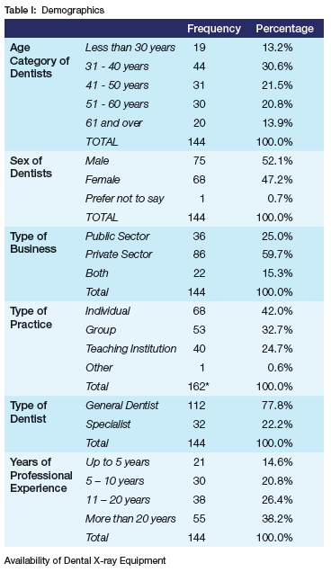
A total of 144 responses were received, with 52.1% of respondents identifying as male and 47.2% as female. The largest age group comprised individuals aged 31 to 40 years (30.6%), while 13.9% of respondents were over 60 years old (Table I). The majority of participants (59.7%) were employed in the private sector, 25% worked in the public sector, and 15.3% practised in both sectors. In terms of workplace setting, 42% of respondents worked in individual practices, 32.7% in group practices, and 24.7% in teaching institutions. General dentists accounted for the majority of participants (77.8%), while 22.2% were dental specialists (Table I).
Availability of Dental X-ray Equipment
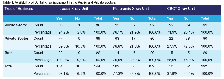
The majority of respondents (93.1%) reported having access to intraoral X-ray units, while 6.9% did not have access to such equipment (Table II). Panoramic radiography was available to 77.3% of respondents. There was no statistically significant difference in the availability of panoramic units between public and private sectors (p=0.699) or between general dentists and dental specialists (phi=0.217). Cone Beam Computed Tomography (CBCT) equipment was accessible to 37.9% of respondents, indicating a moderate level of adoption of advanced imaging technologies (Table II). *The number of responses regarding the type of practice is higher as some dentists work in more than one practice and were therefore not limited to only denoting one response.
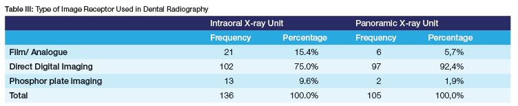
Of the 84.6% of respondents who used digital imaging for intraoral radiography, 75.0% utilised DD, while 9.6% relied on PSP (Table III).
Intraoral Radiography
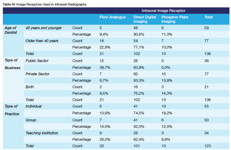
As shown in Table IV, intraoral radiographs were mainly executed using DD imaging systems across all sectors and age groups. However, analogue imaging remained more common among dentists over the age of 40 (22.9%) compared to the younger cohort (9.4%) and in the public sector (38.7%).
Among dentists and dental specialists in the private sector, PSP imaging was used by 13.9% of respondents. The use of digital imaging (PSP and DD) in teaching institutions (73.5%) was less than that of individual (89.1%) and group (85.1%) practices. The use of analogue imaging in individual (10.9%) or group practices (14.0%) was lower than in teaching institutions (26.5%). Conversely, PSP imaging was more frequently used in individual (18.2%) and group practices (12%) compared to teaching institutions (8.8%).
Panoramic Radiography
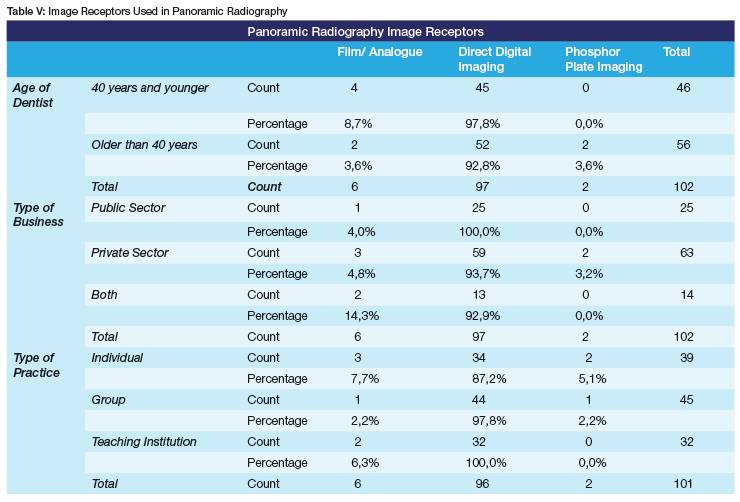
DD imaging was the preferred method for panoramic radiography across all age groups, with 97.8% of younger dentists and 92.8% of older dentists utilising this technology (Table V). The use of analogue imaging for panoramic radiography was limited, with 7.7% of dentists in individual practices using analogue systems, and minimal usage in teaching institutions (6.3%). PSP for panoramic imaging was not used by the teaching institutions. Some respondents reported using more than one imaging method, indicating a possible transitional phase or the use of analogue imaging as a backup system.
DISCUSSION
Demographics of Respondents
There is scarce data regarding the proportion of male and female dentists in South Africa. This study observed a higher proportion of male dentists compared to similar research conducted in the USA.44 Approximately 60% of dentists in South Africa work in the private sector, providing dental services to only 16% of the population, while the majority of the population relies on a limited number of public-sector dentists for treatment. This imbalance is a significant concern and aligns with previous studies predicting a critical shortage of dentists in the public sector by 2030.15,45,46
Intraoral X-ray Equipment
As one of the wealthiest provinces in South Africa, Gauteng has a high number (93.1%) of intraoral X-ray units, comparable to Australia (96.8%), Belgium (94%) and Morocco (87.2%).22,47,48 In this study, intraoral x-ray units were more commonly available among general dentists than among dental specialists. The majority of dentists using analogue film were over the age of 40, likely due to their familiarity with the technology and the significant costs associated with transitioning to digital imaging infrastructure.
Despite the drawbacks of intraoral DD, it was widely adopted in both the public and private sectors in this study. PSP imaging was not used in the public sector and had limited use in the private sector. PSP plates are small, prone to scratches, loss or theft, and costly to replace, making them impractical for large teaching institutions and overcrowded public-sector clinics.29 The continued use of analogue film in teaching institutions may be attributed to its inclusion in the current dental curriculum.
Panoramic Radiography
The availability of panoramic radiography in Gauteng (77%) is comparable to that of developed countries, including Saudi Arabia (90%), Canada (77%), Belgium (76%), Korea (96%), and Sweden (61%).12,22,25,26,48,49 In this study, panoramic X-ray units were equally accessible in both the public (78.1%) and private sectors (78.8%), a higher proportion than in Sweden, where 68% of dentists working in the public sector 32% of dentists working in the private sector have access to these units.26 The findings of this study align with those from Belgium, Pakistan, and Turkey, where a greater proportion of dental specialists, compared to general dentists, had access to panoramic radiography.11,22,50 Similar to findings in Belgium, Korea, and Saudi Arabia, few dentists in this study reported using PSP or analogue film for panoramic imaging.12,22,25 The widespread adoption of DD imaging for panoramic radiography across the public and private sectors, teaching institutions, and various practice types can be attributed to its cost-effectiveness, speed, and efficiency, particularly in high-volume public healthcare settings.51
Digitising the Analogue Film
Analogue radiographic images can be digitised using a film digitiser, digital camera, or smartphone.52 This process enables access to the benefits of digital technology, including electronic archiving, image enhancement, and efficient storage.53 However, the image quality of digitised films remains inferior to that of DD systems.3 Despite the limitations in image quality, digitising analogue films remains a valuable practice, offering many of the advantages inherent to digital imaging systems.
Digital vs analogue imaging
Dental radiographic images can be captured, viewed, and transmitted using either digital or analogue methods. Digital imaging presents several advantages, including reduced radiation exposure, enhanced sensitivity, and increased efficiency in archiving and sharing radiographic images across various platforms.11,29 Digital dentistry also enables the integration of imaging software with the Computer-Aided Design/Computer-Aided Manufacturing (CAD-CAM) technology and intraoral scanners, which allows for precise planning, design, and fabrication of dental restorations with high accuracy and efficiency.54 Digital radiography is widely utilised in most teaching institutions in Gauteng, with limited use of analogue film for intraoral radiography. A possible explanation for this continued use of analogue imaging is that training institutions may include it in their curriculum to illustrate the fundamental principles of image formation and to facilitate the interpretation of older radiographic images.
The use of digital imaging for intraoral radiography was slightly higher among the younger cohort for both intraoral (90.6%) and panoramic (97.8%) imaging. The older cohort demonstrated a stronger preference for intraoral analogue imaging (22.9%) compared to their younger counterparts. These findings are consistent with research conducted in Pakistan and Saudi Arabia,27,52 which also suggests that the competitive nature of the dental profession drives younger dentists to adopt new technologies more readily in response to growing patient expectations.27,52 In contrast, older dentists may not feel the same level of urgency to adopt these technologies into their practice.40 This trend contrasts with findings from Brazil, where the high cost of digital imaging has resulted in a higher prevalence of analogue film use among younger dentists compared to their older counterparts.13
Globally, the ease of digitally communicating patient health records and radiographic images has enabled the efficient sharing and storage of dental radiographs via email, tablets, and smartphones, forming an integral component of a patient's comprehensive dental health record.52 This digital capability has enhanced treatment planning and improved dental healthcare delivery, particularly in rural and remote areas.55 However, in countries such as Belgium and Ecuador, the digital transmission of patient health records and radiographic images has raised serious concerns regarding patient privacy and data security, which remain significant challenges.22,56-58
LIMITATIONS
This study was only conducted in one province, and these results may therefore not be generalised to the rest of South Africa, as the use of analogue and digital dental imaging in other provinces may differ. Despite the low response rate, the data obtained still offers valuable insights into current digital imaging practices and highlights trends that warrant further exploration.
CONCLUSION
The findings of this study highlight the need to revise the dental radiology curriculum to integrate additional competencies, including digital literacy in dental radiography, Teledentistry, and the responsible use of CBCT and specialised dental imaging. The dental radiology curriculum must continually evolve to reflect technological advances and clinical practices, ensuring graduates are competent and adaptable to this dynamic field. As digital imaging becomes the standard, ethical concerns around data security must be addressed, with adoption influenced by factors such as practice size and global collaboration. The use of AI, which is already prevalent in forensic dentistry, is expected to completely transform dentistry due to its efficiency and cost-effectiveness and its ability to classify numerous dental pathologies faster and more easily than humans.59,60
Despite the high cost of digital imaging infrastructure, the enhanced efficiency and patient throughput of digital imaging have made it popular among dentists27 as it enhances diagnostic capabilities and access to care, though challenges in privacy, infrastructure, and educational integration remain. Consequently, while the theoretical understanding of analogue imaging remains relevant, it is no longer necessary to include analogue imaging in clinical training due to the widespread shift towards digital systems. A balanced approach is essential to embrace innovation without compromising core radiographic principles.
This information will benefit the dental profession in its entirety, as the resulting data will inform and guide the dental radiology curriculum and serve as the foundation for further research in this discipline. Consequently, it will also provide insight into some of the skills a newly qualified dentist will need in this burgeoning field of dental radiology.
RECOMMENDATIONS
Based on the results of this study, the inclusion of analogue imaging in the dental radiology curriculum is no longer necessary, given the widespread adoption of digital imaging technologies in current dental practices. Revision of the current dental radiology curriculum must include emerging digital imaging technologies, Teledentistry and AI, notwithstanding digital ethics and data security.
FURTHER RESEARCH
Further research across all South Africa's provinces is necessary to develop a more comprehensive understanding of national dental radiography practices.
Declaration of Interest
The authors declare that they have no competing interests to disclose
Funding
No funding was obtained for this project.
REFERENCES
1. Erdelyi RA., Duma VF, Sinescu C, Dobre GM, Bradu A, Podoleanu A. Dental Diagnosis and Treatment Assessments: Between X-rays, Radiography and Optical. Materials (Basel). 2020;13(4825):1-24. [ Links ]
2. Van Der Stelt PF. Filmless imaging: The uses of digital radiography in dental practice. Journal of the American Dental Association. 2005;136(10):1379-87. doi: 10.14219/jada.archive.2005.0051 [ Links ]
3. Supriyadi S, Prastyarini S, Hidayati L, Lestari P. Dentist Perceptions on Dental-Radiodiagnosis Using Smartphone: Crossectional study. Journal of International Dental and Medical Research. 2022;15(2):783-8. [ Links ]
4. Pauwels R. History of Dental Radiography: Evolution of 2D and 3D Imaging Modalities. Medical Physics International. 2020;8(1):235-77. [ Links ]
5. Sattar MA, Aluaan SH, Alrazzaq WA. Evaluation of KV Reduction on image quality in OPG X-Ray. Ind J Forensic Med & Tox. 2020;14(2)2459-66. [ Links ]
6. Khurshid Z. Digital Dentistry: Transformation of Oral Health and Dental Education with Technology. Eur J Dent. 2023;17(4):943-4. [ Links ]
7. Duong AH, Nguyen TD, Duong GH, Tran TT. Assessing digital transformation readiness: a comprehensive study of local clinics in Northwest Vietnam. International Journal of Information Technology (Singapore). 2025 Apr 1;17(3):1607-17. [ Links ]
8. Fahad Alotaibi K, Mohd Kassim A. A Fully Integrated System for Digital Dentistry Workflow and Manufacturing Management. Journal of Advanced Research Design Journal. 2026; 139:175-86. Available from: https://akademiabaru.com/submit/index.php/ard [ Links ]
9. Gangavati R, Baad R, Vibhute N, Varma S, Kamte S, Sankpal S. An Insight into the Radiographic Practice Among Dentists of Karad City, India. European Journal of Pharmaceutical and Medical Research. 2016;3(3):368-73. [ Links ]
10. Abbas H, Arabi AI, Baroudi B, Makhdom S, Alwadani MA, Akhdar ES Al, et al. Intraoral Radiographic Processing Skills among Dentists in Saudi Arabia. Open Access Maced J Med Sci. 2021; 9:139-42. [ Links ]
11. Samejo I, Kumar B, Musharraf H, Ahmed J, Memon L, Bhatti R. Assessment of Knowledge and Perspective toward Dental Radiography among Dental Practitioners of Sindh Province Pakistan. J Evol Med Dent Sci. 2021;10(29):2186-93. [ Links ]
12. Al Sadhan R. A cross-sectional survey of dentists' use of digital radiographic techniques in Riyadh, Saudi Arabia. Saudi Journal of Oral Sciences. 2021;8(3):139. [ Links ]
13. Moreira-Souza L, de Oliveira Reis L, Nogueira-Reis F, da Costa E, Freitas D. How is the Use of Digital Dental Radiology in a Developing Country? An Overview of Brazil. Int J Dent Sci [Internet]. 2022;24(3):176-90. Available from: https://www.researchgate.net/publication/360230509 [ Links ]
14. Freire DBL, Celeste RK, Vizzotto MB, Nunes LN, Arús NA, Silveira HLDD. Impact of dentists and equipment in the performing dental imaging examinations: a longitudinal analysis. Braz Oral Res. 2022;36(47). doi: 10.1590/1807-3107bor-2022.vol36.0047. [ Links ]
15. Tiwarii R, Bhayat A, Chikte U. Forecasting for the need of dentists and specialists in South Africa until 2030. PLoS One [Internet]. 2021; 16:1-14. doi: 0.1371/journal.pone.0251238 [ Links ]
16. Hasan A, Naz F, Ali A, Khan JA, Ali B. Technical Errors in Intra Oral Radiographs Obtained in Endodontic Department of a Teaching Dental Hospital. J Pak Dent Assoc. 2019;28(2)50-4 [ Links ]
17. Zhang W, Huynh C, Jadhav A, et al. Comparison of Efficiency and Image Quality of Photostimulable Phosphor Plate and Charge-Coupled Device Receptors in Dental Radiography. J Dent Educ. 2019;83(10)1205-12. doi: 10.21815/JDE.019.120. [ Links ]
18. Panwar A, Gupta S, Nagaraju K, Malik S, Goel S, Sharma A. Awareness of radiation protection among dental practitioners in UP and NCR region, India: A questionnaire-based study. Journal of Oral Medicine and Radiology. 2022;(2):13-20. [ Links ]
19. Torresan TT, Rodrigues IC, Poletto MC, et al. Radioprotection in Dentistry: knowledge and practices. Res Soc Dev. 2021;10(14):1-14. [ Links ]
20. Al-Nuaimi N, Mahdee AF, Al-Hashimi R, Mannocci F. Knowledge and Attitude of Iraqi Dentists Towards the Use of Cone Beam Computed Tomography in Endodontics: A Questionnaire Study. International Medical Journal. 2021;28(6):658-63. [ Links ]
21. van der Zande MM, Gorter RC, Aartman IH, Wismeijer D. Adoption and use of digital technologies among general dental practitioners in the Netherlands. PLoS One. 2015;10(3): e0120725. doi: 10.1371/journal.pone.0120725. [ Links ]
22. Snel R, Van De Maele E, Politis C, Jacobs R. Digital dental radiology in Belgium: a nationwide survey. Dentomaxillofacial Radiol. 2018;47(8):20180045. doi: 10.1259/dmfr.20180045. [ Links ]
23. Al-Mousa DS, Alakhras M, AlSa'di AG, Chau M, Hayre C, Mahasneh AM. Attitudes and practices of radiation protection among Jordanian dental radiography practitioners. Radiography (Lond). 2024;30(6):1556-62. doi: 10.1016/j.radi.2024.09.061. [ Links ]
24. Ng AYQ, Lai CWM, Ho CXH, Lim LZ. Dental imaging in Singapore: a survey of 2D radiographic techniques and CBCT practices. Dentomaxillofac Radiol. 2025;54(8):649-58. doi: 10.1093/dmfr/twaf033. [ Links ]
25. An SY, Lee KM, Lee JS. Korean dentists' perceptions and attitudes regarding radiation safety and protection. Dentomaxillofacial Radiology. 2018;47(3):1-8. [ Links ]
26. Svenson B, Ståhlnacke K, Karlsson R, Fält A. Dentists' use of digital radiographic techniques: Part II - extraoral radiography: a questionnaire study of Swedish dentists. Acta Odontol Scand. 2019;77(2):150-7. doi: 10.1080/00016357.2018.1525763. [ Links ]
27. Hasan A, Khan JA, Ali B, Afshan Z, Shakir MN, Shah SYA. Practices of Dentists about Digital Techniques in Dental Radiology and Radiographic Safety. J Pakistan Dent Assoc. 2019;28(04):181-6. DOI: https://doi.org/10.25301/JPDA.284.181. [ Links ]
28. Ahlqvist J, Jäghagen EL, Friedlander-Barenboim S et al. Resources Allocated for Undergraduate Education in Oral Radiology in European Dentistry Programmes: A Survey Study. Eur J Dent Educ. 2025;29(2):384-91. doi: 10.1111/eje.13078. [ Links ]
29. Elkhateeb SM, Aloyouny AY, Omer MMS, Mansour SM. Analysis of photostimulable phosphor image plate artifacts and their prevalence. World J Clin Cases. 2022;10(2):437-47. doi: 10.12998/wjcc.v10.i2.437. [ Links ]
30. Cesur AK, Demirel O, Altan SG, Özcan M. Evaluation of two dental digital imaging systems based on quality scorings, burn-out effects and cervical width determination. Balkan Journal of Dental Medicine. 2020;24(2):71-6. doi: 10.2478/bjdm-2020-0012. [ Links ]
31. Souza-Pinto GN, Santaella GM, Coli AA, Oenning AC, Haiter-Neto F. Analysis of the deterioration of photostimulable phosphor plates. Dentomaxillofacial Radiology. 2020;49(6):20190500. doi: 10.1259/dmfr.20190500. [ Links ]
32. Anu Sushanth A, Srivastava KC, Shrivastava D, Hosni HA, Khan ZA, Al-Johani K, et al. Recommendations, practices and infrastructural model for the dental radiology set-up in clinical and academic institutions in the COVID-19 era. Biology (Basel). 2020;13;334(9). doi: 10.3390/biology9100334. [ Links ]
33. Beshtawi KR, Shaik S, Peck MT, Chetty M. The accuracy of various radiographic modalities for implant therapy. S. Afr. Dent. J. 2021;76(7): 396-403. Doi: 10.17159/2519-0105/2021/v76no7a1. [ Links ]
34. Farrier SL, Drage NA, Newcombe RG, Hayes SJ, Dummer PM. A comparative study of image quality and radiation exposure for dental radiographs produced using a charge-coupled device and a phosphor plate system. Int Endod J. 2009;42(10):900-7. doi: 10.1111/j.1365-2591.2009.01593.x. [ Links ]
35. Saucke K. Analysis of a Quality Assurance Program For Intraoral Photostimulable Phosphor Plates at a Large Academic Institution and Recommendations for Programmatic Enhancements. [dissertation] San Antonio: University of Texas Health Science Center. 2021;(August). Available from: https://uthscsa.edu/biomedical-sciences/programs/radiological-sciences-phd/career-paths/alumni [ Links ]
36. Witwatersrand U of. School of Oral Health Sciences, Department of General Dental Practice, Maxillo-Facial and Oral Radiology Study Guide BDS III, BDS IV. 2024. [ Links ]
37. Bhayat A, Nkambule NR, Madiba TK. A Review of the 2030 Human Resources for Health Strategy: Implications for Dentistry in South Africa. South African Dental Journal. 2021;76(06):367-73. doi: 10.17159/2519-0105/2021/v76no6a7. [ Links ]
38. Winton B, Sabol M. A multi-group analysis of convenience samples: free, cheap, friendly, and fancy sources. Int J Soc Res Methodol. 2022;00(00):1-16. doi: 0.1080/13645579.2021.1961187. [ Links ]
39. Harris PA, Taylor R, Minor BL, et al. The REDCap consortium: Building an international community of software platform partners. J Biomed Inform. 2019;95(May):103208. doi: 10.1016/j.jbi.2019.103208. [ Links ]
40. Masyte V, Sefeldaite S, Venskutonis T. A Questionnaire of Digital Radiography and CBCT Use and Knowledge among Lithuanian Dentists. J Oral Maxillofac Res. 2021;12(1): e2. doi: 10.5037/jomr.2021.12102. [ Links ]
41. Mauthe PW, Eaton KA. An investigation into dental digital radiography in dental practices in West Kent following the introduction of the 2006 NHS General Dental Services contract. Prim Dent Care. 2011;18(2):73-81. doi: 10.1308/135576111795162893. [ Links ]
42. Duckett LJ. Quantitative Research Excellence: Study Design and Reliable and Valid Measurement of Variables. J Hum Lact. 2021;37(3):456-463. doi: 10.1177/08903344211019285. [ Links ]
43. Jung YM. Data analysis in quantitative research. Handb Res Methods Heal Soc Sci. Singapore: Springer Singapore. 2019;(June):955-69. doi: 10.1007/978-981-10-5251-4_109. [ Links ]
44. Surdu S, Mertz E, Langelier M, Moore J. Dental Workforce Trends: A National Study of Gender Diversity and Practice Patterns. Med Care Res Rev. 2021;78(1_suppl):30S-39S. doi: 10.1177/1077558720952667. [ Links ]
45. Bhayat A, Chikte U. Human Resources for Oral Health Care in South Africa: A 2018 Update. Int J Environ Res Public Health. 2019 May 14;16(10):1668. doi: 10.3390/ijerph16101668. [ Links ]
46. Husic JB, Melero FJ, Barakovic S, et al. Aging at work: A review of recent trends and future directions. Int J Environ Res Public Health. 2020;17(20):7659. doi: 10.3390/ijerph17207659. [ Links ]
47. Ihle IR, Neibling E, Albrecht K, Treston H, Sholapurkar A. Investigation of radiation-protection knowledge, attitudes, and practices of North Queensland dentists. J Investig Clin Dent. 2019; 10:e12374. doi: 10.1111/jicd.12374 [ Links ]
48. Elmorabit N, Obtel M, Azougagh M, Marrakchi A, Ennibi OK. Development and validation of a questionnaire on radiation protection knowledge, attitudes, and practices among Moroccan dentists. J Appl Clin Med Phys. 2025;26(1): e14555. doi: 10.1002/acm2.14555. [ Links ]
49. Gillies RC, Quiñonez C, Wood RE, Lam EWN. Radiograph prescription practices of dentists in Ontario, Canada. J Am Dent Assoc. 2021;152(4):284-292. doi: 10.1016/j.adaj.2020.12.007. [ Links ]
50. Vural UK, Gökalp S. Diagnostic methods for dental caries used by private dental practitioners in Ankara. Niger J Clin Pract. 2017;20(3):382-7. doi: 10.4103/1119-3077.181360. [ Links ]
51. Sunilkumar AP, Parida BK, You W. Recent Advances in Dental Panoramic X-Ray Synthesis and Its Clinical Applications. IEEE Access. 2024;12(July):141032-51. [ Links ]
52. Almazrooa SA, Mansour GA, Alhamed SA, Ali SA, Akeel SK, Alhindi NA, et al. The Application of Teledentistry for Saudi Patients' Care: A National Survey Study. J Dent Sci [Internet]. 2021; 16:280-6. Available from: https://doi.org/10.1016/j.jds.2020.04.014 [ Links ]
53. Noorsaeed AS, et al. Overview on Updates on Digital Dental Radiography. J. Pharm. Res. Int. 2021;33(59B):23-8. Available from: https://journaljpri.com/index.php/JPRI/article/view/5010. [ Links ]
54. Tallarico M. Computerization and Digital Workflow in Medicine: Focus on Digital Dentistry. Materials (Basel). 2020;13(9):2172. doi: 10.3390/ma13092172. [ Links ]
55. Iorgulescu, G, et al. Ethical and Medico-Legal Aspects Behind the Use of Digital Technologies in Dentistry. Rom J Leg Med. 2020;28(2):202-7. doi: 10.4323/rjlm.2020.202. [ Links ]
56. Nakajima I, et al. Empirical Study on Medical Information and Communication Technology System in Dentistry in Southeast Asia. Oral Health Care: Tokyo. doi: 10.5772/intechopen.101080. [ Links ]
57. Chérrez-Ojeda I, Vera C, Vanegas E, et al. The Use of Information and Communication Technologies in Latin American Dentists: A Cross-sectional Study from Ecuador. BMC Oral Health [Internet]. 2020;20(146):1-9. Doi: 10.1186/s12903-020-01137-z. [ Links ]
58. Kanani H, Khubchandani M, Dangore-Khasbage S, Pandey R. Teledentistry: A Comprehensive Review and Its Application in Pediatric Dental Care. Cureus. 2024;16(1):1-7. [ Links ]
59. Kumar A, Bhadauria HS, Singh A. Descriptive analysis of dental X-ray images using various practical methods: A review. PeerJ Comput Sci. 2021;7:e620. doi: 10.7717/peerj-cs.620. [ Links ]
60. Putra RH, Doi C, Yoda N, Astuti ER, Sasaki K. Current applications and development of artificial intelligence for digital dental radiography. Dentomaxillofac Radiol. 2022;51(1):20210197. doi: 10.1259/dmfr.20210197. [ Links ]
 Correspondence:
Mrs. Brigitte Walsh
Tel: 011 488 3956; E-mail: Brigitte.Walsh@wits.ac.za
Authors contributions
1. Mrs. Brigitte Walsh, Principal Researcher, 30% Contribution
2. Dr Lynne Janette Hazell, Main supervisor, 23,3% Contribution
3. Dr Louisa Mokoena, Co-supervisor, 23,3% Contribution