



Servicios Personalizados
Revista

Articulo
 Inglés (pdf)
 Articulo en XML
 Referencias del artículo

 Traducción automática
 Enviar articulo por email
Indicadores
Links relacionados
-
 Citado por Google
-
 Similares en Google
Compartir

 Permalink

South African Dental Journal
versión On-line ISSN 0375-1562versión impresa ISSN 0011-8516
S. Afr. dent. j. vol.81 no.2 Johannesburg 2026
https://doi.org/10.17159/sadj.v81i02.24161
RESEARCH
Evaluation of factors associated with gingival enlargement during fixed orthodontic treatment
I PitsoI; M MoshaoaII; TK MadibaIII; A MasengeIV; S ManenzheV
IDepartment of Periodontics and Oral Medicine, School of Dentistry, University of Pretoria, Pretoria, South Africa, E-mail: itumeleng.pitso@up.ac.za. ORCID: https://orcid.org/0009-0006-1748-6010
IIDepartment of Orthodontics, School of Dentistry, University of Pretoria, Pretoria, South Africa, E-mail: mpule.moshaoa@up.ac.za. ORCID: https://orcid.org/0000-0003-0306-3739
IIIDepartment of Community Dentistry, University of Pretoria, Pretoria. E-mail: thommy.madiba@up.ac.za or thommy.madiba@gmail.com. ORCID: https://orcid.org/0000-0002-0171-0595
IVDepartment of Statistics, University of Pretoria, Pretoria, South Africa. E-mail: andries.masenge@up.ac.za. ORCID: https://orcid.org/0000-0001-8372-2356
VDepartment of Periodontics and Oral Medicine, School of Dentistry, University of Pretoria, Pretoria, South Africa, E-mail: shumane.manenzhe@up.ac.za. ORCID: https://orcid.org/0009-0002-6633-4957
ABSTRACT
INTRODUCTION: Gingival enlargement (GE) is a common finding among patients undergoing fixed orthodontic treatment.
OBJECTIVE: To evaluate the prevalence and clinical severity of gingival enlargement (GE) in patients undergoing fixed orthodontic treatment (OT) at the University of Pretoria Oral Health Center (UPOHC). The study also aims to determine the Plaque Index (PI) among these patients and explore the association of gingival enlargement with various factors, such as the different phases of orthodontic treatment, treatment duration, type of archwire used, age, and gender.
METHODS: A cross-sectional study was conducted with patients undergoing fixed OT. Gingival enlargement was assessed using the gingival index proposed by Bokenkamp and Bonhorst (1994), and the plaque index (PI) was measured using O'Leary's method and an examination of clinical features. Variables such as treatment duration, orthodontic stage, type of archwire, and the use of molar bands were evaluated for their potential associations with GE.
RESULTS: Gingival enlargement was found in 94% of participants, with higher prevalence and severity in cases with prolonged treatment and increased PI. However, there was no significant association between GE and variables such as gender, orthodontic stage, or molar bands (p>0.05). A significant link was found between generalised PI and GE severity (p=0.0001).
CONCLUSION: Plaque accumulation emerged as an important factor in GE development during OT, emphasising the importance of optimal oral hygiene practices. Educating patients and implementing preventive strategies may mitigate GE risk and improve OT outcomes.
CLINICAL RELEVANCE: The findings of this study can aid in facilitating the development of treatment protocols aimed at preventing and reducing the occurrence of GE during fixed OT.
Keywords: Gingival enlargement, fixed orthodontic treatment, plaque index, oral hygiene, orthodontic appliances
INTRODUCTION
Gingival enlargement (GE) is a common finding among patients undergoing fixed OT. The occurrence of GE during fixed OT has been attributed to several factors. Gingival enlargement is an abnormal overgrowth of gingival tissues associated with factors like plaque accumulation, orthodontic appliances, allergic reactions, and hormonal changes related to age and gender.1 Gingival enlargement is often erroneously used interchangeably with gingival hyperplasia, hypertrophy, and fibrosis.1 The term GE is preferred because hyperplasia, hypertrophy, and fibrosis are changes observed microscopically in GE but cannot be accurately differentiated clinically. In the initial stages, Gingival enlargement is localized to the interdental papilla and extends to involve marginal and attached gingiva. Eventually, the gingiva may cover part or the whole crown of a tooth.2 GE may cause functional and aesthetic challenges in those affected, which is often the reason why people seek treatment.
The American Academy of Periodontology (AAP) and the European Federation of Periodontology (EFP) have developed a classification for periodontal disease and conditions.3 According to the AAP and EFP classification of 2018, GE can be classified under two broad categories: dental plaque biofilm-induced and non-dental plaque biofilm-induced gingival diseases.3 In the AAP/EFP the classification of GE is based on the causative and/or predisposing factors.3-5 Gingival enlargement is further categorised based on the extent of involvement into localised (to specific teeth) affecting less than 30% of teeth and generalised affecting more than 30% of the teeth present in the mouth.3
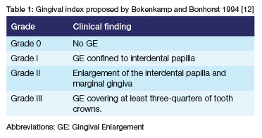
The severity of GE can be determined using an index, of which there are various proposed indices2,6,7 used to assess the severity of GE. The gingival index is important for determining the severity of GE. The use of reliable and reproducible indices for the evaluation of GE is invaluable,1 as they inform treatment. An index for evaluating GE was first described by Kimball in 1939 [8] and later by Harris and Ewalt in 1942.9 In 1972, Angelopoulos and Goaz described a gingival index that measured only the vertical component of GE. Miller et al. then described an index in 1992 that assessed GE in the vertical and horisontal components.10 A variety of other indices have been used to measure and determine the severity of GE, like the photographic GE scoring by Ellis et al.11 The gingival index proposed by Bokenkamp and Bonhorst, 1994 as shown in Table 1,12 which grades enlargement based on the extent of GE in relation to the tooth surface, is widely used. It has the following advantages: it is non-invasive as it is based on visual inspection of the gingival tissues, it can measure GE in the horizontal and vertical components, it does not require many measurements, and is clinically relevant. This index was developed for clinical application and is easy to apply in everyday clinical practice and research settings as it is reproducible.12
The clinical manifestations of GE vary, presenting as red, pink, soft, and shiny gingiva that bleeds easily in inflammatory gingival enlargement; and darker red or purple or pale fibrotic gingiva in non-inflammatory GE.13,14 Inflammatory and non-inflammatory GE can be acute or chronic depending on the causative and predisposing factors. Factors associated with acute forms of GE include: mechanical, chemical, or physical irritation; associated with specific viral, fungal and other bacterial micro-organisms not commonly associated with periodontal disease and mouth breathing.1
Chronic GE is mainly associated with the accumulation of dental plaque-biofilm linked to insufficient oral hygiene practices, dental appliances, including orthodontic appliances or prostheses, and mouth breathing.1,15 Other factors associated with chronic GE include systemic factors encompassing the use of certain systemic drugs, inflammatory and immune conditions, neoplasms, endocranial and nutritional diseases. The drugs associated with GE include; calcium channel blockers (e.g. Nifedipine, Verapamil, Diltiazen, Amlodipine, Felodipine), immunosuppressants (e.g. Cyclosporine), anticonvulsants (e.g. Phenytoin and Sodium Valproate) and high-dose oral contraceptives.16,4 Increased hormonal levels during pregnancy have been shown to lead to more pronounced gingivitis and possible marginal enlargement, depending on the level of plaque control prior to pregnancy.17,18 All factors mentioned promote plaque-induced GE. Thus, plaque-induced GE is the most common form of GE seen clinically, due to the interplay between plaque and several local and systemic factors that modify the plaque-associated disease process.1
Clinical studies suggest that orthodontic treatment (OT) may be associated with a decrease in periodontal health or changes involving periodontal tissues which manifest as GE.6-11 Gingival enlargement is a common finding during OT and is characterised by the formation of true periodontal pockets with clinical attachment loss or pseudo pockets without clinical attachment loss.19 Most of the causative factors of GE during OT are associated with local irritants such as dental plaque biofilm and calculus.20,21 The associated GE seen during OT is considered an inflammatory reaction due to difficulty in performing adequate mechanical plaque control measures, thus inflammatory GE is a more common clinical feature with OT.22
Orthodontic appliance predisposes to increased biofilm establishment and subsequent GE amongst patients undergoing orthodontic treatment which can be linked to insufficient oral hygiene measures.10,11 The duration of fixed OT has been shown to influence GE especially when the treatment time extends to more than 24 months.23 This has been mainly attributed to reduced patients' motivation towards brushing their teeth and maintaining good oral hygiene with lengthy treatment.24,25
The orthodontic appliances that have been shown to inhibit the efficient removal of the dental plaque biofilm include molar bands, brackets, wires, and other orthodontic attachments due to many mechanical retention sites.20,22,26,27 Plaque accumulation associated with orthodontic appliances could result in moderate gingivitis and varying degrees of GE.28 In their study, Almansob and co-workers found the occurrence of GE to be greater in banded molars when compared with bonded molars.23 The GE associated with fixed OT can be alleviated by thorough oral hygiene instructions and practices before and during OT including professional supportive therapy. Patients should also be kept motivated to maintain good oral hygiene and be educated about the negative effects of increased plaque accumulation.
Other factors reported to have a strong influence on the development of GE during OT include age and gender, with males and younger patients reported to present with increased GE compared to females and older patients.23 The development of GE amongst males and younger patients was associated with poor plaque control measures.23,25,27,29
A study by Kasmaei et al.30 also showed that young patients commonly had challenges maintaining good oral hygiene even with regular oral hygiene instructions before and during fixed OT.30 Although an association between plaque accumulation and GE has been shown amongst patients undergoing orthodontic treatment,23,24,31 some studies have found varying results.15,26,27 A study by Zachrisson and Zachrisson did not find a direct correlation between plaque accumulation and GE, suggesting other factors at play during fixed OT.15 In their study, Zachrisson and Zachrisson found that, despite good cleaning and low plaque index, most patients developed generalised moderate GE within one to two months of fixed orthodontic treatment.15
Other studies have shown that GE developed within 1-2 months following placement of fixed orthodontic appliances, even when proper oral hygiene measures were maintained.21,24,25,29 This led to a shift in focus on plaque being the sole risk factor for GE to incorporate other factors, including chemical irritation produced by materials used in orthodontic appliances. During fixed OT, different types of archwires are used depending on the different phases of treatment.32 Titanium Molybdenum Alloy (TMA) archwires have been shown to be most effective and widely used during the finishing stages of fixed OT.33 Nickel, a common alloy in orthodontic appliances, is one of the most commonly implicated material in GE amongst patients undergoing fixed OT.19 It is also a component in stainless steel which is incorporated in brackets and arch wires.34 Nickel is used in the Nickel-Titanium (Ni-Ti) archwires, which are used for fixed OT during the initial leveling and aligning phase. Another study however, found Nickel to influence GE throughout OT, with resultant GE characterized by changes in color, and gingival bleeding upon probing.35 Hence, it has been postulated that Nickel can influence inflammatory reactions leading to GE throughout orthodontic treatment not just in the initial phase of treatment.35,36 The following mechanisms have been considered: the direct interactions between the nickel ions and the gingival tissues resulting in the proliferation of the fibroblasts, leading to GE.36 In addition, Nickel is a strong biological sensitizer associated with two key hypersensitivity reactions that have been described in the literature.37 Type I hypersensitivity reaction which occurs within minutes or hours after direct skin or mucosal contact with Nickel and type IV hypersensitivity reaction which is a delayed-type reaction occurring within hours or days after Nickel exposure.37 The form of GE associated with these reaction does not respond to mechanical dental plaque biofilm removal.19,36
Orthodontic treatment-induced GE shows a specific fibrous and thickened gingival appearance different from fragile gingiva with marginal gingival redness seen in inflammatory gingival lesions not associated with OT.38 There are no clear defined mechanisms involved in OT associated GE initiation and histopathology.38,39 Hence the exact factors leading to GE during orthodontic treatment are not fully understood. This study sought to evaluate the factors associated with GE in patients undergoing fixed OT and also to determine the prevalence of GE in these patients. The results from the study will be valuable in identifying patients at risk for developing GE during fixed OT, enabling better prevention and treatment strategies.
MATERIALS AND METHODS
Study design
This was a cross-sectional descriptive study involving patients undergoing fixed OT from 1 October 2023 to 5 July 2024 at the University of Pretoria Oral Health Centre in the Orthodontic and Periodontic clinics.
Inclusion and Exclusion criteria
Patients undergoing fixed OT at UPOHC, Department of Orthodontics, during the study period were included in the study. The following were excluded from the study: pregnant women and patients taking medications known to cause GE, including calcium channel blockers (e.g., nifedipine, verapamil, diltiazem, amlodipine, felodipine), immunosuppressants (e.g., cyclosporine), anticonvulsants (e.g., phenytoin, sodium valproate), and high-dose oral contraceptives.
Study population and sample size calculation
In the Department of Orthodontics, each registrar is allocated 80 patients for their study program. Based on the combined patient allocation to the three registrars, the initial target sample size was determined to be 240 patients. To account for potential sample loss, the study assumed a 30% loss due to factors such as exclusion criteria, non-attendance, missed appointments, and non-consent to participate in the study.
With an assumed 30% loss, the sample size required to achieve the study's objectives was estimated to be 168 patients.
Data collection
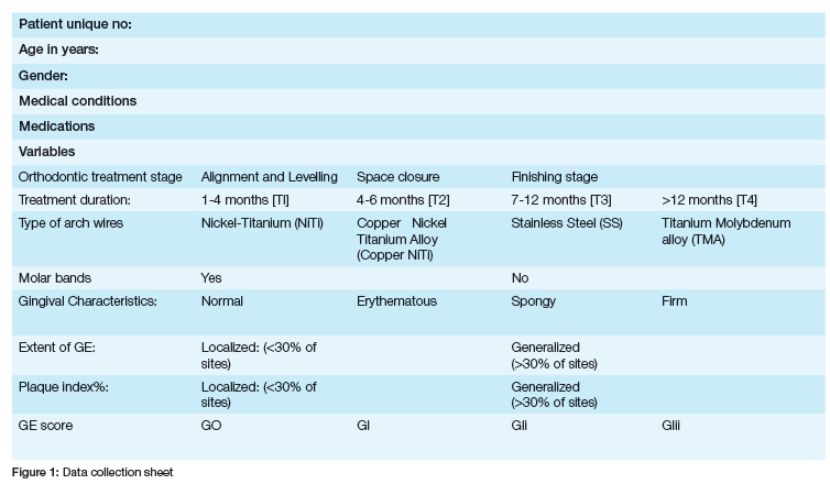
The following data was collected from participants: age, gender, medical history, including medications, type of orthodontic appliances, orthodontic treatment stage, duration, and plaque index were collected as shown in the data collection form Figure 1. In addition, GE was scored and recorded according to an index proposed by Bokenkamp and Bonhorst.12 The extent and distribution of GE were recorded as localised if less than 30% of teeth were involved and as generalised if more than 30% of teeth were involved.3
The gingival characteristics were recorded as either normal, spongy, or erythematous. The presence of dental plaque was assessed using a 2-Tone® disclosing solution and reported using O'Leary's plaque index,40 see Figure 1 above.
Intra-rater and inter-rater reliability:
A single calibrated examiner, Dr Pitso (IR), performed all clinical examinations. Before the examination of the gingiva, calibration was done with Dr Manenzhe (SC). Intra-rater and inter-rater reliability was assessed by repeating measurements of 10% of the patients selected. Intra-rater and inter-rater reliability were analysed using the Intraclass correlation coefficient (ICC).41
Interpretation of the strength of agreement of ICC values was adopted (<0.5 = poor reliability; 0.5-0,75= moderate reliability; 0,75-0,9 = good reliability; >0.9 = excellent reliability).
Inter-rater reliability:
ICC= 0,828, which indicated good reliability and implied consistency in the measurements.
Intra-rater reliability:
ICC value of 0,931 which was an excellent reliability value.
Data Analysis
All data collected was transferred onto a Microsoft Excel spreadsheet. Data analysis was done with SPSS Version 29. Quantitative variables were summarised as proportions frequencies, mean with their standard deviations, range, and percentage. The chi-square and Fisher's exact test were used to evaluate the association between gingival characteristics, the extent of GE, GE score and orthodontic treatment stage, treatment duration, and plaque index. The level of significance was set at p≤0.05.
RESULTS
Study population and prevalence
Out of the estimated representative sample of 168, there was a 59,5% (n=100) response rate. The age groups ranged from 10 to 43 years old, with a mean age of 21.43. Out of the participants, 65(65%) were females and 35 (35%) males. Gingival enlargement was present in 94 (94%) participants, with a mean of 34.7, and 6 (6%) had no GE. Out of the 94% of participants with GE, 32% was found in males and 62% in females.
There were 52 (52%) adolescents and 48 (48%) adults who participated in the study. Out of the 52% of adolescents, 51% presented with GE, whilst 43% out of 48% of adults had GE.
Clinical severity and extent of GE:
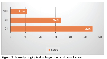
The total number of teeth that presented with GE of varying severity during OT were 825. Figure 2 shows the GE severity scored using the Bokenkamp and Bonhorst index during fixed OT in percentages. The distribution of the different GE grades for the affected teeth was as follows: GI in 453 teeth (55%), GII in 279 (34%), and GIII in 93 teeth (11%).
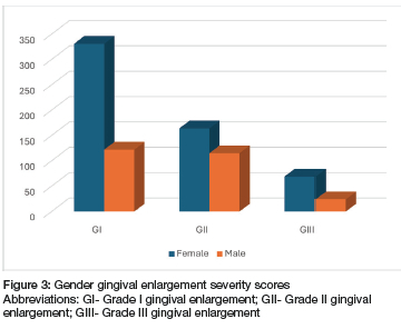
The distribution of the different GE grades for the affected teeth among females and males were as follows: females presented with GI at 40% (331 teeth), GII at 20% (164 teeth), and GIII at 8% (69); and males presented with GI at 15% (122 teeth), GII at 14% (115 teeth), and GIII at 3% (24 teeth), as shown in Figure 3.
The distribution of localised gingival enlargement versus generalised gingival enlargement among the participants revealed a slight predominance of localised gingival enlargement (51%) over generalised GE (49%). It was also observed that participants who have been in treatment for 12 months or more (T4) presented with more generalised GE (57,14 %).
Plaque index
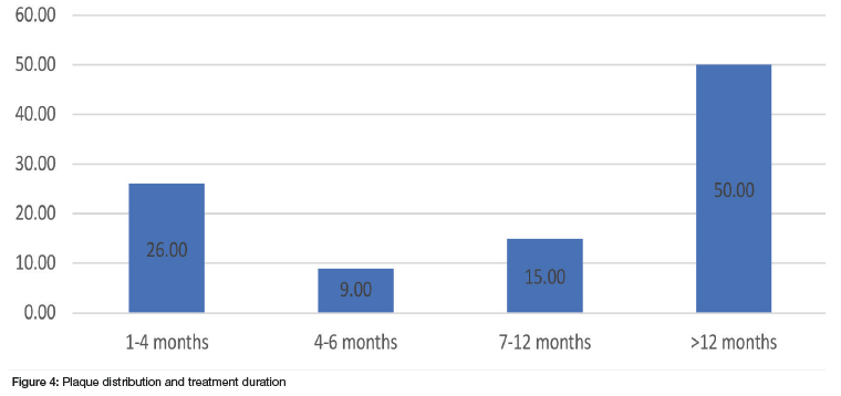
The study found a mean PI percentage of 45.5% . Patients with generalised plaque (83.67%), also exhibited generalised GE as detailed in Table 2. Additionally, adolescents in the study also had a significantly higher generalised plaque distribution (78,8%) compared to their adult counterparts. The participants in the finishing stage and those on treatment for more than 12 months also presented with an increased PI at 53% and 50%, respectively as shown on Figure 4.
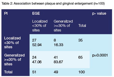
The Pearson chi-squared test was conducted at a level of 5% significance to determine the association between the extent of GE and PI. A significant relationship was found between PI and GE (p=0.0001) as shown in Table 2.
Orthodontic stage:
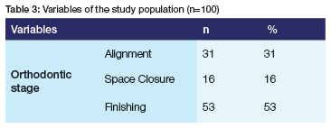
Out of all the fixed OT treatment stages, the majority of participants (53%) were in their finishing stage, 31% in the alignment and levelling stage (31%), and 16% in the space closure stage, as displayed in Table 3. The severity of GE varied across the different treatment stages, as illustrated in Figure 5. All levels of severity were most prevalent in the finishing stage, with 53% of sites presenting with GI, 65% with GII, and 74% with GIII.
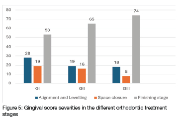
The results, however, showed no statistically significant association between the extent of GE and the different orthodontic stages (p>0.05).
Treatment duration:
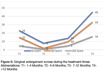
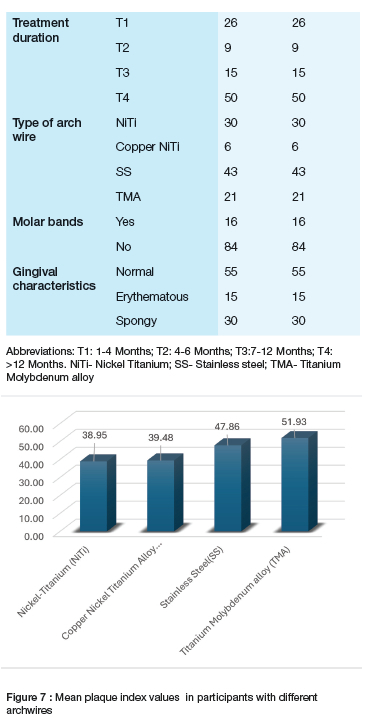
Half (50%) of the participants were at T4 (12 months or more) of fixed OT, with 26% at T1 (1- 4 months), 15% at T3 (7-12 months), and 9% of participants at T2 (4-6 months) treatment duration. The patients who had fixed orthodontic treatment for 12 months or more presented with more generalised GE as illustrated in Figure 6. There was no statistically significant association between the extent of GE and the treatment duration (p>0.05).
Type of archwire:
In terms of the type of archwire material utilised by the participants, it was found that 43% had stainless steel (SS), 30% had NiTi, 21% had TMA, and 6% had Copper NiTi as shown in Table 3 of the summary of frequencies. Among these, those with TMA archwires had the highest mean plaque index (51.93) followed by those who had SS with a mean of (47.86), and mean PI of 39.48 and 38.95 for Copper NiTi and NiTi respectively, as depicted in Figure 7. There was no statistically significant association between the extent of GE and the different types of archwires (p>0.05).
Molar bands:
Participants who had molar bands were 16%, whilst 84% had no molar bands. Within the group of participants with molar bands, a total of 25 teeth were fitted with the molar bands, and only 6% of the sites had GE.
Gingival characteristics:
Majority (55%) of the participants had normal to firm gingival characteristics, while 30% had spongy, and 15 % erythematous gingiva.
DISCUSSION AND CONCLUSION
Discussion
Orthodontic treatment offers patients a functional bite, improved oral health, and a more attractive smile.42 However, the placement of fixed orthodontic appliances frequently leads to some periodontal side effects, such as gingival enlargement (GE), which may conflict with health and aesthetic treatment goals.31 Gingival enlargement in OT is mostly due to challenges in maintaining effective oral hygiene around the brackets, wires, and bands, which provide retention areas for plaque accumulation.43
This study explored the prevalence and factors associated with GE in patients undergoing fixed OT. The study found a high prevalence of GE in the studied population, with a significant association between GE and plaque index. This finding agrees with other previous studies that have linked GE with poor oral hygiene habits and increased plaque accumulation during fixed OT23,24,31 and are in contrast with those of Zachrisson and Zachrisson, who did not find a direct correlation between plaque accumulation and GE,15 suggesting other factors at play other than just plaque. However, in the current study, no statistically significant association between GE and factors studied (age, gender, type of orthodontic appliances, orthodontic treatment stage, and duration) other than plaque were observed.
There was no statistically significant association observed between GE and demographical factors like gender and age. As was the case in other studies where the association between GE, younger and male patients was found to be statistically significant and attributed to insufficient oral hygiene habits.23,25,27,29 Data on the distribution of GE severity grades among females and males however, showed females to have a higher prevalence of GE across all GE severity categories when compared to males as indicated in Figure 3. In addition, adolescents were also found to have a higher prevalence of GE (51%). These results suggest that adolescents are more likely to experience GE during OT in comparison to adults. This corroborates findings from studies that found younger patients to have increased GE during OT.23,25
Treatment duration was also found to influence the presence, extent, and severity of GE, with increased GE severity and more generalised GE found in participants who had been on treatment for more than 12 months. This corroborates the results from other studies where it was shown that when patients are on fixed OT for long, the motivation to maintain optimal oral hygiene diminishes, increasing the risk for more plaque accumulation and, consequently, GE.23-25,43 Treatment fatigue has been cited as an important factor contributing to poor oral hygiene habits and increased plaque accumulation.43 Therefore, it is unsurprising that this study found those patients who were on treatment for longer also presented with more severe and generalised GE. It is, therefore, important that patients understand what the treatment entails, the importance of oral hygiene and the duration of treatment to ensure that when the treatment commences, patients are committed and prepared for the long haul.
Interestingly, participants with TMA archwire presented with higher mean PI (59.3) and GE. This finding was attributed to the fact that TMA was mostly used in the finishing stages of treatment, where motivation for maintaining good oral hygiene may have decreased. The effects of TMA on GE may require further exploration to determine if there are direct effects other than plaque and treatment duration contributing to GE in patients with these archwires. Within our study population, 30% of participants had Nickel Titanium (Ni-Ti) and 6% had Copper Ni-Ti archwires a number higher than participants with TMA 21%. This is significant considering that Nickel has been associated with GE during OT as reported in the literature.35,44 In this study no such associations were found between archwires containing Nickel and GE nor were they found to increase plaque accumulation when compared to TMA and SS. On the contrary, the archwires containing Nickel were associated with lower mean PI. Furthermore, none of the patients with Nickel-containing archwires showed clinical signs of hypersensitivity, such as erythema, erosive or ulcerative lesions, gingival hyperplasia, pain, itching, or burning sensations as reported in the literature.44,45 These symptoms have been documented as potential clinical manifestations of nickel allergy in orthodontic patients. Previous studies have indicated that OT does not directly increase the risk of nickel hypersensitivity unless the patient has a history of nickel allergy.46,47 Hence, it is recommended that clinicians recognise and diagnose potential allergic reactions promptly to facilitate early intervention and minimise patient discomfort.48
Another factor reported to significantly influence the presence of GE during fixed OT is the placement of molar bands.22,26,27 Our findings on plaque retention of molar bands contradict those of other studies. This discrepancy may be primarily due to the fact that only 16% of participants had molar bands. Given the small sample size and limited number of teeth with molar bands, our results should be interpreted with caution and warrant further investigation in future studies.
The GE severity at the tooth level for all 100 participants indicated that the majority of teeth with GE fell under GI, with fewer cases presenting with the more severe GII and GIII. When it came to the extent most participants presented with localised GE. This could explain the majority (55%) of participants presenting with normal to firm gingiva, with the inflammatory reaction limited to the interdental papillae, as seen in GI. Such localised inflammation is expected, given the challenges of effective flossing in patients undergoing fixed orthodontic treatment. Hence, plaque accumulation in this study, remained a significant contributing factor leading to GE during fixed OT. Previous studies have also noted that orthodontic appliances prevent optimal plaque removal, leading to gingival inflammation and GE development during treatment.14,22,23
This finding emphasises the importance of oral hygiene education and regular professional cleaning to mitigate the adverse effects of plaque accumulation associated with orthodontic appliances. Highlighting the importance of maintaining effective oral hygiene before, during, and after fixed OT. It is therefore important to educate patients on what plaque is and its potential impact on gingival health during OT.
Conclusion
Plaque accumulation emerged as the main contributor to GE in this study, highlighting the necessity of optimal oral hygiene maintenance before and throughout fixed OT. Educating patients on the implications of plaque and providing them with practical plaque control techniques, including antimicrobial mouth rinses, interdental brushes, and powered toothbrushes, which have been shown to improve plaque control in orthodontic patients is critical to reducing GE risks associated with orthodontic treatment. Additionally, frequent follow-up and reinforcement of oral hygiene practices can help address the inflammation and prevent GE in patients undergoing fixed OT.
Although the correlation between GE, gender, and age was not found to be statistically significant, the clinical significance cannot be overlooked. As these observations have implications for treatment approaches and prevention strategies for females and adolescents who are considered for orthodontic treatment. The planned supportive oral hygiene care should be tailor-made to aid their oral hygiene practices and should incorporate the use of visual aids to demonstrate the impact of plaque on gingival health. Such intervention before and during OT could aid and assist with patient compliance as treatment duration was also found to influence the presence, extent, and severity of GE. Adolescents and male patients reported to be less compliant with oral hygiene should receive additional education and monitoring to help them maintain good oral hygiene to prevent GE during OT. Female patients on OT with GE require further exploration, and given the high prevalence and increased severity, they should also be given attention. Furthermore, the timing of OT for females and adolescents may be an important determinant of GE initiation and progression, given the impact of hormonal changes on the gingiva in the different life stages.
Study limitations
This study was limited by the cross-sectional study design and causality cannot be inferred. The study was also limited by the small sample, due to issues pertaining to patient availability. The total number of patients assessed for the study was 100 instead of 168. Notwithstanding the limitations, the study has contributed to the literature concerning this important topic.
Recommendations
Given the strong association between plaque accumulation and GE, comprehensive oral hygiene instructions for patients undergoing fixed OT are essential. This should include techniques for thorough cleaning around brackets, wires, and bands, which are common plaque-retention areas. Regular dental cleanings should be scheduled throughout OT to manage plaque levels effectively and prevent GE. Additional cleanings and referral to Periodontics should be considered for patients with extended treatment duration.
Establish a protocol for assessing GE risk before the initiation of OT, factoring in patients' age and gender, medical history, and oral hygiene habits. This protocol could help in providing individualised preventive care and in identifying patients who may need intensified follow-up during OT.
Future studies should prioritse the development and implementation of standardised protocols for capturing pre-treatment photographs to enhance the accuracy and consistency of baseline imaging. Further research should explore salivary biomarkers, inflammatory cytokines, potential periodontal pathogens, and genetic predispositions, and their associations with gingival enlargement during fixed orthodontic treatment.
Author contribution
All the authors contributed to the study conception and design (Dr Itumeleng Pitso (IR), Dr Mpule Moshaoa (MM), Andries Masenge (AM), Dr Shumane Manenzhe (SM) and Prof Thomas K. Madiba (TK). Material preparation and data collection was performed by IR. TK analysed the data. Writing of the original draft manuscript, reviewing and editing of the final manuscript was done by all authors (IR, MM, AM, SM and TK). All the authors read and approved the final manuscript (IR, MM, AM, SM and TK).
Funding
The authors declare that no funds, grants, or other support were received during the preparation of this manuscript.
Declarations
Ethical approval for the study was granted by the University of Pretoria, Faculty of Health Sciences Research Ethics Committee-Ethics Reference number: 510/2023.
This study was performed in line with the principles of the Declaration of Helsinki.
Consent to participate
Informed consent was obtained from all individual participants included in the study. Written informed consent was obtained from the parents.
Consent for publication
The authors affirm that the participants provided informed consent for publication.
Competing interest
The authors declare there are no competing interests.
REFERENCES
1. Kantarci A CF, Hogan E. Gingival Enlargement. In: Newman MG TH, Carranza FA, editor. Newman and Carranza's Clinical Periodontology. Philadelphia,PA: Elsevier; 2019. p. 256-67. [ Links ]
2. Miranda J, Brunet L, Roset P, Farré M, Mendieta C. Reliability of two measurement indices for gingival enlargement. Journal of Periodontal Research. 2012; 47(6):776-82. [ Links ]
3. Caton JG, Armitage G, Berglundh T, Chapple IL, Jepsen S, Kornman KS, et al. A new classification scheme for periodontal and peri-implant diseases and conditions-Introduction and key changes from the 1999 classification. Journal of Periodontology. 2018; 89:S1-S8. [ Links ]
4. Murakami S, Mealey BL, Mariotti A, Chapple IL. Dental plaque-induced gingival conditions. Journal of Clinical Periodontology. 2018; 45:S17-S27. [ Links ]
5. Holmstrup P, Plemons J, Meyle J. Non-plaque-induced gingival diseases. Journal of Clinical Periodontology. 2018; 45:S28-S43. [ Links ]
6. Dubey S, Gattani D, Deotale S, Quazi M. A contemporary review on indices for gingival enlargement. Journal of Advanced Medical and Dental Sciences Research. 2016; 4(5):62. [ Links ]
7. Ahmad AS. Evaluation of index systems measuring gingival overgrowth: Marmara University; 2019. [ Links ]
8. Kimball O. The treatment of Epilepsy with Sodium diphenyl hydantoinate. Journal of the American Medical Association. 1939; 112(13):1244-5. [ Links ]
9. Harris T. Complications following the use of sodium diphenylhydantoinate (Dilantin) therapy. The Journal of the Oklahama State Medical Association. 1942; 35:365-70. [ Links ]
10. Miller CS, Damm DD. Incidence of Verapamil-induced gingival hyperplasia in a dental population. Journal of Periodontology. 1992; 63(5):453-6. [ Links ]
11. Ellis J, Seymour R, Robertson P, Butler T, Thomason J. Photographic scoring of gingival overgrowth. Journal of Clinical Periodontology. 2001; 28(1):81-5. [ Links ]
12. Bökenkamp A, Bohnhorst B, Beier C, Albers N, Offner G, Brodehl J. Nifedipine aggravates cyclosporine A-induced gingival hyperplasia. Pediatric Nephrology. 1994; 8(2):181-5. [ Links ]
13. Srinivasan S, Mahendra J, Anilkumar Kanakamedala A. A comprehensive review of literature on gingival enlargement. Annals of the Romanian Society for Cell Biology. 2020:248-53. [ Links ]
14. Agrawal AA. Gingival enlargements: differential diagnosis and review of literature. World Journal of Clinical Cases. 2015; 3(9):779. [ Links ]
15. Zachrisson S, Zachrisson BU. Gingival condition associated with orthodontic treatment. The Angle Orthodontist. 1972; 42(1):26-34. [ Links ]
16. Moffitt ML, Bencivenni D, Cohen RE. Drug-induced gingival enlargement: an overview. Compendium of Continuing Education in Dentistry. 2013; 34(5):330-6. [ Links ]
17. Kapoor A, Malhotra R, Grover V, Saxena D. Pregnancy associated gingival enlargement. Journal of Oral Health and Community Dentisty. 2010; 4(2):48-51. [ Links ]
18. Buddiga V, Ramagoni NK, Mahantesh H. Gingival enlargement-a case series. Annals and Essences of Dentistry. 2012; 1:73-6. [ Links ]
19. Gorbunkova A, Pagni G, Brizhak A, Farronato G, Rasperini G. Impact of orthodontic treatment on periodontal tissues: a narrative review of multidisciplinary literature. International Journal of Dentisty. 2016; [ Links ]
20. Hosadurga R, Althaf MN, Hegde S, Rajesh KS, Kumar MA. Influence of sex hormone levels on gingival enlargement in adolescent patients undergoing fixed orthodontic therapy: A pilot study. Contemporary Clinical Dentistry. 2016; 7(4):506. [ Links ]
21. Kudirkaite I, Lopatiene K, Zubiene J, Saldunaite K. Age and gender influence on oral hygiene among adolescents with fixed orthodontic appliances. Stomatologija Baltic Dental and Maxillofacial Journal. 2016; 18(2):61-5. [ Links ]
22. Al-Abdaly MMA, Asiri AMA, Al-Abdaly GMM, Ghabri MA, Alqaysi MAH, Aljathnan AMS, et al. Evaluation of the Influence of fixed Orthodontic treatment duration on the severity of inflammatory gingival enlargement (fixed Orthodontic induced gingival enlargements) and some properties of saliva. International Journal of Clinical Medicine. 2022; 13(3):132-46. [ Links ]
23. Almansob Y, Alhammadi M, Luo X, Alhajj M, Zhou L, Almansoub H, et al. Comprehensive evaluation of factors that induce gingival enlargement during orthodontic treatment: A cross-sectional comparative study. Nigerian Journal of Clinical Practice. 2021; 24(11):1649-55. doi:10.4103/njcp.njcp_69_21 [ Links ]
24. Gong Y, Lu J, Ding X. Clinical, microbiologic, and immunologic factors of orthodontic treatment-induced gingival enlargement. American Journal of Orthodontics and Dentofacial Orthopedics. 2011; 140(1):58-64. [ Links ]
25. Zanatta FB, Ardenghi TM, Antoniazzi RP, Pinto TMP, Rösing CK. Association between gingivitis and anterior gingival enlargement in subjects undergoing fixed orthodontic treatment. Dental Press Journal of Orthodontics. 2014; 19:59-66. [ Links ]
26. Erbe C, Hornikel S, Schmidtmann I, Wehrbein H. Quantity and distribution of plaque in orthodontic patients treated with molar bands. Journal of Orofacial Orthopedics. 2011; 72(1):13-20. [ Links ]
27. Kouraki E, Bissada NF, Palomo JM, Ficara AJ. Gingival enlargement and resolution during and after orthodontic treatment. New York State Dental Journal. 2005; 71(4):34. [ Links ]
28. Maudhah AA, Almansoub HA, Mao J. The effects of Orthodontic appliances components on gingival enlargement. International Journal of Dental and Health Sciences. 2019; 06(01):28-38. [ Links ]
29. Khamar PB BS, Dave HB, Yadav TEJ. Recurrent localized chronic gingival enlargement in fixed orthodontic treatment-a novel case report. European Journal of Dental Therapy and Research. 2014; 2(1):192-4. [ Links ]
30. Kasmaei P, Amin Shokravi F, Hidarnia A, Hajizadeh E, Atrkar-Roushan Z, Karimzadeh Shirazi K, et al. Brushing behavior among young adolescents: does perceived severity matter. BMC Public Health. 2014; 14:1-6. [ Links ]
31. Krishnan V, Ambili R, Davidovitch Ze, Murphy NC, editors. Gingiva and orthodontic treatment. Seminars in Orthodontics; 2007: Elsevier. [ Links ]
32. Kusy RP. A review of contemporary archwires: their properties and characteristics. The Angle Orthodontist. 1997; 67(3):197-207. [ Links ]
33. Keim RG, GOTTLIEB EL, NELSON AH, VOGELS III DS. 2002 JCO Study of Orthodontic Diagnosis and Treatment Procedures. Age (years). 2002; 1996:1990. [ Links ]
34. Pinto AS, Alves LS, do Amaral Zenkner JE, Zanatta FB, Maltz M. Gingival enlargement in orthodontic patients: effect of treatment duration. American Journal of Orthodontics and Dentofacial Orthopedics. 2017; 152(4):477-82. [ Links ]
35. Pazzini CA, Júnior GO, Marques LS, Pereira CV, Pereira LJ. Prevalence of nickel allergy and longitudinal evaluation of periodontal abnormalities in orthodontic allergic patients. The Angle Orthodontist. 2009; 79(5):922-7. [ Links ]
36. Bharadwaj P, Gupta A, Harsha NSS, Malik N. A case report on gingivectomy, for the management of Orthodontic treatment induced gingival enlargement. Journal of Orofacial Research. 2021:49-52. [ Links ]
37. Salve RS, Khatri JM. Allergies and its management in orthodontics. International Journal of Applied Dental Sciences. 2022; [ Links ]
38. Simon CP, Bratu DC, Motoc AGM, Pop G, Pop IS, Mederle OA. Immunohistochemical analysis of gingival proliferative processes associated with fixed orthodontic therapy. Revista De Chimie. 2020; 71(2):320-6. [ Links ]
39. Gursoy UK, Sokucu O, Uitto V-J, Aydin A, Demirer S, Toker H, et al. The role of nickel accumulation and epithelial cell proliferation in orthodontic treatment-induced gingival overgrowth. The European Journal of Orthodontics. 2007; 29(6):555-8. [ Links ]
40. Jung Ih, Yeon KH, Song HR, Hwang YS. Cytotoxicity of dental disclosing solution on gingival epithelial cells in vitro. Clinical and Experimental Dental Research. 2020; 6(6):669-76. [ Links ]
41. Koo TK, Li MY. A guideline of selecting and reporting Intraclass Correlation Coefficients for reliability research. Journal of Chiropractic Medicine. 2016; 15(2):155-63. [ Links ]
42. Mahjoub DT, AlJabri RK, Bifari NE, Najjar RS. Oral hygiene awareness and practice in orthodontic patients in Makkah city: A cross sectional study. Journal of Orthodontic Science. 2023; 12(1):32. [ Links ]
43. Rajkanikar J, Vaidya S, Piya A. Influence of Fixed Orthodontic Treatment Duration on the Prevalence and Severity of Gingival Enlargement. Nepal Medical College Journal. 2024; 26(1):5-9. [ Links ]
44. Pazzini CA, Marques LS, Ramos-Jorge ML, Júnior GO, Pereira LJ, Paiva SM. Longitudinal assessment of periodontal status in patients with nickel allergy treated with conventional and nickel-free braces. The Angle Orthodontist. 2012; 82(4):653-7. [ Links ]
45. Noble J, Ahing S, Karaiskos N, Wiltshire W. Nickel allergy and orthodontics, a review and report of two cases. British Dental Journal. 2008; 204(6):297-300. [ Links ]
46. Thyssen JP, Linneberg A, Menné T, Johansen JD. The epidemiology of contact allergy in the general population-prevalence and main findings. Contact Dermatitis. 2007; 57(5):287-99. [ Links ]
47. Hosoki M, Bando E, Asaoka K, Takeuchi H, Nishigawa K. Assessment of allergic hypersensitivity to dental materials. Bio-medical Materials and Engineering. 2009; 19(1):53-61. [ Links ]
48. Di Spirito F, Amato A, Di Palo MP, Ferraro R, Cannatà D, Galdi M, et al. Oral and Extra-Oral Manifestations of Hypersensitivity Reactions in Orthodontics: A Comprehensive Review. Journal of Functional Biomaterials. 2024; 15(7):175. [ Links ]
 Correspondence:
Prof TK Madiba
Address: Oral and Dental Hospital, C/o Steve Biko and Dr. Savage Road, Pretoria, 0001
Tel: +2712 319 2417; Cell: +0845036175; Email: thommy.madiba@up.ac.za or thommy.madiba@gmail.com