



Services on Demand
Journal

Article
 English (pdf)
 Article in xml format
 Article references

 Automatic translation
 Send this article by e-mail
Indicators
Related links
-
 Cited by Google
-
 Similars in Google
Share

 Permalink

South African Dental Journal
On-line version ISSN 0375-1562Print version ISSN 0011-8516
S. Afr. dent. j. vol.80 n.8 Johannesburg Sep. 2025
https://doi.org/10.17159/sadj.v80i07.17552
RESEARCH
Maturation Staging of the Midpalatal Suture using Cone Beam Computed Tomography
R CarimI; S ShaikII; A ShaikhIII
IFaculty of Dentistry, University of the Western Cape, Cape Town, South Africa. ORCID: 0000-0003-4678-8836
IIFaculty of Dentistry, University of the Western Cape, Cape Town, South Africa. ORCID: 0000-0002-4898-3005
IIIFaculty of Dentistry, University of the Western Cape, Cape Town, South Africa and Orthodontist in Private Practice. ORCID: 0000-0002-4898-3005
ABSTRACT
INTRODUCTION: Rapid maxillary expansion (RME) in a young adult is unpredictable because of individual variability in the fusion of the midpalatal suture (MPS) that may be independent of chronologic age.
AIMS AND OBJECTIVES: To determine if the maturation stage of the midpalatal suture can be predicted by using a classification based on its morphology as observed on cone-beam computed tomography (CBCT) images.
DESIGN: A single-centre, retrospective study.
METHODS: 216 CBCT images were analysed; 125 female and 91 males, aged 7 to 78 years. The CBCT images were exported to the OnDemand3DR software program. The most central axial cross-sectional slices were used to evaluate the suture morphology according to Ave maturational stages; namely stages A-E.
RESULTS: There was a statistically significant association between ossification of the MPS and increasing chronological age. The proportion of those aged <12y was higher in stage A/B while the proportion of those aged >=30y was higher in stage E. However, large variations in the degree of closure of the MPS among subjects of the same age group were observed.
CONCLUSIONS: In a patient where age might be a deterrent against the use of conventional RME, CBCT may assist in determining prospectively which patients could benefit from conventional RME.
Keywords: Midpalatal suture, cone beam computed tomography, maxillary expansion, maxillary constriction, transverse dimension, suture synostosis, suture maturation, suture morphology, skeletal maturity indicator.
BACKGROUND
Rapid maxillary expansion (RME) or rapid palatal expansion (RPE) is a commonly used procedure in orthodontics to correct a transverse deficiency of the maxilla and to increase the maxillary arch perimeter in patients who present with mild crowding (crowding of < 5mm) of the dental arches.1 RME has also been advocated in the spontaneous correction of Angle Class II malocclusions2 and to facilitate protraction facemask treatment for the early correction of skeletal Class III malocclusions.3
The concept of maxillary expansion was first introduced by Angell in 1860.4 RME remained controversial for nearly a century until Haas published the results of a study on the rapid expansion of the maxillary dental arch by opening the mid-palatal suture.5 This validated the use of RME in routine orthodontic practice.6
The incidence of maxillary transverse constriction in adolescents is 7.9% and in adults it is 10% .7 The successful use of RME has largely been limited to growing patients and entails the splitting of the midpalatal suture with an appliance that applies high forces of 15-50 newtons to the maxillary and circummaxillary sutures.8 In patients with residual or surplus skeletal growth this treatment results in a combination of dental and orthopaedic expansion. On the contrary, when it is used for the skeletally mature patient, more dento-alveolar tipping as opposed to skeletal expansion with a more unstable outcome occurs. Excessive dental tipping is not desirable as it may lead to periodontal complications like loss of attachment, fenestrations, dehiscences and root resorption.1
An understanding of the growth, development and ossification of the MPS and circummaxillary sutures is essential in predicting the response to RME in a young child, adolescent or young adult. Growth of the nasomaxillary complex follows a cephalo-caudal pattern; hence skeletal maturation involves progressive closure of the midpalatal and circummaxillary sutures. This leads to increased impedance to RME and eventual failure to separate the hemimaxillae.9 During the infantile period, the suture is a broad gap between the maxillary bones and is filled with connective tissue which appears radiolucent on radiographs as it is not mineralised. As maturation and ossification progresses into the juvenile stage, bony spicules begin to form from the margins of the suture, along with "islands" in the middle of the sutural gap. This results in a mixture of non-mineralised connective tissue and calcified bone. During the adolescent period, these bony spicules begin to interdigitate leading to calcification and fusion of the suture.10
Patency of the MPS has been reported in patients aged 27, 54 and 7111,12,24 years respectively. These findings indicate that there is individual variability in the fusion of the midpalatal suture that may be independent of chronologic age. Furthermore, sutural ossification occurs from posterior to anterior and the timing of ossification varies between the genders.11,12
To circumvent the complications associated with conventional RME in skeletally mature patients, miniscrew-assisted rapid palatal expansion (MARPE) may be used to increase orthopaedic changes in the midface especially in post-pubertal patients, and to negate the negative consequences that may occur with conventional RME.13 MARPE is essentially another method of RME that is bone supported as opposed to conventional RME appliances.
The surgical approach is another medium used to expand the maxilla. This method is generally employed in young adults and older patients when the midpalatal suture has already fused or when the maxillary transverse constriction is greater than 5mm. This method entails an intraoperative widening of the maxilla through a multipiece LeFort I osteotomy or a surgically assisted rapid palatal expansion (SARPE).14,15 The midsagittal osteotomy line is favoured for SARPE; the rationale being the considerable ossification that has to be weakened in this region.12 Proffit et al. have reported that surgical expansion of the maxilla is the least stable of all orthognathic procedures.9 Furthermore, SARPE is associated with a large range of adverse surgical complications and morbidity.14
The clinical decision between RME or SARPE to treat maxillary deficiency has traditionally been based on the chronological age of the patient. However, the individual variability in the ossification and fusion of the midpalatal suture has led to a lack of consensus in the literature regarding the age at which transverse maxillary deficiency should be treated surgically14,16 as successful nonsurgical RME has been reported in young adults as well.17,18
In addition to chronological age, other indicators of midpalatal suture maturation (MPSM) that have been proposed include sutural morphology as assessed on occlusal radiographs,19 skeletal maturity indicators (SMI) as appraised on a hand-wrist radiograph (HWR),20 cervical vertebral maturation (CVM) indicators evaluated on lateral cephalograms 21 as well as the five-stage classification of MPSM by Angelieri and colleagues.22
SMI and CVM were developed to predict the timing of facial growth, and not to directly quantify facial skeletal maturity.23
Revelo and Fishman20 demonstrated that there is an association between SMI and midpalatal suture maturation, however, only fifty percent of the total MPS was ossified by the end of puberty.
The occlusal radiograph which is often used for assessing the palatal suture has proven to be unreliable. The limitations encountered includes inadequate visualization of the posterior aspect of the intermaxillary suture because of the superimposition of the vomer and the external structure of the nose on the midpalatal suture. This can lead to misinterpretation of the fusion stage of the midpalatal suture. Furthermore, it is surmised that the midpalatal suture is a straight-running oronasal suture and that the radiographic path projects through this suture. However, the midpalatal suture may not always run in a straight path. If the midpalatal suture is not visible on an occlusal radiograph, it may be because the suture runs in an oblique direction relative to the x-ray path. Therefore a "radiologically closed" midpalatal suture is not analogous to a histologically closed suture.24
To circumvent the limitations associated with conventional radiographs, Angelieri et al.22 described a 5-stage classification system for the midpalatal suture with the use of CBCT images. The authors used the histological classification to define MPSM on CBCT images. These researchers were able to show that the midpalatal suture was not fused in 12% of adult patients.22 Clinically, it is this subgroup of patients who are difficult to identify. The results of the latter study also validated earlier studies that showed no fusion of the mid-palatal suture in subjects of ages 27 years, 32 years, 54 years and even 71 years.11,12,25
Considering the variation observed in MPSM in the literature, the aim of this study was to determine whether the maturation stage of the MPS can be predicted in a South African sample by using a classification based on its morphology as observed on CBCT images.
Research on age-specific characteristics of sutural morphology is an important factor to consider for orthodontic treatment planning in patients with constricted maxillary widths. Should maxillary expansion be needed, the use of conventional RME appliances may not produce the desired skeletal effects after synostosis of the MPS since the resultant effects of RME would largely be dental and unstable. Therefore, a reliable and reproducible tool is needed to determine a more valid diagnostic method for these patients so that an appropriate approach to treatment can be implemented. This will prevent undesired dento-alveolar movement and the associated iatrogenic periodontal problems that may ensue.
AIMS AND OBJECTIVES
The aim of this study was to evaluate MPSM in young children (<12 years), older children (12-<15 years and 15-<18 years), young adults (18-<30 years) and older adults (>=30 years) as observed on CBCT images.
The objectives of this study were to:
1. Classify the maturational stage of the midpalatal suture in young children (<12 years), children (12-<15 years and 15-<18 years), young adults (18-<30 years) and older adults (>=30 years) on CBCT images.
2. Determine the inter and intra-rater reliability (reclassification reliability).
3. Determine the relationship between the maturational stage of the midpalatal suture and chronological age.
4. Determine the relationship between the maturational stage of the midpalatal suture and gender.
MATERIALS AND METHODS
This was a cross-sectional retrospective study based on CBCT images obtained from the archives of the dental faculty, spanning the period March 2011 to December 2018. Permission to conduct the study was obtained from the Biomedical Research Ethics Committee (BM18/2/2).
For the purposes of this study 250 CBCT scans were required. The sample size estimation was based on the key research question to be answered, in this case the determination of the relationship between chronological age, gender and the classification of the MPS which consists of five stages according to Angelieri et al,22
This required the use of multinomial logistic regression with the estimation of five parameters (one for gender, and four for age category). The rule of thumb given by Peduzzi etal.26 states that the smallest outcome category (stage) should have at least the number of cases = 10* the number of parameters to be estimated, i.e. 10*5 parameters = 50. Thus, a minimum sample size of 50*5 stages = 250 was required. To ensure adequate representation of all age groups and both genders in the sample, the total sample was divided equally into each age group-gender combination; i.e. 25 scans per group as follows: young children from below the age of 12, young children aged 12 to 15 years, teenagers from age 15 to age 18, younger adults from 18 to 30 years of age and older adults (> 30 years of age). The stratification of the sample population was based on previous studies of MPSM.6,10-12 After excluding patients with prior expansion appliances, orthognathic surgery and craniofacial syndromes including cleft lip and palate, the final sample size was 216 CBCT scans. Every CBCT volume was assigned a random numerical identifier.
A Newtom® (VGI®, Verona, Italy) CBCT scanner was used to obtain full field-of-view scans (15 x 15 cm). The images were saved in DICOM and transferred to the OnDemand3D® software package (Cybermed Inc, South Korea).
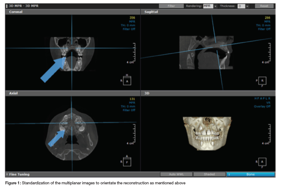
Images for sutural evaluation were obtained in a standardized way. Slice thickness was set at 0.3mm for evaluation and 1mm for identification. The image analysis software cursor was positioned as follows (Figure 1):
1. The head was oriented in the natural head position in all three planes of space. In the sagittal view, the patient's head was adjusted so that the anteroposterior long axis of the palate was horizontal. The vertical and horizontal cursors were positioned in the centre of the palate in the coronal and axial views.
2. Sagittal plane - the horizontal axis of the cursor passed through the centre of the supero-inferior dimension of the hard palate, with the line corresponding to ANS and PNS or a parallel to ANS and PNS.
3. Coronal plane - the cursor was positioned over the midpalatal suture and nasal spine.
4. Axial plane - the vertical axis passed through the anterior and posterior nasal spine.
The most central axial cross-sectional slices were used for sutural assessment. The slice thickness was set at 1mm to get a 1:1 ratio (Figures 2-4).
For subjects who exhibited a curved palate, two sagittal slices were used. In the first sagittal slice the horizontal axis of the cursor passed through the suture in the most posterior region, and in the second slice, the cursor passed through the most anterior region. The posterior and anterior region of the midpalatal suture were then identified separately but both were used to evaluate the maturation stage.
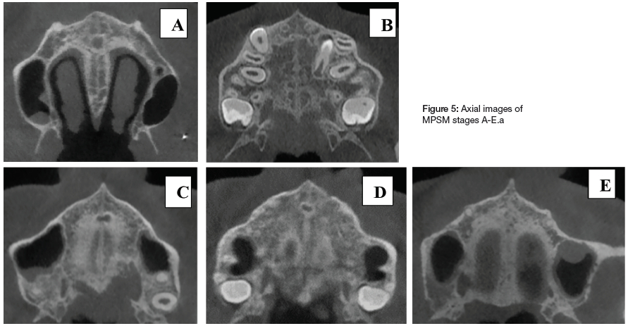
Prior to staging of suture maturation, the two examiners, one an expert in radiology and one a registrar in orthodontics, were calibrated. The maturational stages of the MPS were classified according to the morphologic parameters established by Angelieri and colleagues (Figure 5).22
1. Stage A: The MPS is characterized by one relatively straight high-density midpalatal suture line with little or no interdigitation.
2. Stage B is observed as one scalloped, high-density line at the midline, or by a scalloped high-density line in some areas and, in other areas, as 2 parallel, scalloped, high-density lines close to each other and separated by small low-density spaces.
3. Stage C is visualized as two parallel, scalloped, high-density lines that are close to each other, separated in some areas by small low-density spaces. The suture can be arranged in either a straight or an irregular pattern.
4. Stage D is visualized as two scalloped, high-density lines at the midline on the maxillary portion of the palate, the MPS cannot be identified in palatine bone.
5. Stage E, sutural fusion has occurred in the maxilla. The MPS cannot be identified.
After the calibration process, staging of suture maturation on the CBCT images was performed by both examiners. Each examiner arrived at a diagnosis separately. The diagnoses were then compared to assess the level of concordance. If there was disagreement regarding the suture stage, this was followed by a robust discussion until consensus was reached. The consensus diagnosis formed the "ground truth" for this thesis. To assess the reliability of classifying the maturational stages of the midpalatal suture (stages A-E), staging and reclassification of the midpalatal suture was repeated by both examiners who randomly assessed and reclassified every tenth CBCT scan after a two-week period. A total of twenty-two images were reclassified.
STATISTICAL ANALYSIS
Categorical variables (age group, gender and stage) were summarised by frequency and percentage tabulation. Continuous variables (age and gender) were summarised by the mean, standard deviation, median and interquartile range. The association between age group and gender, gender and stage and age group and stage, was determined by the chi-squared test. The relationship between age, gender and stage was determined by ordinal logistic regression with maturation stage as the dependent variable and age group and gender as the independent variables.
Interrater and reclassification reliability (intra-rater) were quantified by Cohen's weighted kappa and the Stuart-Maxwell test for bias.27 The magnitude of the kappa was interpreted according to the classification by Landis and Koch.28 Data analysis was carried out in SAS. The 5% significance level was used for all statistical tests.
RESULTS
The intra- rater reliability was 0.94 (95% confidence interval/CI 0.91-0.97) and the inter-rater reliability (reclassification) was equal to 0.92 (95% CI 0.83-1.00). There was no significant intraobserver bias (p>0.99). Thus, the staging categorisation was reliable.
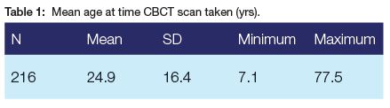
Based on the research design, 250 CBCT scans were required. This sample size would have ensured an equal representation of all age groups and both genders in the sample. However, we were only able to obtain 216 CBCT studies. Overall, the mean age at the time the CBCT images were taken was 24.9 years with a standard deviation (SD) of 16.5 years and an age range of 7.1-77.5 years (Table 1).
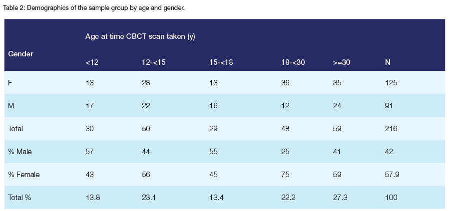
From the available images, females were overrepresented in the sample and accounted for 57.9% of the final CBCT images. The <12year and the 15-<18year groups were under-represented, while the oldest age group (>=30) was over-represented (Table 2). Furthermore, males were under-represented in the 18-<30year age group (p=0.033).
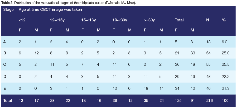
MPSM stages B to E were approximately equally represented in the sample, while the prevalence of stage A was very low (Table 3).
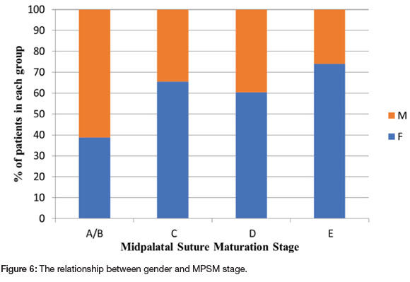
Stage A was too small (n=13) to be used as an outcome group on its own and was combined with stage B. There was a statistically significant association between gender and stage (p=0.0010, Cramer's V=0.27; small effect size). The proportion of males in stages A/B was higher than in the other stages, while the proportion of females in stage Ε was higher than in the other stages. Overall, a higher proportion of females displayed the later stages of maturation when compared to males (Figure 6).
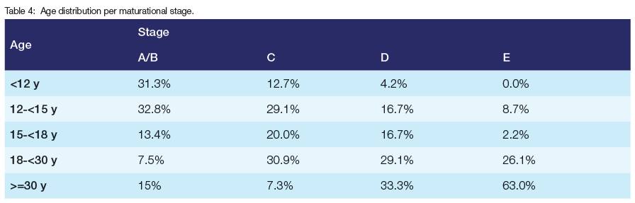
The association between age group and MPSM stage was significant (p<0.0001; Cramer's V=0.36; moderate effect size); however, great variability was frequently noted. The proportion of those aged <12y was higher in stage A/B, however, stage D was observed in 4.2% of children (2/48) in this age group.
A high prevalence of stage E (63%) was observed in older adults (>=30 yrs). In addition, 14.9% of older adults (>= 30 yrs) had no fused suture (Table 4).
6. DISCUSSION
In the present study, the morphology of the MPS as assessed on CBCT images was correlated to chronological age and gender to ascertain a relationship between age, gender and MPSM stage.
The CBCT images were staged according to the classification proposed by Angelieri et al.22 The classification is based on the evaluation of the most central cross-sectional axial slice. However, should this slice not be accurately located, the MPSM stage may be misconstrued on the CBCT image. Therefore, in our study, the slice thickness was set to 1mm to allow for a 1:1 ratio. The central most cross-sectional slice was determined by identifying the slice number corresponding to the most superior aspect of the palate; the slice number corresponding to the most inferior aspect of the palate and then subtracting the two. The median of this number was selected as the most central cross-sectional slice.
For this study to be reliable and reproducible there had to be agreement between the examiners. The examiners selected the consensus method as the "ground truth" to minimize the probability of diagnostic errors. When evaluating radiographs, the term "ground truth" is more appropriate than "gold standard" as a gold standard will necessitate a histological specimen of the midpalatal suture as a frame of reference. 22
The drawbacks that were experienced with the classification proposed by Angelieri and colleagues were differentiating stage A from stage B where stage A is defined as "relatively straight high density line".22 Relative is a broad term and is subject to opinion. Stage A should rather have been defined in terms of percentage of straight versus scalloped. Stage A and B infer early suture development, prior to suture ossification. The transition from stage A to stage B will not necessitate a change in the treatment plan or prognosis. In both stage A and B, the suture is patent and conventional RME is possible. Therefore, disagreements between examiners regarding the staging between stage A and stage B is not critical.
Furthermore, the demarcation between stage D and stage E was also sometimes obscure. Identification of sutural stage D or E is suggestive of ossification of the MPS.
This study showed that the association between gender and stage was significant; it was observed that the proportion of males in stages A/B was higher than in the other stages whereas MPSM stages C, D and E displayed a greater prevalence in females. This inferred that maturation between the genders differed, with females maturing earlier than males. These observations are in concordance with previous studies on MPSM. 22,29,30 In a sample aged 11 to 15 years stage C was observed in 56.8% of females and 42.5% of males.29 Similarly, de Miranda Ladewig et al. evaluated CBCT images of MPSM in subjects aged 16 to 20 years and reported that a greater percentage of females displayed the late maturational stages of D and E (53.0%), when compared with the males (38.6%).30
The findings of this study validate previous reports that chronological age is not analogous with MPSM. Ossification of the MPS increased with chronological age; the proportion of those aged <12y was higher in stage A/B when the MPS is patent compared to the other stages while the proportion of those aged >=30y was higher in stage E (ossification has occurred in both the maxillary and palatine portions of the MPS). However, large variations in ossification staging of the MPS among subjects of the same age group were observed (Table 4). The results showed that stage A/B was observed In 14.9% of the >=30 yr age groups. This is an important finding, as a fairly sizable proportion of older patients presented with patent sutures at an age where conventional RME is deemed unsuccessful. An observation that was also mirrored by Angelieri et al.22 was the presence of stage D (fusion of the palatine area of the MPS) in 4.2% of subjects <12 years of age.
Stages A and B are characterized by a patent MPS, therefore RME carried out during this stage will have greater skeletal effects than dental effects. Stage C is visualized as two parallel, scalloped, high-density lines that are close to each other, separated in some areas by small low-density spaces. During stage C there is increased interdigitation of bone resulting in greater resistance to separation. Despite the increased sutural resistance encountered during stage C, RME carried out at this stage is still possible. However, RME should be initiated immediately because of the impending start of fusion of the palatine portion of the MPS.31
The MPS fuses from posterior to anterior 11,12 Therefore, the posterior region of the MPS will show an increased resistance to expansion. Stage D is characterized by fusion of the palatine bone and stage E is characterised by fusion of the entire MPS (palatine and maxillary areas). These patients are more suitably treated with SARPE or MARPE.31 However, when MPSM was used as a predictor for MARPE success in patients aged 15 years to 37 years, it was found that stages A and B showed successful MPS opening while limited MPS opening was observed in subjects in stages D and E. However, failure of MPS opening appeared in only individuals with MPS stage E.32 Therefore, the success of MARPE in stages D and E is variable. Clinicians should be cognisant that the greater the area of MPS patency, the greater the potential success of orthopaedic expansion.
Sutural morphology may be a reflection of the loading conditions under which the suture is subjected to.33 It has been postulated that the age at which sutural synostosis occurs is directly determined by extrinsic functional demands affecting that specific suture area.34 If a suture functions to reduce stresses transmitted throughout the facial skeleton, the bone at the sutural margins should show more deflection than the surrounding bone.35 The complexity of sutural interdigitations is proportional to the magnitude of the functional load. Sutures become more interdigitated in areas of high stress to prevent the bones from becoming disarticulated.36
Studies have ascribed the patent suture in adults to a decrease in the functional forces from the muscles of mastication. This may be due to the natural processes of aging with the resultant loss of teeth or to the refined, softer diet of western civilizations. Therefore, the maturation and ossification of the midpalatal suture depends on functional forces exerted on the maxillary bone.37 As a result, chronological age alone is not the sole determinant of the developmental stage of the suture. Consideration should therefore be given to the reciprocal effects from functional forces on suture maturation.
The assessment of the MPS on the CBCT may be a valid tool to aid in the clinical decision between conventional RME, MARPE and SARPE in young adult patients in whom the prognosis of conventional RME is doubtful.22,28,29 It has been recommended that a report should be requested from a radiologist as MPS assessment showed "potential reliability and reproducibility" but extensive training is required to increase reliability and reproducibility.38
LIMITATIONS
There are several limitations to this study:
It was a single centre study; therefore, the ideal sample size was not reached. According to the research design there should have been a 1:1 ratio of male:female patients. After applying the inclusion and exclusion criteria the actual sample size was 216. No patient was exposed to radiation for the purpose of this research. CBCT images were obtained from the archives of the University spanning the period March 2011 to December 2018.
Secondly there was no control group. The control group; namely autopsy specimens should have been the "gold standard".
With respect to the resistance that clinicians encounter when transverse force is applied to the MPS, the maturation and ossification of the circummaxillary sutures are as important as the maturation and interdigitation of the midpalatal suture in decreasing the skeletal response to RME.39 Only the morphology and maturation of the midpalatal suture was considered in our study.
CONCLUSION
The association between gender and suture stage was significant. The proportion of males in stages A/B was higher than in the other stages, while the proportion of females in stage E was higher than in the other stages. Overall, a higher proportion of females displayed the later stages of maturation when compared to males.
In a patient where age may be a potential limiting factor for the use of conventional RME as a treatment option, the classification proposed by Angelieri et al. (2013) may assist in determining prospectively which late adolescent or young adult patient could benefit from such an intervention.
REFERENCES
1. Bishara, S.E. and Staley, R.N. Maxillary expansion: clinical implications. American Journal of Orthodontics and Dentofacial Orthopedics. 1987;91(1):3-14. [ Links ]
2. Guest, S.S., McNamara Jr, J.A., Baccetti, T. and Franchi, L., 2010. Improving Class II malocclusion as a side-effect of rapid maxillary expansion: a prospective clinical study. American Journal of Orthodontics and Dentofacial Orthopedics, 738(5):582-591. [ Links ]
3. de Silva Fo, O.G., Boas, C.V. and Capelozza, L.F., 1991. Rapid maxillary expansion in the primary and mixed dentitions: a cephalometric evaluation. Amercan Journal of Orthodontics and Dentofacial Orthopedics, 100(2):171-179. [ Links ]
4. Angell, D.H., 1860. Treatment of irregularity of the permanent or adult teeth. Dental Cosmos,1:540-544. [ Links ]
5. Haas, A.J., 1961. Rapid expansion of the maxillary dental arch and nasal cavity by opening the midpalatal suture. The Angle Orthodontist, 37(2):73-90. [ Links ]
6. Timms, D.J., 1999. The Dawn of rapid maxillary expansion. The Angle Orthodontist, 69(3): 247-250. [ Links ]
7. Harrison JE, Ashby D. Orthodontic treatment for posterior crossbites. Cochrane Database Syst Rev. 2000;12(2):CD000979. [ Links ]
8. Lagravere, M.O., Major, P.W. and Flores-Mir, C., 2005. Long-term dental arch changes after rapid maxillary expansion treatment: a systematic review. The Angle Orthodontist, 75(2):155-161. [ Links ]
9. Proffit, W.R., Sarver, D.M. and Ackerman, J.L., 2013. Contemporary Orthodontics. 5th ed. St Louis: Mosby. [ Links ]
10. Melsen B. Palatal growth studied on human autopsy material: a histologic microradiographic study. American Journal of Orthodontics. 1975; 68(1):42-54. [ Links ]
11. Persson, M. and Thilander, B. Palatal suture closure in man from 15 to 35 years of age. American Journal of Orthodontics. 1977; 72(1):42-52. [ Links ]
12. Knaup, B., Yildizhan, F. and Wehrbein, H. Age-related changes in the midpalatal suture. Journal of Orofacial Orthopedics/Fortschritte der Kieferorthopädie. 2004;65(6):467- 474. [ Links ]
13. Cantarella, D., Dominguez-Mompell, R., Moschik, C., Sfogliano, L., Elkenawy, I., Pan, H.C., Mallya, S.M. and Moon, W., 2018. Zygomaticomaxillary modifications in the horizontal plane induced by micro-implant-supported skeletal expander, analyzed with CBCT images. Progress in orthodontics, 19(1):41. [ Links ]
14. Suri, L. and Taneja, P., 2008. Surgically assisted rapid palatal expansion: a literature review. American Journal of Orthodontics and Dentofacial Orthopedics, 733(2): 290-302. [ Links ]
15. Yao, W., Bekmezian, S., Hardy, D., Kushner, H.W., Miller, A.J., Huang, J.C. and Lee, J.S., 2015. Cone-beam computed tomographic comparison of surgically assisted rapid palatal expansion and multipiece Le Fort I osteotomy. Journal of Oral and Maxillofacial Surgery, 73(3): 499-508. [ Links ]
16. Wertz, R. and Dreskin, M., 1977. Midpalatal suture opening: a normative study. American Journal of Orthodontics, 71(4): 367-381. [ Links ]
17. Handelman, C.S., Wang, L., BeGole, E.A. and Haas, A.J., 2000. Nonsurgical rapid maxillary expansion in adults: report on 47 cases using the Haas expander. The Angle Orthodontist, 70(2):129-144. [ Links ]
18. Stuart, D.A. and Wiltshire, W.A., 2003. Rapid palatal expansion in the young adult : time for a paradigm shift ? Journal Canadian Dental Association, 69(6): 374-377. [ Links ]
19. Lehman, J.J., Haas, A.J. and Haas, D.G. Surgical orthodontic correction of transverse maxillary deficiency: a simplified approach. Plastic and reconstructive surgery. 1984;73(1): 62-68. [ Links ]
20. Revelo, B. and Fishman, L.S. Maturational evaluation of ossification of the midpalatal suture. American Journal of Orthodontics and Dentofacial Orthopedics. 1994; 105(3):288-292. [ Links ]
21. Baccetti, T., Franchi, L., Cameron, C.G., McNamara, J.A. Treatment timing for rapid maxillary expansion. The Angle Orthodontist. 2001; 71:343-350. [ Links ]
22. Angelieri, F., Cevidanes, L.H., Franchi, L., Gonçalves, J.R., Benavides, E. and McNamara Jr, J.A. Midpalatal suture maturation: classification method for individual assessment before rapid maxillary expansion. American Journal of Orthodontics and Dentofacial Orthopedics.2013; 144(5):759-769. [ Links ]
23. Baccetti, T., Franchi L., McNamara Jr, J.A., 2002. An improved version of the cervical vertebral maturation (CVM) method for the assessment of mandibular growth. The Angle Orthodontist, 72:316-323. [ Links ]
24. Wehrbein, H. and Yildizhan, F., 2001. The mid-palatal suture in young adults. A radiological-histological investigation. The European Journal of Orthodontics, 23(2): 105-114. [ Links ]
25. Korbmacher, H., Schilling, A., Püschel, K., Amling, M. and Kahl-Nieke, B. Agedependent three-dimensional microcomputed tomography analysis of the human midpalatal suture. Journal of Orofacial Orthopedics/Fortschritte der Kieferorthopädie. 2007; 68(5):364-376. [ Links ]
26. Peduzzi, P., Concato, J., Kemper, E., Holford, T.R. and Feinstein, A.R., 1996. A simulation study of the number of events per variable in logistic regression analysis. Journal of clinical epidemiology, 49(12):1373-1379. [ Links ]
27. Shoukri, M. M. Measures of Interobserver Agreement and Reliability.2nd edition. Boca Raton, FL: Chapman & Hall/CRC Press;2010. [ Links ]
28. Landis, J.R. and Koch, G.G. The measurement of observer agreement for categorical data. Biometrics.1977; 33 (1): 159-17. [ Links ]
29. Tonello, D.L., de Miranda Ladewig, V., Guedes, F.P., Conti, A.C.D.C.F., Almeida- Pedrin, R.R. and Capelozza-Filho, L. Midpalatal suture maturation in 11-to 15-yearolds: A cone-beam computed tomographic study. American Journal of Orthodontics and Dentofacial Orthopedics. 2017;152(1): 42-48. [ Links ]
30. de Miranda Ladewig, V., Capelozza-Filho, L., Almeida-Pedrin, R.R., Guedes, F.P., de Almeida Cardoso, M. and Conti, A.C.D.C.F. Tomographic evaluation of the maturation stage of the midpalatal suture in postadolescents. American Journal of Orthodontics and Dentofacial Orthopedics. 2018;153(6):818-824. [ Links ]
31. Angelieri F, Cevidanes LH, Franchi L, McNamara Jr JA. Evaluation of facial suture maturation on CBCTs: a predictor of maxillary orthopedic treatment response. In: Kapila S, Nervina J, Hatch N. Expedited Orthodontics: Improving the efficiency of orthodontic treatment through novel technologies. Ann Arbor: University of Michigan. Craniofacial Growth Series 2015; 51: 257-80. [ Links ]
32. Oliveira, C.B., Ayub, P., Angelieri, F., Murata, W.H., Suzuki, S.S., Ravelli, D.B. and Santos-Pinto, A., 2021. Evaluation of factors related to the success of miniscrew- assisted rapid palatal expansion. The Angle Orthodontist, 91(2), pp.187-194. [ Links ]
33. Mao, J.J. Mechanobiology of Craniofacial Sutures. Journal of Dental Research.2002; 81(12):810-816. [ Links ]
34. Kokich, V.G. Age changes in the human frontoxygomatic suture from 20 to 95 years. American Journal of Orthodontics.1976; 69(4): 411-30. [ Links ]
35. Herring, S.W. and Teng, S. Strain in the braincase and its sutures during function. American Journal of Physical Anthropology.2000;112(4):575-593. [ Links ]
36. Herring, S.W. Formation of the vertebrate face epigenetic and functional influences. American Zoologist.1993; 33(4):472-483. [ Links ]
37. Katsaros, C., Zissis, A., Bresin, A. and Kiliaridis, S. Functional influence on sutural bone apposition in the growing rat. American Journal of Orthodontics and Dentofacial Orthopedics. 2006;129(3):352-357. [ Links ]
38. Barbosa, N.M.V., Castro, A.C.D., Conti, F., Capelozza-Filho, L., Almeida-Pedrin, R.R.D. and Cardoso, M.D.A., 2019. Reliability and reproducibility of the method of assessment of midpalatal suture maturation: A tomographic study. The Angle Orthodontist, 89(1), pp.71-77. [ Links ]
39. Zimring, J. and Isaacson, R.J., 1965. Forces Produced by Rapid Maxillary Expansion. The Angle Orthodontist, 35(3): 178-186. [ Links ]
 Correspondence:
Name: Dr Ridwaana Carim
Email address: Ridwaana.carim@gmail.com
Funding
The research was self-funded.
Conflict of Interest
No conflict of interest to declare.
Acknowledgement
The authors wish to acknowledge Dr P.Gaylard of Data Management and Statistical Analysis (DMSA) for her help with the statistical analysis.