



Servicios Personalizados
Revista

Articulo
 Inglés (pdf)
 Articulo en XML
 Referencias del artículo

 Traducción automática
 Enviar articulo por email
Indicadores
Links relacionados
-
 Citado por Google
-
 Similares en Google
Compartir

 Permalink

South African Dental Journal
versión On-line ISSN 0375-1562versión impresa ISSN 0011-8516
S. Afr. dent. j. vol.80 no.2 Johannesburg mar. 2025
https://doi.org/10.17159/sadj.v80i02.20656
CASE SERIES
Dentine thicknesses of first molar roots: A review of the literature with illustrative cases
M MeyerI; AC OettleII; CH JonkerIII; S Rajbaran-SinghIV
IAnatomy and Histology Department, School of Medicine, Sefako Makgatho Health Sciences University, Pretoria, 0204, South Africa. Maxillofacial and Oral Radiology Department, School of Dentistry, Sefako Makgatho Health Sciences University, Pretoria, 0202, South Africa. Email: marisca.meyer@smu.ac.za Orchid number: https://orcid.org/0000-0002-3519-5018
IIMBChB, DTE, MSc, PhD Anatomy and Histology Department, School of Medicine, Sefako Makgatho Health Sciences University, Pretoria, 0204, South Africa. Email: profoettle@gmail.com Orchid number: https://orcid.org/0000-0002-9389-057X
IIIFaculty of Health, Peninsula Dental School, University of Plymouth Ground, Truro Dental Education Facility, Knowledge Spa, Royal Cornwall Hospital, Truro, TR1 3HD, United Kingdom Email: casper.jonker@plymouth.ac.uk. https://orcid.org/0000-0002-9110-5208
IVMaxillofacial and Oral Radiology Department, School of Dentistry, Sefako Makgatho Health Sciences University, Pretoria, 0202, South Africa. Email: sandeepa.singh@smu.ac.za. Orchid number: https://orcid.org/0000-0003-0658-2542
ABSTRACT
Thin dentinal walls of first molars can significantly impact the success and outcomes of endodontic procedures, including root canal treatment. Root canal treatment is commonly performed on first molars, which are prone to decay due to their early eruption and complex occlusal structure. The treatment entails a series of mechanical and chemical disinfection techniques where the root canal spaces are enlarged and can be compromised by perforations leading to infection, inflammation and eventual tooth loss. The thinner the dentine, the greater the likelihood of perforation. "Danger zones" have been identified as the distal aspect of mesial/mesiobuccal roots and the mesial aspect of distal roots. Knowledge of dentine thickness patterns in dental roots and understanding factors that influence dentine thickness can help reduce the risk of perforation and improve treatment outcomes. This literature review gives an overview of various aspects of the dentine thickness of the maxillary and mandibular first permanent molar. The content is supported by illustrative micro-CT images and clinical cases. Treating physicians should take note that variations in dentine thicknesses have been reported between sexes, with ageing and among populations, but unfortunately no studies on dentine thickness could be found specifically for the South African context.
Keywords: Danger zone, safety zone, iatrogenic perforation, tertiary dentine, mesial root, distal root, mesiobuccal root, mandibular first molar, maxillary first molar, root length.
INTRODUCTION
The thickness of dentine is of particular importance in root canal treatment. Root canal treatments are very commonly done on first molar teeth as these teeth are the first to erupt and be exposed to the oral environment. In addition, first molars also have a complex occlusal plane with numerous Assures and pits causing a potential risk for carious lesions.1,2
The success of root canal treatment relies on various factors, including perforations which involve pathological communications between the root canal system and the surrounding periodontium.3 Perforations can occur during access preparation as well as during cleaning and shaping in some areas and, in particular, the "danger zone" due to thin dentine. As it is very difficult for a treating clinician to accurately predict the estimated dentine thickness of the tooth before treatment commences4-6, about 10% of root canal treatment failures are caused by perforation alone.3 Perforations may lead to adverse consequences such as peri-radicular inflammation, endodontic re-infection, destruction of periodontal fibres, bone resorption, the development of a periodontal defect or eventual tooth loss.7,8 especially when bacterial infection is allowed to establish. Perforations may occur due to pathological processes or treatment consequences. Various dental materials have been proposed over the years for perforation repair with varying degrees of success. The use of bioactive materials, such as mineral trioxide aggregate (MTA It is therefore not surprising that the estimated pooled success rates of primary root canal treatment with a minimum one-year follow-up ranged from only 68% to 85%, according to a study executed by Ng et al (2011).9
The aim of this paper is to provide an overview of the available literature on the dentine thicknesses supported by illustrative clinical cases involving root canal treatment. Knowledge of the variations and patterns of dentine thickness of the molar teeth roots may lower the risk for perforation which ultimately improves the success of the treatment.
Pivotal anatomical aspects
Despite the variations in both functional roles and morphological features of human dentition all teeth share a common anatomical blueprint.10 The following structures all play an important role in tooth health and damage to any of these structures may increase the probability of needing endodontic treatment. Positioned superior to the gingiva, the tooth displays a visible portion known as the crown which is encased externally by a layer of enamel and internally by dentine.10 The root(s) extend/s beyond the gingiva into their sockets in the alveolar bone of either the mandible or the maxilla and is securely held in place by a layer of cementum.11 The enamel layer of the crown is continuous with the cementum which extends down into the roots. The cemento-enamel junction (CEJ) serves as a continuous junction between the crown and root(s) and is situated on the same plane as the gingiva. The dentine, on the other hand, extends beyond the crown, traversing into the root, and encapsulates a central cavity, namely the pulp chamber.10 This chamber houses an intricate network of blood vessels, nerves and other organic matter, collectively known as the dental pulp, which is essential for preserving the tooth's vitality.10

Endodontic treatment
Where the pulpal tissue is irreversibly inflamed due to, for example, trauma or carious exposure, the need for endodontic treatment arises which entails a series of mechanical and chemical disinfection techniques.12,13
During the initial stage of treatment, a series of dedicated burs or root canal instruments are used to create access through the crown area of the tooth into the pulp chamber to expose the orifices of the root canals and refine the access.14
Traditionally, the root canal space is enlarged through a series of manual stainless-steel instruments.15 Throughout the cleaning and shaping process of endodontic treatment, a sequence of root canal instruments is introduced at the orifice level and worked apically until the working length is reached. The diameter and size of traditional root canal instruments increase with progression through the traditional cleaning and shaping sequence and a variable amount of tooth structure is removed with each instrument.
In modern times, operators have mainly been using rotary instruments due to their benefits which include reduced clinical chair time and more predictable outcomes.16 This rotary instrument is usually made of nickel-titanium and eases the process by allowing the canal to be prepared more accurately and evenly. Ultimately, the prepared root canal spaces are filled with an obturation material (gutta percha). Cuspal coverage (for example a crown or onlay) is also advocated as the final restoration to ensure a proper coronal seal and preservation of the tooth after treatment.17 Figure 2 exhibits a number of teeth restored with indirect restorations (crowns): the right mandibular second premolar and first molar as well as the right maxillary first and second molars.
Following root canal treatment, in preparation for the placement of a crown, a metal band called a dental ferrule may be positioned around the coronal surface of the tooth to support the crown up to 2mm supra-gingivally.18 However, in certain cases the remaining tooth structure might not be sufficient to support a crown despite the use of an adequate dental ferrule. In these cases, a prosthetic device in the form of a small rod called a post is fitted and cemented inside the root to accommodate a core to serve as a base for a crown.17 An indirect cast post and core can be cemented prior to crown placement or, alternatively, a suitable core with or without a post (depending on the amount of remaining tooth structure) can be created chairside to facilitate crown placement.19 A post and core can also be manufactured outside the mouth by a laboratory (indirect method). Figure 3A depicts a virtual illustration using cone beam computed tomography (CBCT) of a post and core placed in a right mandibular first molar (Fig. 3A). Figure 3B illustrates an indirect cast post and core with a covering crown that was placed after root canal treatment had been completed on the right maxillary second molar. The right maxillary first molar was restored using a direct method to fabricate the core with eventual crown placement. The teeth are viewed through a portion of a panoramic radiograph.

Unfortunately, the preparation of a post space is not without the risk of perforation.17 The root canal space in some areas can be extremely vulnerable with high risk of perforation causing a communication between root canal walls and the external tooth surface, known as the periodontal space.20
According to Kvinnsland et al (1989),21 53% of iatrogenic perforations occur during post insertion, while the remaining 47% occur during routine root canal treatment. As perforations are severe complications that reduce the success rate of root canal procedures8 and may result in chronic infection and ultimately tooth loss,7 focus should be on prevention of perforations rather than the management thereof.8 Modern guided endodontic techniques can facilitate endodontic procedures with a reduced risk of perforation, but a perfect outcome is not always guaranteed. To increase the predictability of an endodontic treatment outcome, knowledge of dentinal factors including dentine thickness patterns in dental roots is needed to guide clinicians to safely execute root canal treatments and avoid perforations.
DENTINAL FACTORS THAT COULD CONTRIBUTE TO PERFORATIONS
Types of dentine
Dentine is a calcified tissue substance which constitutes the bulk of the tooth, providing structure and protection to the more sensitive pulp beneath.22 The composition of human dentine includes a primary layer which originates in early childhood during the initial stages of tooth development, known as primary dentine, as well as a secondary layer that forms upon tooth eruption and persists throughout an individual's lifetime, known as secondary dentine.23 Another layer may also grow in reaction to a stimulus such as carious attack or dental wear, which is known as tertiary dentine (Fig. 4).22

Although dentine is composed of a combination of both organic and inorganic matter, hydroxyapatite, an inorganic mineral, forms the main component.23,24 Organic matter such as collagen and fluid constitute the lesser half of dentine,24 with odontoblasts being present in this component.22 Odontoblasts are specialised cells for unidirectional secretion of dentine and may persist for the lifespan of the individual in the absence of trauma or disease.22 Through pulp exposure due to a traumatic event or caries, primary odontoblasts are often irreversibly injured.25 Because they are differentiated cells, odontoblasts cannot proliferate when they are permanently damaged. However, odontoblast-like cells possess the ability to secrete reparative dentine which is known as tertiary dentine.25 Additionally, orthodontic treatment creates alterations in the blood flow and leads to the release of inflammatory mediators that stimulate odontoblasts. This causes tertiary dentine deposition, reducing pulpal space and increasing dentine thickness, causing the teeth to become darker in colour.26 This may be investigated as a method to diagnose a potential tooth with thicker dentine and roots.
As a mineralised connective tissue, dentine is well adapted to its functional role as a major structural component of the tooth. However, dentine has been shown to decrease in elasticity and increase in hardness with increasing age.24 This may be due to the decrease of collagen which main function is to provide elasticity to the tooth, as well as the deposition of precipitation of mineral salts associated with the ageing process.24 An increase in hardness may result in the practitioner pressing down harder during post preparation, which in turn may increase the risk of perforation.
Dentine thickness in the danger and safety zones of the tooth roots
A site for possible perforation, namely a danger zone, has been identified in the coronal area of the root canal system (Fig. 5).27-29 The danger zone is known as the thin area of root dentine extending from the outer surface of the root canal wall to the furcation area between the roots of multi-rooted teeth (inner aspect of the roots).30 In contrast, the safety zone is known as the thicker area of root dentine extending from the outer surface of the root canal walls to the external aspect of the roots.

In the mesial root of the first mandibular molar, the danger zone will be situated on the distal side of the root while the safety zone will be situated on the mesial side of the root (Fig. 5). In the distal root of the first mandibular molar, the danger zone will be situated on the mesial side of the root while the safety zone will be situated on the distal side of the root (Fig. 5).
In the mesiobuccal root of the first maxillary molar, the danger zone will be situated on the distal side of the root while the safety zone will be situated on the mesial side of the root (Fig. 6). In the distobuccal root of the first maxillary molar, the danger zone will be situated on the mesial side of the root while the safety zone will be situated on the distal side of the root (Fig. 6). Lastly, in the palatal root of the first maxillary molar, the danger zone will be situated on the buccal side of the root while the safety zone will be situated on the palatal side of the root.

Mandibular molars
Most of the literature reviewed on the dentine thickness has been performed on the mesial root of the first mandibular molar (Table 1). Although it has been established that the danger zone in the mesial root of the first mandibular molar is situated on the distal side of the root, it is not agreed at what level the smallest dentine thickness will be found and what the exact dentine thickness is. Various modalities have been used in the past involving divergent populations to determine the level and the dentine thickness. Although most researchers describe the level with regard to the furcation2,28,31-36 (Table 1), some use the distance below the canal orifices,37,38 one from the junction between the apical and middle thirds of the root39 and another from the root apex.30 The thinnest part of dentine in first mandibular molars ranged from 1.5mm to 4.37mm below the furcation, while Chang et al found the smallest dentine thickness at 5mm below the canal orifice and Tabrizizadeh et al at 4mm.37,38 As Micro-CT has been proposed as the most suitable method to describe fine morphological detail in dental studies,40 it is noteworthy that the Micro-CT studies referred to in Table 1 report the smallest dentine thickness of MB: 1.13mm; ML: 1.10mm, 4.37mm below furcation34 which was similar to another study reporting MB: 1.14mm; ML: 1.11mm; MM: 0.97mm at 5mm below furcation,35 while Harris et al. found the smallest distance 3mm from apex MB/ML: MB/ML: 0.22mm-1.13 mm.30
These average thicknesses can be considered as crucial information considering the amount of tooth structure being removed during cleaning, shaping and post preparation, as well as the size of endodontic instruments rotating inside the root canal space.
Maxillary molars
Considering the maxillary first molar, the morphology of the crown shows a distinct lingual groove and root depression on the palatal side, an 8.0mm to 11.0mm zone of significantly thinner dentine. Root canal instruments with large tapers should be carefully operated or avoided, if possible, when creating coronal flaring and canal shaping in these high-risk areas.41 A study conducted by Ordinola-Zapata et al (2019)42 showed that at least 50% of the samples in the study exhibited dentine thickness of less than 1mm in the danger zone of the second MB canal (Table 1). For the first maxillary molar Ordinola-Zapata et al reported the smallest dentine thickness as MB1: 1.24mm; MB2: 0.99mm at 3mm from the furcation42 (Table 1). In accordance, Azimi et al (2020)43 also showed that the dentine thickness in the second MB root canal was significantly lower than that in the first MB root canal across all specimens in the study (Table 1).
The safety zone, as indicated in Fig. 6, exhibits a thicker dentine layer,42 which is often minimally instrumented by endodontic instruments. In short, Abou-Rass et al (1980)27 pointed out the importance of this anatomical area during canal shaping and, since then, many studies have evaluated the safety of various preparation techniques in mesial canals of mandibular molars.29,31,35,44-46
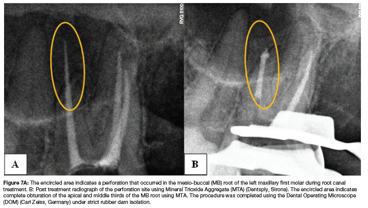
However, preparation of the safety zone is not guaranteed to be without error. Figure 7A indicates a perforation on the mesial side (safety zone) which occurred during the cleaning and shaping of the canal with a rotary instrument. The restoration thereof can be noted in Figure 7B.
DENTINAL THICKNESS FROM THE PULPAL CHAMBER FLOOR TO ROOT FURCATION
Although dentinal thickness in tooth roots is of extreme importance, the dentinal thickness from the pulpal chamber floor to the furcation is also important to note (Fig. 8). During endodontic treatment, undesirable results such as iatrogenic perforation of the furcation while gaining access to the pulp chamber of molar teeth may occur.45,47

This complication could arise due to the procedure being qualitative in nature and having the dentist rely on their previous experience, tactile perception and knowledge of dental anatomy to successfully perform the procedure.48 Furthermore, calcification of the pulp cavity may reduce tactile perceptions leading to treatment failure.49
Thorough understanding of the pulpal space anatomy may assist dentists in attaining successful outcomes during endodontic treatment.47
Majzoub and Kon (1992)50(d reported that in maxillary first molars, the mean distance from the pulp chamber floor to the furcation was equal to or less than 3mm in 86% of the teeth used in their study. In accordance, Sterrett et al51 reported in 1996 that the mean distance ranged from 2.7mm to 3mm for both mandibular and maxillary first and second molars. Deutsch and Musikant (2004)47 also measured anatomical landmarks in the pulp chambers of maxillary and mandibular molars and concluded that in maxillary molars, the mean distance is 3.05mm while in mandibular molars, the mean distance is 2.96mm. Chang et al (2007)37 also measured dentine thickness and estimated that in mandibular first molars, the mean distance between the canal orifice of the mesial root and the furcation is 2.4mm (Table 1).
More recently, Dabawala et al (2015)49 reported thinner pulp chamber floor measurements of 1.70±0.38mm for the maxillary first molar and 1.59±0.31mm for the mandibular first molar. In 2018, Leite Pinto et al33 established that the average distance from the pulp chamber floor to the furcation in first mandibular molars was 2.23mm.
Several studies suggest that secondary dentine apposition occurs primarily on the pulp chamber floor instead of the ceiling.52 Philippas (1961)53 conducted a study comparing ancient and contemporary populations revealing no significant changes in the dentine thickness at the roof of the pulp chamber with increasing age but showed a significant increase in the dentine thickness at the floor of the pulp chamber with increasing age. This may explain the difference in dentinal thicknesses displayed by the abovementioned studies, as Dabawala et al49 used a much younger study group (four to eight years) than the other studies, whose study groups were all classified as adults.
Factors associated with dentine thickness variations
Several variants exist in the root morphology that could have an effect on dentine thickness.46 According to Martin et al (2021),46 individuals with C-shaped rooted mandibular molars exhibit thinner dentine layers than those who do not exhibit C-shaped rooted molar teeth. Furthermore, length of the tooth may also play a role in dentine thickness. Sauaia et al (2010)32 established that the narrowest walls occurred in the longest teeth, whereas shorter teeth exhibited thicker dentine walls (Table 1). Chang et al (2007)37 also concluded that the distal wall of the mesial root is thinner than the mesial wall and that the wall thickness decreases as the distance from the canal orifice increases (Table 1). Similarly, Dwivedi et al (2014)39 also established that in longer mandibular molars, the root wall on the distal surfaces of the mesial roots is thinner compared to shorter teeth (Table 1). In contrast, Zhou et al (2020)2 found that short teeth exhibited thinner dentine thickness values at all locations (Table 1). With regard to maxillary molars specifically, the morphological differences observed are greatly influenced by individual patient characteristics. Dentine thickness in maxillary molars with fused roots showed significantly thinner dentinal walls than maxillary molars with unfused roots.54
Treating physicians should take note that variations in the morphology of the roots and canals have been reported between sexes, with ageing and among populations.2,55 Zhou et al (2020)2 established that dentine thickness increases with age in both men and women (Table 1). An increase in age indicates an increase in possible dental injury due to trauma, caries or cavity preparation in both maxillary and mandibular molars, which will lead to the secretion of tertiary dentine to protect the tooth from further infection.56 In another study executed by Mireku et al (2010),57 their findings indicated that the resistance to vertical root fractures (VRF) of root filled teeth restored with posts decreases with increasing patient age and low dentine thickness. Moreover, Hakami et al (2023)26 also concluded that males in their study exhibited darker tooth shades compared to females of the same age group, suggesting that males have thicker dentine layers than females. The findings of a study by Zhou et al (2020)2 also demonstrate thicker dentine layers in the first mandibular roots of men when compared to women (Table 1). This may indicate a decreased risk of perforation in males.
Although studies regarding the root canal morphology of mandibular and maxillary first permanent molars have been done on various populations, studies regarding dentine thickness are limited to certain geographical regions, teeth and dental roots. From the 15 studies in Table 1 conducted since 2003, six originated from Brazil, three from Iran and one each from Korea, US, India, Türkiye and China. Ten studies were conducted on the first mandibular molar, mesial root, while two studies looked at the mesial root but do not mention from which mandibular molar, and two other studies incorporated the mesial root of the second molar tooth. Only two studies were performed each on the mesiobuccal root of the first maxillary molar and pulpal floor thickness respectively, while only one study looked at the distal root of the first mandibular molar tooth.33,37
Although root morphology of mandibular and maxillary molars of a South African population has been investigated,55 to date no research studies have specifically focused on the dentine thickness of a South African population of different ages and both sexes. There is a clear need to identify and describe crucial morphological characteristics including dentine thickness of the maxillary and mandibular first molar teeth to reduce complications during root canal procedures.
The measurement modalities used in the literature reviewed have a large range of resolution from photographs with a six and 10 times magnification to a microscope with 10 times magnification and a stereomicroscope with a 36 times magnification as well as CBCT and Micro-CT (Table 1). The findings of Berutti and Fedon (1992) showed that the dentine thickness observed on histological sections is one fifth less than that appearing on radiograms. The impact of modality on the observed distances is important as even differences as small as one or two decimal places of a millimetre can be critical.28
CONCLUSION
Improved knowledge of the pattern of dentine thickness along the molar roots may lower the risk for perforation which ultimately could improve the success of the treatment. The thinnest dentine layer in the mesiobuccal canal in the mesial root of the first mandibular molar is reported as 1.13mm from the root canal outline to the distal surface of the mesial root and 1.10mm in the mesiolingual canal. These sites are located 4.37mm below the furcation, while the root surface 3mm from the apex revealed an even thinner dentine layer. On the other hand, the smallest dentine thickness in the first mesiobuccal canal for the first maxillary molar is 1.24mm from the root canal outline to the distal surface of the mesiobuccal root and 0.99mm for the second mesiobuccal canal, both sites located 3mm from the furcation. Treating physicians should take note that variations in dentine thickness have been reported between sexes, with ageing and populations, among others, but unfortunately no studies on dentine thickness in first molars could be found specifically for the South African context.
Authors' declaration
The authors declare that there is no financial interest in this paper and that this paper has not been submitted elsewhere for publication. All authors agree with the content of the manuscript. This manuscript did not receive any funding from funding agencies in the public, commercial or not-for-profit sectors.
Conflict of interest
The authors declare there is no conflict of interest.
REFERENCES
1. Bjørndal L, Laustsen MH, Reit C. Root canal treatment in Denmark is most often carried out in carious vital molar teeth and retreatments are rare. Int Endod J. 2006; 39: 785-90. DOI: 10.1111/j.1365-2591.2006.01149.x [ Links ]
2. Zhou G, Leng D, Li M, et al. Root dentine thickness of danger zone in mesial roots of mandibular first molars. BMC Oral Health. 2020; 20: 1-6. DOI: 10.1186/s12903-020-1026-8 [ Links ]
3. Sarao SK, Berlin-Broner Y Levin L. Occurrence and risk factors of dental root perforations: a systematic review. Int Dent J. 2021; 71: 96-105. DOI: 10.1111/idj.12602 [ Links ]
4. Davis GR, Tayeb RA, Seymour KG, et al. Quantification of residual dentine thickness following crown preparation. J Dent. 2012; 40: 571-6. DOI: 10.1016/j.jdent.2012.03.006 [ Links ]
5. Lee K-W, Kim Y Perinpanayagam H, et al. Comparison of alternative image reformatting techniques in micro-computed tomography and tooth clearing for detailed canal morphology J Endod. 2014; 40: 417-22. DOI: 10.1016/j.joen.2013.09.014 [ Links ]
6. Tabassum S, Khan FR. Failure of endodontic treatment: the usual suspects. Eur J Dent. 2016; 10: 144-7. DOI: 10.4103/1305-7456.175682 [ Links ]
7. Saed SM, Ashley MP Darcey J. Root perforations: aetiology, management strategies and outcomes. The hole truth. Br Dent J. 2016; 220: 171-80. DOI: 10.1038/sj.bdj.2016.132 [ Links ]
8. Clauder T. Present status and future directions - managing perforations. Int Endod J. 2022; 55: 872-91. DOI: 10.1111/iej.13748 [ Links ]
9. Ng Y-L, Mann V Gulabivala K. A prospective study of the factors affecting outcomes of nonsurgical root canal treatment: part 1: periapical health: Outcome of nonsurgical root canal treatment. Int Endod J. 2011; 44: 583-609. DOI: 10.1111/j.1365-2591.2011.01872.x [ Links ]
10. Lease LR. Anatomy of individual teeth and tooth classes. In: Irish J, Scott G, eds. A Companion to Dental Anthropology. John Wiley and Sons, Inc. 2015: 94-107. DOI: 10.1002/9781118845486.ch1 [ Links ]
11. Bhargavi A, Ajay S, Rohit B, et al. Comparative tooth anatomy - A review. IJDSR. 2013; 1: 34-7. DOI: 10.1016/j.ijdsr.2013.03.003 [ Links ]
12. Chourasia HR, Meshram GK, Warhadpande M, et al. Root canal morphology of mandibular first permanent molars in an Indian population. Int J Dent. 2012: 1-6. DOI: 10.1155/2012/745152 [ Links ]
13. Nicholson JW, Czarnecka B. The clinical repair of teeth using direct Alling materials: engineering considerations. Proc Inst Mech Eng H. 2006; 220: 635-45. DOI: 10.1243/09544119H07704 [ Links ]
14. De Moor RJG, Deroose CAJG, Calberson FLG. The radix entomolaris in mandibular first molars: an endodontic challenge. Int Endod J. 2004; 37: 789-99. DOI: 10.1111/j.1365-2591.2004.00870.x [ Links ]
15. Vertucci FJ. Root canal morphology and its relationship to endodontic procedures. Endod Topics. 2005; 10: 3-29. DOI: 10.1111/j.1601-1546.2005.00129.x [ Links ]
16. Jonker C, van der Vyver PJ. Factors influencing the life span of modern root canal instruments - a literature review. SADJ. 2013; 68: 14-23 [ Links ]
17. Cheung GSP Survival of first-time nonsurgical root canal treatment performed in a dental teaching hospital. Oral Surg Oral Med Oral Pathol Oral Radiol Endod. 2002; 93: 596-604. DOI: 10.1067/moe.2002.120254 [ Links ]
18. Stankiewicz NR, Wilson PR. The ferrule effect: a literature review. Int Endod J. 2002; 35: 575-81. DOI: 10.1046/j.1365-2591.2002.00557.x [ Links ]
19. Magne P, Goldberg J, Edelhoff D, et al. Composite resin core buildups with and without post for the restoration of endodontically treated molars without ferrule. Oper Dent. 2016; 41: 64-75. DOI: 10.2341/14-258-L [ Links ]
20. Estrela C, Decurcio DDA, Rossi-Fedele G, et al. Root perforations: a review of diagnosis, prognosis and materials. Braz Oral Res. 2018; 32: 133-46. DOI: 10.1590/1807-3107bor-2018.vol32.0073 [ Links ]
21. Kvinnsland I, Oswald RJ, Halse A, et al. A clinical and roentgenological study of 55 cases of root perforation. Int Endod J. 1989; 22: 75-84. DOI: 10.1111/j.1365-2591.1989.tb00509.x [ Links ]
22. Smith AJ, Scheven BA, Takahashi Y, et al. Dentine as a bioactive extracellular matrix. Arch Oral Biol. 2012; 57: 109-21. DOI: 10.1016/j.archoralbio.2011.07.008 [ Links ]
23. Nudel I, Pokhojaev A, Bitterman Y, et al. Secondary dentin formation mechanism: the effect of attrition. Int J Environ Res Public Health. 2021; 18: 1-10. DOI: 10.3390/ijerph18199961 [ Links ]
24. Senawongse P, Otsuki M, Tagami J, et al. Age-related changes in hardness and modulus of elasticity of dentine. Arch Oral Biol. 2006; 51: 457-63. DOI: 10.1016/j. archoralbio.2005.11.006 [ Links ]
25. Orhan EO, Maden M, Senguüven B. Odontoblast-like cell numbers and reparative dentine thickness after direct pulp capping with platelet-rich plasma and enamel matrix derivative: a histomorphometric evaluation. Int Endod J. 2012; 45: 317-25; DOI: 10.1111/j.1365-2591.2011.01977.x [ Links ]
26. Hakami Z, Marghalani HY, Hedad I, et al. Comparison of tooth color and enamel and dentinal thickness between orthodontically treated and untreated individuals. Diagnostics. 2023; 13: 1-12. DOI: 10.3390/diagnostics13122066 [ Links ]
27. Abou-Rass M, Frank AL, Glick DH. The anticurvature filing method to prepare the curved root canal. J Am Dent Assoc. 1980; 101: 792-4. DOI: 10.14219/jada.archive.1980.0427 [ Links ]
28. Berutti E, Fedon G. Thickness of cementum/dentin in mesial roots of mandibular first molars. J Endod. 1992; 18:545-8. DOI: 10.1016/S0099-2399(06)81211-2 [ Links ]
29. Sant'Anna Júnior A, Cavenago BC, Ordinola-Zapata R, et al. The effect of larger apical preparations in the danger zone of lower molars prepared using the mtwo and reciproc systems. J Endod. 2014; 40: 1855-9. DOI: 10.1016/j.joen.2014.06.020 [ Links ]
30. Harris SP, Bowles WR, Fok A, et al. An anatomic investigation of the mandibular first molar using micro-computed tomography. J Endod. 2013; 39: 1374-8. DOI: 10.1016/j.joen.2013.06.034 [ Links ]
31. Garcia Filho PF, Letra A, Menezes R, et al. Danger zone in mandibular molars before instrumentation: an in vitro study. J Appl Oral Sci. 2003; 11: 324-6. DOI: 10.1590/S1678-77572003000400009 [ Links ]
32. Sauáia TS, Gomes BPFA, Pinheiro ET, et al. Thickness of dentine in mesial roots of mandibular molars with different lengths. Int Endod J. 2010; 43: 555-9. DOI: 10.1111/j.1365-2591.2010.01694.x [ Links ]
33. Leite Pinto S, Lins R, Videira Marceliano-Alves M, et al. The internal anatomy of danger zone of mandibular molars: A cone-beam computed tomography study. J Conserv Dent. 2018; 21: 481-4. DOI: 10.4103/JCD.JCD_271_18 [ Links ]
34. De-Deus G, Rodrigues EA, Belladonna FG, et al. Anatomical danger zone reconsidered: a micro-CT study on dentine thickness in mandibular molars. Int Endod J. 2019; 52: 1501-7. DOI: 10.1111/IEJ.13141 [ Links ]
35. Keles A, Keskin C, Alqawasmi R, et al. Evaluation of dentine thickness of middle mesial canals of mandibular molars prepared with rotary instruments: a micro-CT study. Int Endod J. 2020; 53: 519-28. DOI: 10.111 1/iej. 13247 [ Links ]
36. Sousa VCD, Alencar AHGD, Bueno MR, et al. Evaluation in the danger zone of mandibular molars after root canal preparation using novel CBCT software. Braz Oral Res. 2022; 36: 1-12. DOI: 10.1590/1807-3107bor-2022.vol36.0038 [ Links ]
37. Chang Y-R, Choi Y-S, Choi G-W, et al. Evaluation of danger zone in mesial root of mandibular first molar by cone beam computed tomography (CBCT). Korean Journal of Oral and Maxillofacial Radiology. 2007; 40: 103-10 [ Links ]
38. Tabrizizadeh M, Reuben J, Khalesi M, et al. Evaluation of radicular dentin thickness of danger zone in mandibular first molars. J Dent (Tehran). 2010; 7: 196-9 [ Links ]
39. Dwivedi S, Dwivedi CD, Mittal N. Correlation of root dentin thickness and length of roots in mesial roots of mandibular molars. J Endod. 2014; 40: 1435-8. DOI: 10.1016/j. joen.2014.02.011 [ Links ]
40. Jonker CH, Van Der Vyver PJ, Oettlé AC. Root and canal morphology of the mandibular first molar: A micro-computed tomography-focused observation of literature with illustrative cases. Part 1: External root morphology. SADJ. 2023; 78: 449-56. DOI: 10.17159/sadj. v78i09.16861 [ Links ]
41. Bolbolian M, Ramezani M, Valadabadi M, et al. Dentin thickness of the danger zone in the mesial roots of the mandibular molars: a cone beam computed tomography analysis. Front Biosci (Schol Ed). 2023; 15: 1-9. DOI: 10.31083/j.fbs1501003 [ Links ]
42. Ordinola-Zapata R, Martins JNR, Versiani MA, et al. Micro-CT analysis of danger zone thickness in the mesiobuccal roots of maxillary first molars. Int Endod J. 2019; 52: 524-9. DOI: 10.1111/iej.13025 [ Links ]
43. Azimi V Comparison of dentinal wall thickness in the furcation area (danger zone) in the first and second mesiobuccal canals in the maxillary first and second molars using cone-beam computed tomography. Eur Endod J. 2020; 2: 81-5. DOI: 10.14744/eej.2020.18189 [ Links ]
44. Akhlaghi NM, Kahali R, Abtahi A, et al. Comparison of dentine removal using V-taper and K-Flexofile instruments. Int Endod J. 2010; 43: 1029-36. DOI: 10.1111/J.1365-2591.2010.01769.x [ Links ]
45. Silva EJNL, Rover G, Belladonna FG, et al. Impact of contracted endodontic cavities on fracture resistance of endodontically treated teeth: a systematic review of in vitro studies. Clin Oral Invest. 2018; 22: 109-18. DOI: 10.1007/s00784-017-2268-y [ Links ]
46. Martin G, Arce Brissón G, Chen B, et al. Root dentine thickness in C-shaped lower second molars after instrumentation: A CBCT and micro-CT study. Aust Endod J. 2021; 47: 122-9; DOI: 10.1111/aej.12509 [ Links ]
47. Deutsch A, Musikant B. Morphological measurements of anatomic landmarks in human maxillary and mandibular molar pulp chambers. J Endod. 2004; 30: 388-90. DOI: 10.1097/00004770-200406000-00003 [ Links ]
48. Gaba C, Gurtu A, Bansal R, et al. Morphological measurements of anatomical landmarks in human maxillary first molar pulp chambers and evaluation of number of pulp canal orifices using spiral computed tomography: An in vitro study. J Conserv Dent. 2019; 22: 233-6. DOI: 10.4103/JCD.JCD_568_18 [ Links ]
49. Dabawala S, Chacko V, Suprabha BS, et al. Evaluation of pulp chamber dimensions of primary molars from bitewing radiographs. Pediatr Dent. 2015; 37: 361-5 [ Links ]
50. Majzoub Z, Kon S. Tooth morphology following root resection procedures in maxillary first molars. J Periodontol. 1992; 63: 290-6; DOI: 10.1902/jop.1992.63.4.290 [ Links ]
51. Sterrett JD, Pelletier H, Russell CM. Tooth thickness at the furcation entrance of lower molars. J Clin Periodontal. 1996; 23: 621-7. DOI: 10.1111/j.1600-051X.1996.tb00585.x [ Links ]
52. Lokade J, Rawlani S, Baheti R, et al. Morphological measurements of anatomic landmarks in human mandibular molar pulp chambers - an in vivo study. Journal of Korean Dental Science. 2011; 4: 1-5. DOI: 10.5856/JKDS.2011.4.1.001 [ Links ]
53. Philippas GG. Influence of occlusal wear and age on formation of dentin and size of pulp chamber. J Dent Res. 1961; 40: 1186-98. DOI: 10.1177/00220345610400061301 [ Links ]
54. Keskin C, Toplu D, Keleş A. Dentine thickness in maxillary fused molars depends on the fusion type: An ex vivo micro-computed tomography study. Int Endod J. 2023; 56: 637-46. DOI: 10.1111/iej.13891 [ Links ]
55. Buchanan GD, Gamieldien MY Tredoux S, et al. Root and canal configurations of maxillary premolars in a South African subpopulation using cone beam computed tomography and two classification systems. J Oral Sci. 2020; 62: 93-7. DOI: 10.2334/josnusd.19-0160 [ Links ]
56. Spencer P, Wang Y, Katz JL. Dentin. In: M Akay, editor. Wiley Encyclopedia of Biomedical Engineering. New Jersey: Wiley, 2006: 1051-60. DOI: https://doi.org/10.1002/9780471740360.ebs0346 [ Links ]
57. Mireku AS, Romberg E, Fouad AF, et al. Vertical fracture of root filled teeth restored with posts: the effects of patient age and dentine thickness. Int Endod J. 2010; 43: 218-25. DOI: 10.1111/j.1365-2591.2009.01661.x [ Links ]
 Correspondence:
Name: Marisca Meyer
Email: marisca.meyer@smu.ac.za