



Servicios Personalizados
Revista

Articulo
 Inglés (pdf)
 Articulo en XML
 Referencias del artículo

 Traducción automática
 Enviar articulo por email
Indicadores
Links relacionados
-
 Citado por Google
-
 Similares en Google
Compartir

 Permalink

South African Dental Journal
versión On-line ISSN 0375-1562versión impresa ISSN 0011-8516
S. Afr. dent. j. vol.80 no.2 Johannesburg mar. 2025
https://doi.org/10.17159/sadj.v80i02.20670
SCOPING REVIEW
Cone beam computer tomography imaging of temporomandibular joint for forensic identification: A scoping review
L Vio-AllelI; GM FonsecaII
IDDS (c), School of Dentistry and Centro de Investigación en Odontología Legal y Forense (CIO), Faculty of Dentistry, Universidad de La Frontera (Temuco, Chile)
IIDDS, PhD, Centro de Investigación en Odontología Legal y Forense (CIO), Faculty of Dentistry, Universidad de La Frontera (Temuco, Chile)ORCID: 0000-0001-7397-5907
ABSTRACT
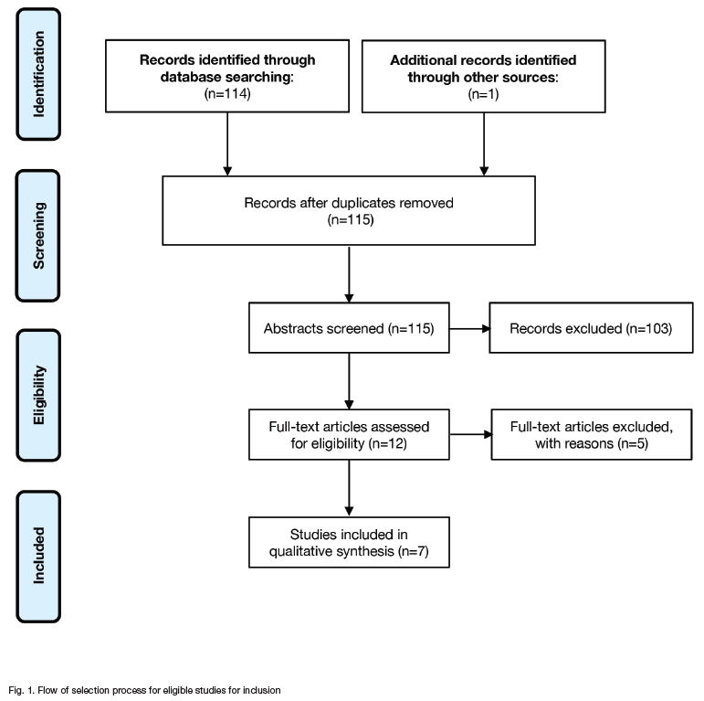
The temporomandibular joint (TMJ) is one of the most complex joints in the human body and it has been demonstrated to possess morphological features that are potentially valuable for forensic identification, both comparative and reconstructive. Cone beam computed tomography (CBCT) has exhibited increasing utilisation in dentistry due to its lower radiation dose, cost-effectiveness and expedited processing time. Given the substantial challenges encountered in forensic identification, we present a scoping review of TMJ studies for forensic identification through CBCT. From a total of 115 articles identified, seven were selected, all published between 2013 and 2024 by authors from Asian countries and without international collaborations. Several proposals for identification were ascertained, predominantly focused on reconstructive identification. Only one article postulated a comparative identification method. This indicates there is interest in utilising CBCT to study the TMJ for forensic identification; however, as there are no population validations, standardised protocols or technical capabilities for field work, further research is warranted.
Keywords: Cone beam computed tomography, forensic identification, forensic dentistry, temporomandibular joint
INTRODUCTION
The temporomandibular joint (TMJ) is one of the most complex and mobile joints in the human body. The TMJ articulates the mandibular condyles with the mandibular fossa and the articular tubercle of the temporal bone to, in conjunction with the soft tissues, perform functions such as mastication, deglutition and speech, among others.1 The mandible is the largest and most robust bone of the facial skeleton. Consequently, in the absence of the pelvis or cranium, the mandible is highly valuable for forensic identification purposes.2 It has been observed that the mandibular condyle exhibits sexual and age-related morphological variability and adaptive changes resulting from anomalies, trauma and malocclusion.3 These variations are of significant interest in forensic sciences as they may facilitate the identification process in individuals with minimal prior interventions.4-6 Furthermore, the utilisation of the TMJ in forensic facial reconstruction procedures in traditional environments in combination with virtual modelling has been documented.7
Two distinct types of identification exist: "comparative" identification, wherein information collected from the deceased individual (postmortem or PM) is compared with information from the suspected individual in life (antemortem or AM) and "reconstructive" identification (wherein, in the absence of AM information, PM data facilitates the reconstruction of the individual's biological profile). Comparative identification is consistently preferred as it is the sole method that can definitively establish an individual's identity.8 While visual identification can resolve various circumstances of medico-legal interest,9 significant cadaveric alterations (such as putrefaction, carbonisation, fragmentation or skeletonisation) not only impede the process but also necessitate the application of scientifically validated methodologies.10 In these contexts, imaging/radiological identification methods have demonstrated their utility, efficiency and fundamentally non-invasive nature for both comparative and reconstructive identification purposes.11
In the field of dentistry, while conventional and panoramic radiographs provide information for the morpho pathological study of the TMJ,12 three-dimensional images, such as those obtained through computed tomography (CT), offer a substantially greater level of detail.13 Nevertheless, CT presents drawbacks, including high cost and significant radiation exposure.14 Conversely, cone beam computed tomography (CBCT) has gained increasing prevalence in dentistry due to its lower radiation dose, affordability and faster processing time.15 Moreover, the data acquired from CBCT can be transformed into conventional radiographic images enabling, for instance, the extraction of information about a specific tooth, the generation of a panoramic radiograph and the performance of volumetric analysis of the skull. These factors contribute to the consideration of CBCT as a valuable and accurate tool for human identification.16
Given the significant challenges faced in forensic identification, where both technological advancements and a comprehensive approach are essential for resolving routine and extraordinary circumstances,17 we present a scoping review of TMJ studies for forensic identification through CBCT. The aim is to establish analysis variables by considering proposals that incorporate technical, logistical and procedural elements, while taking into account the standards, scope and limitations of current identification paradigms.
METHODOLOGY
A scoping review was conducted following the PRISMA (Preferred Reporting Items for Systematic Reviews and Meta-Analyses) protocol18 of the PubMed/Medline database utilising the search strategy "(CBCT OR "Cone Beam") AND (condyle OR TMJ) AND forensic". The search was executed on September 2 2024 with the categorisation, analysis and evaluation of relevance of the titles and abstracts conducted between September 3 and September 9 2024. Subsequently, the full articles were reviewed on September 10 2024. As a supplementary search, a review of the articles cited in the references of the latter was performed. Original full articles and case reports were included, while reviews, letters to the editor, theses and animal studies were excluded. The results of interest encompassed forensic identification methods using CBCT in TMJ. The selected articles were grouped and categorised according to their proposed methodologies.
RESULTS
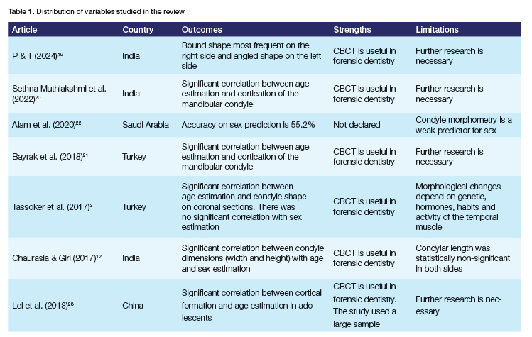
From a total of 115 articles identified, seven were ultimately selected that met the pre-established criteria (Fig. 1). The results of the various variables identified are presented in Table 1, which demonstrates an absolute predominance of Asian countries (India: 3 articles,12,19,20 Turkey: 2 articles,3,21 Saudi Arabia22 and China23: 1 article each). The publication years ranged from 2013 to 2024. No international collaborations, multicentred studies or applications of artificial intelligence (AI) technologies were identified.
The methods proposed in the reviewed articles were categorised into three groups of potential applications of CBCT in TMJ for forensic identification purposes:
I. Sex and/or age estimation through analysis of TMJ morphological patterns
Three articles3,12,22 proposed this reconstructive methodology for human identification, which involved analysing the morphological variations of the mandibular condyle using CBCT to estimate sex and age. CBCT provides measurements of condylar height, width and length as variables to estimate age using linear regression analysis equations. All articles concurred that there is a significant relationship between the shape of the condyle and the age of the individual, but not with sex, where the authors do not reach a consensus. According to Alam et al 22 condyle morphometry is an unreliable indicator for sex estimation.
II. Age estimation through assessment of the mandibular condyle bone cortex status
Three articles20,21,23 proposed the analysis of the degree of bone cortication of the mandibular condyle through CBCT to estimate the age of an individual. Three types of cortication are described from CBCT images: "Type I: No bone cortex is observed in the condyle"; "Type II: The bone on the surface of the condyle appears to have a lower density than the structures around the condyle"; "Type III: The surface of the condyle appears to have a density equal to or greater than the surrounding cortical areas". It was established that the age of complete bone cortical formation is 22 years in females and 21 years in males.23 A significant relationship was determined between the level of bone cortication and the chronological age of the individual.
III. AM-PM comparison of condylar morphological patterns
This comparative identification method was proposed by authors from India,19 which involved comparing the information from an AM CBCT with one from a PM CBCT. Morphological patterns can be identified in the mandibular condyle, which can be classified as round, angled, convex or flat condyle. The round shape is the most prevalent, representing 38.7% of cases. Additionally, differences are observed between the right and left condyle: the round shape predominates in the right, while the angled shape is more frequent in the left. These findings demonstrate that CBCT enables the identification of significant morphological diversity for human identification.
DISCUSSION
A significant limitation of this review is its potential contribution to the notion that the use of an TMJ CBCT in human identification procedures constitutes an "extraordinary" situation, either due to the fortuitous discovery of an AM CBCT or through the deliberate implementation of this technological approach in a disaster victim identification (DVI) procedure. Nevertheless, this review is grounded on the premise that in cases of identification involving fragmented human remains, which cannot be analysed using primary identifiers (according to INTERPOL, fingerprints, comparative dental analysis or DNA24), such situations are not "extraordinary" and necessitate not only these technologies but also forensic practitioners trained in their application.25 TMJ has demonstrated its significance as a focus of attention for forensic investigation using PM CT;26,27 furthermore, substantial evidence in the literature corroborates the potential utility of CBCT in field identification procedures.28,29 We concur with Nickell & Fischer that the available AM information will determine the type of identification procedure, and even in the absence of such information, any anatomical peculiarity could prove decisive for reconstructive identification or corroborate information obtained through other sources.30 We consider that there exists a knowledge gap regarding the applicability of CBCT in TMJ within forensic contexts, thus warranting further investigation.
CBCT has demonstrated significant utility in contemporary dental practice, with notable applications in diagnostic clinics, precise indications and substantial potential in the forensic field.31,32 This review corroborates the interest in studying the TMJ in the forensic scene through CBCT, which presents new avenues for the development of scientific research in both comparative and reconstructive identification. Nevertheless, we consider that there exist limitations which, when applied to field conditions, continue to preclude this tool from being a viable alternative. We concur with de Boer et al (2020) that "incidents with severely fragmented, compromised or commingled human remains pose complex logistical, practical and ethical challenges that may prolong the DVI operation or even preclude identification of some individuals altogether";33 substantial evidence in the literature indicates that CBCT requires specific protocols, methodological considerations and technical expertise that present significant challenges in domestic situations,34 and even more so in circumstances of forensic identification in the field. To our knowledge, there are no reported techniques or protocols for standardising CBCT imaging of TMJ in cadavers under field conditions. Although there are studies using PM CBCT,35,36 these have been conducted in ideal laboratory environments and without the recognised challenges of a DVI procedure. Of particular note is the technical note published by Leite Beaini et al regarding a device designed to position skulls for extraoral radiographic recordings and CBCT.37 We agree with the authors that designs of this nature increase the feasibility of implementing these techniques in forensic environments, thus warranting further studies to provide the forensic scientific community with appropriate methods to standardise and validate the procedure in situations of tissue vulnerability. As early as 2004, Rötzscher et al demonstrated that the positive identification of injured individuals using dental morphological and radiological methods is only feasible if the fragile remains of a charred human skull can be adequately stabilised.38
This review demonstrates that the predominant focus of CBCT utilisation in TMJ research has been in reconstructive identification. Age estimation through evaluation of the mandibular condyle's cortical bone presents a promising technique for forensic identification. This method is particularly valuable in circumstances where cadaveric deterioration precludes the application of established methods documented in the literature.21 However, this optimism appears to contrast with the findings of Alam et al, who reported the non-feasibility of sex estimation through evaluation of TMJ morphological patterns.22 Several researchers posit that morphological variables in TMJ structures may be influenced by genetic, ethnic, hormonal and degenerative factors, as well as by habits, trauma or socioeconomic status - all of which hold significant identifying value in the medico-legal field.3,39-41 It is noteworthy that all studies identified in this review were conducted on Asian populations with limited additional information, necessitating cautious interpretation of results when considering application in other contexts.
It is noteworthy that, while it has been asserted that comparative identification methods are the primary and priority methods,8 only one article in this review proposed CBCT as a viable tool in these kind of methods.19 It has been suggested that in medico-legal procedures, resources may not align with needs, and certain technological requirements may only be feasible for specific agencies or services, due to their costs and accessibility.42 Possessing an AM CBCT would be a clearly advantageous but potentially problematic situation (although Mowafey et al indicate that US officers assigned to international missions in high-risk areas undergo a CBCT as an AM record,43 this practice is far from being standard in developing countries). Even if such an AM CBCT is available, forensic agencies should obtain a comparable PM image in order to compare and match both sets of data, which is the foundation for comparative identifications.8 Some authors have already proposed that the comparison of skeletal and interventional findings on medical imaging should be considered as a "primary biological identifier" rather than a secondary one, thus enhancing AM information gathering strategies and best practice guidelines for quantifying identification coincidences would improve successful results, even in circumstances such as circumstantial or random findings of identifiers in TMJ.17,25 It would be advantageous for these agencies and services to have previously established agreements to ensure the availability of these technological resources when necessary. It is evident that this approach, beyond conforming to international standards, would facilitate the identification process and provide greater scientific support for it. The use of CBCT for comparative identifications has been minimally reported in the literature, enabling three-dimensional AM-PM superposition and comparison, assisting in establishing an approximate age range by evaluating the bone cortex, and addressing the situation of insufficient other information.44 We posit that challenging the methodology in all its possibilities and scenarios would allow it to be validated in accordance with the new international scientific and legal standards.45 The interest of Asian countries in forensic dental research has been previously documented, as well as their propensity toward research in reconstructive identification.46 We consider that international collaboration and multicentred studies would facilitate the expansion of the database for corresponding population validations in cases of reconstructive identifications. Similarly, AM-PM simulation exercises would enable the development of skills necessary for matching in DVI environments in comparative identifications. The emerging challenges in the application of AI tools would potentially enable new avenues of research and validation in CBCT of the TMJ, considering that forensic identification through AI has already become a widely disseminated reality.47
It is evident that in DVI procedures, each operation is unique, rendering generic recommendations challenging. Prior planning is essential, and agreements on technical roles both at the scene and in the morgue must be precise yet flexible. The appropriate management of AM and PM imaging information is integrated into a chain of key procedures where strategies for remains recovery, reconciliation and quality control must align with validated technical standards and, fundamentally, demonstrate utility in their cost-benefit analysis.33 Although the utilisation of CBCT in TMJ for human identification demonstrates significant potential, it continues to face considerable challenges that require addressing. Adserias-Garrica et al assert that further research is necessary regarding the frequency of certain anatomical variations within a population, a crucial element for establishing hierarchies of identifying value according to their frequency, as well as the interpretation of these variations in images generated by novel technologies.25 Moreover, the implementation and validity of these techniques in legal settings necessitate a robust scientific foundation, including peer review, knowledge of the error rate and acceptance by the scientific community. In the absence of a standardised protocol for obtaining images in compromised postmortem conditions, the practical implementation of CBCT remains unsystematic and contingent on conditions that are excessively specific to be considered a reliable resource in the field at present.
CONCLUSION
This review demonstrates that there is considerable interest in investigating the potential of CBCT imaging of TMJ for forensic identification, although the focus is primarily on reconstructive rather than comparative identification. While the utilisation of TMJ CBCT in human identification procedures may be considered an exceptional circumstance, cases involving fragmented human remains are not uncommon and necessitate not only these advanced technologies but also forensic practitioners trained in their application. It is recommended to further explore the proposed methods, implement AI tools, develop networks and international collaborations to enhance and validate multicentre studies, and establish protocols for the technical application of these technologies in the field.
Conflict of interest
The authors declare that the manuscript was created without any commercial or financial associations that may give rise to a conflict of interest.
Funding
None
REFERENCES
1. Gutiérrez-Tiznado P, López-Lázaro S, Fonseca GM. Use of "bicondylar" as a descriptive term for the temporomandibular joint: A scoping review. Int J Morphol 2022; 40: 1054-9 [ Links ]
2. Suzuki K, Nakano H, Inoue K, Nakajima Y Mizobuchi S, Omori M, et al. Examination of new parameters for sex determination of mandible using Japanese computer tomography data. Dentomaxillofac Radiol 2020; 49: 20190282 [ Links ]
3. Tassoker M, Kabakci ADA, Akin D, Sener S. Evaluation of mandibular notch, coronoid process, and mandibular condyle configurations with cone beam computed tomography. Biomed Res 2017; 28: 8327-35 [ Links ]
4. Shanin KA, Chatra L, Shenai P. Dental and craniofacial imaging in forensics. JOFRI 2013; 1: 56-62 [ Links ]
5. Coelho J, Armelim Almiro P, Nunes T, Kato R, Garib D, Miguéis A, et all . Sex and age biological variation of the mandible in a Portuguese population -forensic and medico-legal approaches with three-dimensional analysis. Sci Justice 2021; 61: 704-13 [ Links ]
6. Pretty IA. Forensic dentistry: 1. Identification of human remains. Dent Update 2007; 34: 621-2, 624-6, 629-30 passim [ Links ]
7. Benazzi S, Fantini M, De Crescenzio F, Mallegni G, Mallegni F, Persiani F, et al. The face of the poet Dante Alighieri reconstructed by virtual modelling and forensic anthropology techniques. J Archaeol Sci 2009; 36: 278-83 [ Links ]
8. Fonseca GM, Cantín M, Lucena J. Odontología Forense II: La Identificación Inequívoca. Int J Odontostomat 2013; 7: 327-34 [ Links ]
9. Blau S, Graham J, Smythe L, Rowbotham S. Human identification: a review of methods employed within an Australian coronial death investigation system. Int J Legal Med 2021; 135: 375-85 [ Links ]
10. Zimmerman HA, Meizel-Lambert CJ, Schultz JJ, Sigman ME. Chemical Differentiation of Osseous, Dental, and Non-skeletal Materials in Forensic Anthropology using Elemental Analysis. Sci Justice 2015; 55: 131-8 [ Links ]
11. Manigandan T, Sumathy C, Elumalai M, Sathasivasubramanian S, Kannan A. Forensic radiology in dentistry. J Pharm Bioall Sci 2015; 7: S260-4 [ Links ]
12. Chaurasia A, Giri S. Evaluation of mandibular condyle morphology in Indian ethnics. J Oral Med Oral Surg Oral Pathol Oral Radiol 2017; 3: 17-22 [ Links ]
13. Omami G, Miller CS. Imaging Evaluation of the Temporomandibular Joint. Dent Clin North Am 2024; 68: 357-73 [ Links ]
14. Dieckmeyer M, Sollmann N, Kupfer K, Löffler MT, Paprottka KJ, Kirschke JS, et al. Computed Tomography of the Head: A Systematic Review on Acquisition and Reconstruction Techniques to Reduce Radiation Dose. Clin Neuroradiol 2023; 33: 591-610 [ Links ]
15. Nardi C, Talamonti C, Pallotta S, Saletti P, Calistri L, Cordopatri C, et al. Head and neck effective dose and quantitative assessment of image quality: a study to compare cone beam CT and multislice spiral CT. Dentomaxillofac Radiol 2017; 46: 20170030 [ Links ]
16. Alam MK, Ganji KK, Munisekhar MS, Alanazi NS, Alsharif HN, Iqbal A, et al. Application of CBCT technology in forensic odontology: A narrative review. Saudi Dent J 2022; 34: 88-96 [ Links ]
17. Cunha E, Obertová Z. Forensic identification in a multidisciplinary perspective focusing on big challenges. Forensic Sci Res 2024; 9: owae063 [ Links ]
18. Tricco AC, Lillie E, Zarin W, O'Brien KK, Colquhoun H, Levac D, et al. PRISMA Extension for Scoping Reviews (PRISMA-ScR): checklist and explanation. Ann Intern Med 2018; 169: 467-73 [ Links ]
19. P JE, T N U. Distinctive Anatomical Patterns of the Mandibular Coronoid Process, Condyle, and Sigmoid Notch: Cone Beam Computed Tomography (CBCT) Imaging for Advanced Personal Identification. Cureus 2024; 16: e60978 [ Links ]
20. Sethna Muthlakshmi KS, Krithika CL, Asokan K. Evaluation and correlation of condylar cortication by cone-beam computed tomography: A retrospective study. Contemp Clin Dent 2022; 13: 30-4 [ Links ]
21. Bayrak S, Halıcıoglu Sıı, Kose Gü, Halıcıoglu K, Evaluation of the relationship between mandibular condyle cortication and chronologic age with cone beam computed tomography. J Forensic Leg Med 2018; 55: 39-44 [ Links ]
22. Alam MK, Ganji KK, Munisekhar MS, Alanazi NS, Alsharif HN, Iqbal A, et al. A 3D cone beam computed tomography (CBCT) investigation of mandibular condyle morphometry: Gender determination, disparities, asymmetry assessment and relationship with mandibular size. Saudi Dent J 2021; 33: 687-92 [ Links ]
23. Lei J, Liu MQ, Yap AUJ, Fu KY. Condylar subchondral formation of cortical bone in adolescents and young adults. Br J Oral Maxillofac Surg 2013; 51: 63-8 [ Links ]
24. I NTERPOL. Disaster Victim Identification Guide. 2023. [cited 2 November 2024]. Available from: https://www.interpol.int/en/How-we-work/Forensics/Disaster-Victim-Identification-DVI [ Links ]
25. Adserias-Garriga J, Feirstein S, Bell D, Skropits H, Dirkmaat DC. Human identification through forensic skeletal analysis: three case reviews. Forensic Sci Res 2024; 9: owae053 [ Links ]
26. Glengarry J, Beaugeois M, Bugeja L, Huggins R, O'Donnell C. Suspension-associated dislocation of the jaw in hanging. Int J Legal Med 2023; 137: 1489-95 [ Links ]
27. Beaugeois M, Glengarry J, O'Donnell C, Bugeja L. What is the presence and role of dislocation of the temporomandibular joint in the injury profile of hanging deaths? Pathology 2024; 56: S91-2 [ Links ]
28. Silva RF, Picoli FF, Botelho TL, Resende RG, Franco A. Forensic Identification of Decomposed Human Body through Comparison between Ante-Mortem and Post-Mortem CT Images of Frontal Sinuses: Case Report. Acta Stomatol Croat 2017; 51: 227-31 [ Links ]
29. Kumagai A, Izumisawa M, Takahashi N, Biwasaka H. Effectiveness and limitations of human identification from cremains: A report of two cases. Leg Med (Tokyo) 2021; 53: 101933 [ Links ]
30. Nickell J, Fischer JF. Crime Science: Methods of forensic detection. Lexington: University Press of Kentucky, 1999 [ Links ]
31. Munhoz L, Okada S, Hisatomi M, Yanagi Y Arita ES, Asaumi J. Are computed tomography images of the mandible useful in age and sex determination? A forensic science meta-analysis. J Forensic Odontostomatol 2024; 42: 38-57 [ Links ]
32. Danforth RA, Herschaft EE, Weems RA. Dental, oral, and maxillofacial radiographic features of forensic interest. In: Senn DR, Weems RA, eds. ASFO's Manual of Forensic Odontology, 5th ed. Boca Raton: CRC press, 2013 [ Links ]
33. de Boer HH, Roberts J, Delabarde T, Mundorff AZ, Blau S. Disaster victim identification operations with fragmented, burnt, or commingled remains: experience-based recommendations. Forensic Sci Res 2020; 5: 191-201 [ Links ]
34. Kim NH, Yang BE, Kang SH, Kim YH, Na JY, Kim JE, et al. Preclinical and Preliminary Evaluation of Perceived Image Quality of AI-Processed Low-Dose CBCT Analysis of a Single Tooth. Bioengineering (Basel) 2024; 11: 576 [ Links ]
35. Kamburoğlu K, Kolsuz E, Kurt H, Kiliç C, Özen T, Paksoy CS. Accuracy of CBCT measurements of a human skull. J Digit Imaging 2011; 24: 787-93 [ Links ]
36. Kamburoğlu K, Kiliç C, Ozen T, Yüksel SP Measurements of mandibular canal region obtained by cone-beam computed tomography: a cadaveric study. Oral Surg Oral Med Oral Pathol Oral Radiol Endod 2009; 107: e34-42 [ Links ]
37. Beaini TL, Dias PE, Melani RF. Dry skull positioning device for extra-oral radiology and cone-beam CT. Int J Legal Med 2014; 128: 235-41 [ Links ]
38. Rötzscher K, Grundmann C, Benthaus S. The effects of high temperatures on human teeth and dentures. Int Poster J Dent Oral Med 2004; 6: Poster 213 [ Links ]
39. Schmeling A, Olze A, Reisinger W, Geserick G. Forensic age estimation and ethnicity Leg Med 2005; 7: 134-7 [ Links ]
40. Burt NM, Semple D, Waterhouse K, Lovell NC. Identification and interpretation of joint disease in paleopathology and forensic anthropology. Springfield: Charles C Thomas, 2013 [ Links ]
41. Silver WE, Souviron RR. Dental autopsy. Boca Raton: CRC Press, 2009 [ Links ]
42. Fonseca GM, Rojas-Torres J. "To do more with less". Forensic Sci Int 2024; 361:112118 [ Links ]
43. Mowafey B, Van de Casteele E, Youssef JM, Zaher AR, Omar H, Politis C, et al. Can mandibular lingual canals be used as a forensic fingerprint? J Forensic Odontostomatol 2015; 33: 26-35 [ Links ]
44. Kumagai A, Izumisawa M, Takahashi N, Biwasaka H. Effectiveness and limitations of human identification from cremains: A report of two cases. Leg Med (Tokyo) 2021; 53: 101933 [ Links ]
45. Rivera-Mendoza F, Martín-de-Las-Heras S, Navarro-Cáceres P, Fonseca GM. Bite Mark Analysis in Foodstuffs and Inanimate Objects and the Underlying Proofs for Validity and Judicial Acceptance. J Forensic Sci 2018; 63: 449-59 [ Links ]
46. Espinoza-Silva PV López-Lázaro S, Fonseca GM. Forensic odontology and dental age estimation research: a scoping review a decade after the NAS report on strengthening forensic science. Forensic Sci Med Pathol 2023; 19: 224-35 [ Links ]
47. Mohammad N, Ahmad R, Kurniawan A, Mohd Yusof MYP. Applications of contemporary artificial intelligence technology in forensic odontology as primary forensic identifier: A scoping review. Front Artif Intell 2022; 5: 1049584 [ Links ]
 Correspondence:
Name: Gabriel M Fonseca
Email: gabriel.fonseca@ufrontera.cl
Author's contribution
1. Lorena Vio-Allel - conceptualisation, draft preparation (40%)
2. Gabriel M Fonseca - draft preparation, review and editing (60%)