



Services on Demand
Journal

Article
 English (pdf)
 Article in xml format
 Article references
 Automatic translation
 Send this article by e-mail
Indicators
Related links
-
 Cited by Google
 -
 Similars in Google
Share

 Permalink
SA Orthopaedic Journal
On-line version ISSN 2309-8309Print version ISSN 1681-150X
SA orthop. j. vol.25 n.2 Centurion 2026
https://doi.org/10.17159/2309-8309/2026/v25n1a8
CURRENT CONCEPTS REVIEW
Surgical management of the hallux valgus: a current concepts review
Duduzile SigodiI, ; Ruvyn FrankII; Phakamani G MthethwaI
IDepartment of Orthopaedic Surgery, University of KwaZulu-Natal, Nelson R Mandela School of Medicine, Durban, South Africa
IIMaritzburg Orthopaedic Centre, Life Hilton Private Hospital, Pietermaritzburg, South Africa
ABSTRACT
Hallux valgus (HV) remains among the most prevalent forefoot deformities encountered in orthopaedic practice, characterised by complex multiplanar deformity involving lateral deviation, pronation and associated joint pathology. Its aetiology is multifactorial, encompassing extrinsic factors such as footwear and genetic predisposition, as well as intrinsic biomechanical alterations including ligamentous laxity and abnormal foot architecture. The pathogenesis involves deviations in the first metatarsophalangeal joint (MTPJ) biomechanics, destabilised by a dysfunctional windlass mechanism and contributing to cartilage degeneration, bony prominences and sesamoid subluxation.
Clinical assessment integrates detailed history, physical examination and radiographic evaluation of deformity parameters such as the HV and intermetatarsal angles. Conservative management includes orthoses, exercise therapy, footwear modification, and adjunct modalities, which alleviate symptoms and may delay surgical intervention.
Surgical correction remains the definitive treatment, with over 100 procedures described. Techniques are tailored based on deformity severity, including distal soft tissue procedures, various osteotomies (chevron, scarf, Mitchell, Lapidus) and arthrodesis, each with specific indications, advantages and complications. Recent advancements in minimally invasive surgery (MIS) have shown promising results, including superior deformity correction, reduced pain and quicker recovery, though long-term outcomes are comparable to traditional open techniques. Comparative studies highlight benefits of MIS but emphasise the importance of surgeon experience and patient-specific factors. Overall, the evolution of surgical techniques offers a wide spectrum of options for halting deformity progression and restoring function, underscoring the importance of individualised treatment strategies guided by deformity severity, radiographic findings and patient preferences.
LEVEL OF EVIDENCE: 5
Keywords: hallux valgus, osteotomies, minimally invasive surgery, deformity correction
Introduction
Hallux valgus (HV) remains one of the most common forefoot deformities encountered in orthopaedic practice. Contemporary imaging studies, particularly computed tomography (CT), have revealed that up to 87% of cases exhibit a pronation component of the first metatarsal, highlighting the deformity's multiplanar configuration.1 The term 'hallux abducto-valgus' was introduced by the German surgeon Carl Hueter, who described the condition as a lateral deviation of the great toe at the metatarsophalangeal (MTP) joint.2,3
Aetiology
Multiple factors are implicated in the aetiology of HV.3-10 These include footwear choices - especially tight, narrow and high-heeled shoes - as well as genetic predisposition, ligamentous hyperlaxity, pes planus, neuromuscular disorders such as cerebral palsy, and Achilles tendon tightness. The condition predominantly affects women, with a female-to-male ratio of approximately 15:1, and women are more likely to require surgical intervention. In addition, habitual footwear habits and ligamentous laxity predisposes females to hallux valgus.3-10
Pathogenesis of HV
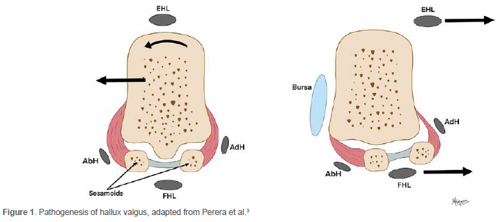
A central element in the development of HV involves alterations in the anatomy and biomechanics of the first MTP joint2,3 (Figure 1). The windlass mechanism, which maintains plantar fascia tension during push-off in gait, plays a vital role in stabilising the medial aspect of the first ray. Dysfunction of this mechanism can result in abnormal joint kinematics, facilitating progressive deformity.7 Conditions such as pes planus (flatfoot) and genetic predisposition further contribute to instability and uneven load distribution across the forefoot.6 Recent research suggests that anatomical variations, including torsional abnormalities of the metatarsals and the presence of calcaneal spurs, may influence both the severity and progression of the deformity.7
Extrinsic factors, particularly footwear, are also crucial. Tight, narrow shoes with high heels increase pressure on the first MTP joint contributing to lateral deviation of the hallux. The association between restrictive footwear, especially styles that constrict the toes, and HV development is well established.7,8 Deformity progression involves not only biomechanical alteration but also joint pathology. Cartilage degeneration within the first MTP joint correlates with abnormal loading patterns, often leading to pain and reduced joint mobility. Structural changes such as medial eminence formation on the first metatarsal and lateral subluxation of the sesamoids complicate both the deformity and its surgical management.9
Clinical evaluation
A comprehensive clinical assessment includes detailed symptom history and physical examination. Family history of bunions and the localisation of tenderness are important. Deep, aching or plantar pain commonly suggests sesamoid pathology; medial discomfort may reflect bursal inflammation. Pain elicited while barefoot indicates possible joint involvement at the MTP or first tarsometatarsal (TMT) joint, as opposed to pressure-related discomfort over the medial eminence. Typical presenting concerns include footwear intolerance, pain, cosmetic deformity, and metatarsalgia affecting the lesser toes.10 Physical examination findings may include associated planovalgus deformity, a tight Achilles tendon, rigid or flexible HV, first toe pronation, callosities or corns on the lesser toes, second MTP joint synovitis, interdigital neuromas, and hypermobility of the first TMT joint.10
Radiographic evaluation
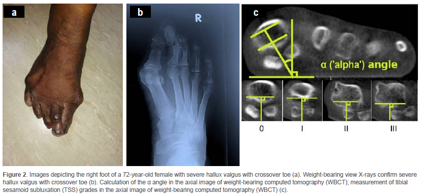
Radiographs are essential for diagnosis, preoperative planning and postoperative follow-up, providing a quantitative assessment of deformity and joint congruency.1 Key parameters comprise the hallux valgus angle (HVA), intermetatarsal angle (IMA), sesamoid position, and joint alignment.2 The HVA, defined as the angle between the longitudinal axes of the first metatarsal and the proximal phalanx of the hallux, is diagnostic when exceeding 15°. Accurate measurement of HVA assists in severity grading and outcome prediction, with recurrence often defined as an increase of more than 10° postoperatively.11 The IMA, formed between the axes of the first and second metatarsals, typically exceeds 9° in HV deformity.12 Evaluation of the first TMT joint, including the metatarsal-medial cuneiform angle, provides additional detail on the deformity's structural basis.13 Assessment of sesamoid position, based on weight-bearing radiographs, offers insight into deformity severity and guides surgical planning14 (Figures 2a and b).
To mitigate the HV recurrence, Kim et al. introduced the CT based method, which involved using semi-weight-bearing coronal CT axial views to measure the α angle and tibial sesamoid grades15 (Figure 2c). The patients were positioned supine with their upper bodies stabilised on the CT platform. The foot is placed in 45° of plantarflexion at the ankle level, keeping the first metatarsophalangeal joint (MTPJ) parallel to the platform. The patient is instructed to push their foot downward to generate a semi-weight-bearing environment. The plantar aspect of the foot is firmly applied to the CT table under the supervision of a senior radiologist. The α angle is subtended by a) an inferior line was drawn between the lateral edge of the lateral sulcus and the medial edge of the medial sulcus; b) a superior line was drawn between the points of the medial and lateral corners of the first metatarsal head; c) bisections of the above two lines were connected to a straight line perpendicular to the horizontal ground axis; d) the α angle was measured between the straight line and a vertical line perpendicular to the ground axis. In addition, Kim et al. graded the tibial sesamoid position using a modified evaluation tool in the semi-weight-bearing CT axial view (CT 4 position). The grading was based on the position of the tibial sesamoid relative to the intersesamoid ridge, ranging from grade 0 (entirely medial) to grade 3 (entirely lateral). Patients with HV deformities have a more pronated first metatarsal, with an α angle > 15.8° and pseudo subluxation of the sesamoids.15 MRI can add valuable information regarding joint degeneration, plantar plate integrity and sesamoid pathology, particularly in complex cases, thereby enriching the standard radiographic workup.16
Conservative management of HV
Conservative treatment strategies aim to alleviate symptoms, improve foot function, and potentially delay or eliminate the need for surgical intervention in HV. Various modalities have demonstrated efficacy in managing this common deformity, characterised by medial deviation of the hallux and associated joint pain.
Orthotic devices and toe spacers
Custom foot orthoses and toe separators represent primary conservative measures. Clinical evidence indicates these interventions can significantly reduce pain and accommodate deformity progression. Toe spacing devices have also been shown to improve toe alignment and decrease discomfort in moderate cases.17
Exercise therapy
A systematic review by Ying et al. identified various exercise programmes, including toe-spread-out exercises and resistive training, that effectively reduced the HVA and strengthened intrinsic foot muscles.18 Furthermore, the Japanese Orthopaedic Association clinical practice guidelines suggest that manual therapy combined with structured home exercise programmes has demonstrated improvements in pain, joint mobility and function.19 These interventions target muscular imbalances and biomechanical deficiencies contributing to deformity.
Footwear modification
Appropriate footwear remains central in conservative management. Shoes with wide toe boxes and flexible soles can reduce pressure on the first MTPJ, alleviating pain and potentially slowing deformity progression. Emphasising proper fit is critical in preventing exacerbation of the deformity.19
Taping and adjunct modalities
Kinesiotaping may provide short-term symptomatic relief, with some evidence suggesting it can enhance toe alignment and reduce pain.19,20 Additional therapies, such as cryotherapy and nonsteroidal anti-inflammatory drug (NSAID) use, can be employed during episodes of acute inflammation to manage pain effectively.21
Surgical options for HV
The choice of surgical technique is informed by the deformity's severity, specific radiographic assessments such as the HVA, IMA, distal metatarsal articular angle (DMAA), and individual patient characteristics, including joint stability and associated foot pathologies.22 The overarching goal is to achieve anatomical realignment while maintaining or enhancing foot function. Over 100 surgical procedures have been documented, which can be categorised into the following main types: a) distal soft tissue procedures; b) first metatarsal osteotomies (distal, shaft, or proximal); c) proximal phalangeal osteotomies; d) arthrodesis of the first MTPJ; e) arthrodesis of the first metatarsocuneiform (MTC) joint; f) excision arthroplasty (Keller's procedure).22
Simple bunionectomy
The procedure involving simple excision of the medial eminence and subsequent medial capsular plication has fallen out of favour due to a significant risk of recurrence and generally poor patient satisfaction outcomes.23
Distal soft tissue procedures
A refined approach, the modified McBride procedure, entails excising the medial eminence, performing a lateral capsulotomy, adductor tendon release, and medial capsular plication.23 Studies by Mann and Coughlin have demonstrated improvements in the HVA by approximately 14° and the IMA by 5° with these procedures.24
First metatarsal osteotomies
These procedures generally involve lateral translation of the metatarsal head to correct the IMA and properly reposition the metatarsal head over the sesamoids. The choice of osteotomy is determined by the severity of the deformity: a) distal metatarsal osteotomy, suitable for mild-to-moderate deformities; b) metatarsal shaft osteotomy, applied in moderate-to-severe cases; c) proximal metatarsal osteotomy or Lapidus fusion, referred for severe deformities.25 Safeguarding the vascularity of the first metatarsal head is crucial to prevent avascular necrosis (AVN), as its blood supply is largely from the plantar vessels entering at the metatarsal neck. Additionally, maintaining the first metatarsal's length is critical to avoid causing transfer metatarsalgia. Effective dorsiflexion during osteotomy can mitigate loading on the first metatarsal, preventing transfer lesions.1
Distal chevron V-shaped osteotomy
The distal chevron osteotomy is the subtype of the boat-shaped osteotomy used in HA, characterised by its V-shaped cut, and is a well-established procedure for correcting mild-to-moderate HV deformities. This osteotomy can be combined with lateral soft tissue releases, including release of the lateral metatarsal sesamoid ligament and, optionally, adductor hallucis tenotomy. Typically, displacement of up to 50% of the metatarsal head is achievable while maintaining stability. Fixation methods vary and include screws, Kirschner wires (K-wires), or bioabsorbable fixation devices. Some surgeons advocate for an extended plantar limb of the osteotomy, which offers increased contact surface area, thereby enhancing union rates and fixation stability.26 In a randomised controlled trial, Resch et al. compared distal chevron osteotomy with or without adjunctive adductor tenotomy in 106 feet.27 Results indicated superior radiographic and clinical outcomes in the group receiving adductor release; however, patient satisfaction was similar across both cohorts. A significant complication associated with chevron osteotomy is osteonecrosis of the metatarsal head and deformity recurrence. These risks can be mitigated by placing the plantar limb proximal to the plantar branches of the metatarsal artery and by avoiding saw passage beyond the lateral cortex and capsule.27
Mitchell osteotomy
First described by Hawkins et al.,28 the procedure involves a double-step osteotomy through the neck of the first metatarsal. The technique displaces the head laterally and plantarly to correct HV deformity.28,29 While historically widely used, Mitchell osteotomies are gradually declining in popularity among surgeons due to concerns regarding significant shortening of the first metatarsal. Excessive shortening can predispose to transfer metatarsalgia, which may negatively impact patient satisfaction.30
Scarf osteotomy
The 'Z'-shaped scarf osteotomy (boat-shaped) consists of a longitudinal transverse cut with a distal dorsal limb and a proximal plantar limb inclined at approximately 60° to the longitudinal axis. This osteotomy is inherently stable and allows for substantial lateral and plantar translation of the metatarsal head, effectively reducing the IMA. Fixation is typically achieved with two screws placed across the shaft of the metatarsal. The procedure is technically demanding and requires significant surgical expertise, with reported complication rates of up to 11%.31,32 Notably, there is a risk of subluxation, which can dorsiflex the metatarsal head. The incidence of guttering can be minimised by ensuring the transverse cut is parallel to the floor rather than along the long axis of the metatarsal, or by employing a rotational scarf osteotomy when appropriate, according to the metatarsal DMAA.32
MIS for HV
The first-generation MIS for HV correction, known as the Reverdin-Isham procedure, involved a medial oblique closing wedge osteotomy of the first metatarsal without internal fixation.33 This approach was associated with a high recurrence rate and did not effectively address the IMA. Second-generation techniques were exemplified by the Bösch procedure, performed with a 2 mm burr, and the SERI (simple, effective, rapid, inexpensive) procedure, which utilised either a burr or a small oscillating saw.34 These involved a transverse subcapital osteotomy fixed with percutaneous K-wires. Although these techniques reported good clinical outcomes and patient satisfaction, they were limited by up to 61% incidence of dorsal or plantar malalignment, often due to inadequate fixation, necessitating K-wire removal and carrying a risk of infection.34
Third-generation MIS techniques addressed these issues by employing percutaneous screw fixation to provide rigid stability.33,35 Such methods, including minimally invasive chevron and Akin osteotomies (MICA) or percutaneous chevron and Akin (PECA), enable earlier weight-bearing, quicker return to footwear, and improved range of motion. Consequently, various osteotomy methods and stabilisation options were developed, initially using K-wires (second-generation), then screw fixation (third generation), and more recently, bevelled screw heads for enhanced stability.33-36
Recently, the field has progressed into a 'fourth generation' characterised by percutaneous techniques capable of correcting multiplanar rotational deformities. This is achieved through manipulation of an unstable extra-articular distal osteotomy held rigidly in place with two screws, one of which must be bicortical to ensure rotational and biomechanical stability36-38 (Tables I and II).
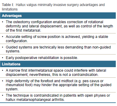
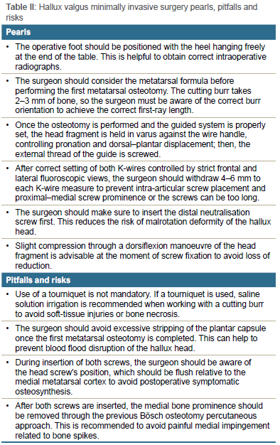
The modern fourth-generation MIS technique for HV correction was initially described by Redfern and Vernois,37 utilising a chevron osteotomy, which was subsequently modified to a transverse osteotomy by Robinson and Lam et al.39 This technique is performed under fluoroscopic guidance, and both osteotomy and fixation are achieved without direct visualisation of the bone or screws, using percutaneous incisions less than 3 mm.37-39
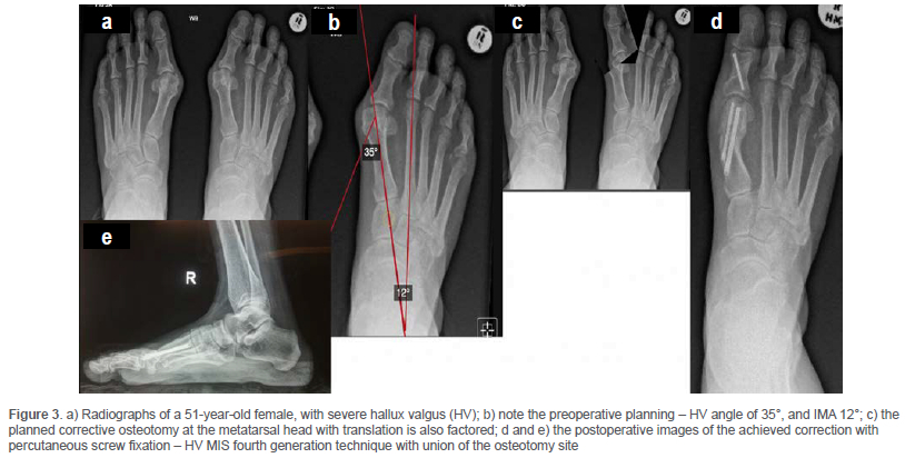
An extracapsular chevron or transverse osteotomy is created through a percutaneous incision at the metatarsal neck. After lateral displacement of the metatarsal head and three-dimensional correction of the deformity, the osteotomy is stabilised with two parallel (or divergent) screws, with the proximal screw being bicortical to enhance fixation stability.37-39 Medial ledge excision and removal of any prominent medial exostosis are performed, and a percutaneous Akin osteotomy, along with lateral release of the first MTP joint (including division of the lateral sesamoid-phalangeal ligament), can be added to ensure joint congruency37-39 (Figures 3 and 4).
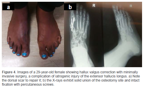
This technique allows for multiplanar rotational correction of the deformity by manipulating an unstable, extra-articular distal transverse osteotomy held rigidly with two screws, one of which must be bicortical to ensure biomechanical stability.37-39 The fourth-generation MIS aims to address all deformity components - coronal, sagittal and rotational - and correct severe deformities with greater displacement of the metatarsal head while maintaining joint stability. The principle of metatarsal head correction in this approach parallels that of open scarf and Akin osteotomies, but with added correction of rotation. The fixation's stability permits immediate weight-bearing, early hallux mobilisation, and accelerated rehabilitation.37-39
Open versus MIS HV
The evidence supporting third- and fourth-generation MIS techniques for HV correction is predominantly derived from large single-surgeon case series, with few randomised controlled trials available.36-39 Systematic reviews comparing open and MIS approaches generally report no significant differences in radiological correction, patient-reported outcome measures (PROMs), or complication rates.40 These reviews suggest that MIS techniques achieve comparable, and often non-inferior, clinical and radiological outcomes relative to traditional open procedures, with potential benefits including reduced pain scores, smaller scars, lower analgesia consumption, and possibly faster recovery.40,41 It is important to acknowledge that some patient-seeking benefits from MIS, such as decreased intraoperative bleeding, earlier return to activity, and reduced perioperative pain and scarring, are rarely systematically measured or reported, which may create a gap between patient expectations and reported outcomes.40,41 Open scarf and Akin osteotomy procedures have been associated with excellent short-term patient satisfaction and low complication rates.42
Recurrence remains one of the most significant complications following HV correction with both open and MIS techniques.43-47 It is multifactorial in origin, influenced by patient-specific factors such as anatomy, preoperative deformity, soft tissue characteristics, comorbidities and compliance with postoperative instructions, as well as surgical factors including the choice of technique, implant and surgeon expertise.43-47
The optimal management of severe deformities remains contentious, with many surgeons recommending proximal procedures such as fusion or osteotomy over distal first metatarsal osteotomies. Although these complex corrections can be performed via MIS, there is no definitive evidence favouring MIS over open techniques in such cases.48-50
General complications include wound issues, infection, transfer metatarsalgia, AVN of the first metatarsal head, fracture, hardware prominence, postoperative neuralgia, and complex regional pain syndrome. Systematic reviews report overall complication rates of approximately 7-8% for open techniques, with metatarsal fracture being the most common (4.6%) in a review of 1 189 patients over a decade.51 In MIS, hardware-related issues are most prevalent, with about 40% requiring screw removal due to symptomatic irritation, despite the use of low-profile implants.52,53 Other MIS-specific complications include thermal skin injury (13%), tendon injury (1.5%), and nerve injury (2.4%), attributable to limited direct visualisation.53 The overall complication rate for MIS approaches is higher (~16.6%) than open procedures, mainly due to the prominence of hardware, though rates of fracture, AVN and nerve injury are comparable.51,53 Despite the volume of data, there is a paucity of high-quality randomised controlled trials (RCTs) directly comparing open and MIS techniques with regard to complications. Ongoing research is necessary to clarify whether one approach offers superior safety or efficacy.54
Combined osteotomies for severe or complex HV
The treatment of severe or complex HV, particularly in cases presenting with significant deformity, often necessitates combined surgical interventions to achieve optimal correction. Such approaches may include multiple osteotomies, such as double first metatarsal osteotomies combined with Akin osteotomies, modifications of the Lapidus procedure, or minimally invasive distal osteotomies.55
Cost-effectiveness and resource utilisation in HV surgery - MIS and open surgery
While both methods aim to correct the deformity, disparities in postoperative outcomes, pain management and associated costs are crucial in informing surgical decisions. MIS has been associated with multiple benefits that contribute to overall cost effectiveness.56 Fluoroscopy time, an aspect that can substantially affect operation costs, also shows promising trends in MIS. Procedures such as percutaneous chevron and Akin osteotomies have demonstrated efficient utilisation of fluoroscopy, leading to reduced operational times, as noted by Acevedo et al.57 The efficient use of imaging techniques can minimise both financial expenditure and potential risks associated with prolonged exposure to radiation.
Operating room costs also involve the assessment of implant costs. Studies indicate that while the costs of certain implants may be comparable, the overall requirement for additional materials and longer direct healthcare personnel involvement in open surgery can elevate overall expenditure.58 In terms of postoperative management, effective pain control is paramount. Research on postoperative analgesia indicates that MIS procedures have shown significant advantages, with lower postoperative pain levels necessitating less intense analgesic regimens, further minimising additional costs and enhancing patient satisfaction.59
Rehabilitation protocols and return to activity
Open procedures such as scarf or Akin osteotomies often necessitate a more conservative rehabilitation protocol. Patients are usually advised to abstain from weight-bearing activities for a period of six weeks, reflecting the greater soft tissue trauma involved in these techniques.60 During this phase, physical therapy tends to focus on gentle range-of-motion exercises and progressive resistance training to facilitate healing.
According to a meta-analysis by Ji et al., the differences in rehabilitation timelines are attributable to the extent of soft tissue injury: open surgery induces more extensive trauma, necessitating longer recovery periods, whereas MIS allows for earlier mobilisation due to its targeted and minimally invasive nature.60 Patients undergoing MIS frequently return to normal activities and light sporting pursuits within six to eight weeks postoperatively.53 Full recovery and return to high-impact activities, such as running, generally exceed three months.
Proximal first metatarsal osteotomies
Proximal first metatarsal osteotomies are crucial surgical options for correcting moderate to severe HV deformities. These procedures are associated with various complications, notably metatarsal shortening, especially in proximal closing wedge osteotomies. The extent of shortening reported across studies varies, with potential reductions ranging around 5 mm in some series.61,62 The medial opening wedge osteotomy can achieve lengthening of the metatarsal and tightening of medial soft tissues but often requires bone grafting due to the osteotomy's nature. However, this technique carries a significant risk of dorsal malunion, which can impact clinical outcomes.63
Proximal oblique (Ludloff) osteotomy
The modified Ludloff osteotomy is a valuable surgical technique used to correct HV deformities. This procedure involves making an oblique cut commencing approximately 2 mm distal to the MTC joint on the dorsal aspect, extending distally and inferiorly, with an angle of the cut around 30° relative to the long axis of the metatarsal. Keeping the distal plantar cortex intact until the completion of the distal cut enhances stabilisation during the procedure, particularly when utilising a proximal screw to create rotation around a single axis.64,65 Neufeld et al. have demonstrated that immediate weight-bearing postoperatively is feasible with dorsal locking plate fixation, which may enhance patient recovery and satisfaction.66 Moreover, the use of dorsal plating has been recommended for patients with compromised bone quality, such as those with rheumatoid arthritis or low bone mineral density, as this approach adds stability while minimising the risk of dorsal malunion, although this complication remains a significant concern with such procedures.64
Proximal crescentic osteotomy
Proximal crescentic osteotomies have been described as effective surgical procedures for the correction of HV deformities. Coughlin and Jones reported that crescentic osteotomies are typically performed approximately 1 cm distal to the MTC joint, utilising a crescent blade with the concavity oriented proximally.10 This design facilitates the rotation of the metatarsal shaft during the procedure.67 The fixation of these osteotomies with internal devices is generally considered straightforward, contributing to their widespread adoption.68
Akin osteotomy
In cases of HV interphalangeus, the Akin osteotomy of the proximal phalanx is frequently combined with a medial closing wedge osteotomy of the proximal phalanx base to effectively correct the deformity. The Akin osteotomy is particularly indicated when the interphalangeal angle (IPA) exceeds 10°, enhancing the alignment of the first toe.69 This osteotomy is rarely executed as a standalone procedure; rather, it is typically used as an adjunctive procedure in conjunction with other corrective methods for HV.70 The surgical placement of the osteotomy is crucial; it must be sufficiently distal to avoid intra-articular extension into the MTPJ due to the concave anatomy of the proximal phalanx base. This consideration is vital as it minimises the risk of joint dysfunction postoperatively.71
Arthrodesis
First MTPJ arthrodesis is often indicated in cases of HV associated with rheumatoid arthritis, recurrent deformities, severe cases, hallux rigidus, or neurological conditions. This surgical procedure effectively reduces the IMA while preserving the adductor pull on the proximal phalanx, which is crucial for maintaining functional alignment in the forefoot.72 The choice between an arthrodesis and an osteotomy may ultimately depend on individual patient factors and specific clinical scenarios, as well as considerations regarding long-term functional outcomes.73
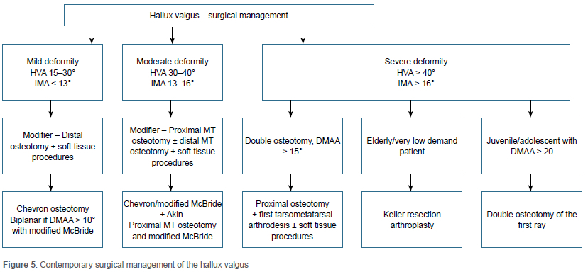
First MTC joint arthrodesis, commonly referred to as the Lapidus procedure, is indicated for severe HV, first MTC instability, and recurrent deformities. According to Klouda et al., the modified Lapidus arthrodesis has demonstrated a success rate of approximately 75% when employed as a primary corrective measure.74 This procedure is associated with changes in metatarsal length; it typically results in a decrease of about 5 mm when no bone graft is utilised, whereas the use of a graft may lead to slight lengthening. These alterations can help restore alignment of the first ray, but they can present technical challenges during the surgical intervention.74 Complications arising from this procedure include HV, recurrence of deformities, deep vein thrombosis and fixation failure. Moreover, stiffness is a prevalent postoperative concern affecting a significant number of patients, with some studies reporting rates around 58%.75 Recent studies have shown that the modified Lapidus procedure effectively decreases the pronation of the first metatarsal, contributing to improved clinical outcomes and lower recurrence rates in patients with HV76 (Figure 5).
Conclusions
HV remains a common and multifaceted forefoot deformity with numerous surgical options. Open procedures, particularly distal osteotomies such as chevron and scarf, have demonstrated high union rates and durable correction, though recurrence remains a concern, particularly with under-correction or severe deformities. Minimally invasive techniques, especially third- and fourth-generation approaches, are increasingly supported by evidence, offering benefits such as reduced soft tissue trauma and faster recovery; however, long-term data are limited. Proximal osteotomies and joint-preserving procedures like the Lapidus are crucial for high-grade or recurrent deformities, though they carry increased risks of complications. Overall, successful management requires careful patient assessment, deformity characterisation, and surgical expertise. Further high-quality, long-term studies and standardised outcome measures are needed to define the optimal approach, improve durability, and minimise recurrence in this widely encountered condition.
Ethics statement
The authors declare that this submission is in accordance with the principles laid down by the responsible research publication position statements as developed at the 2nd World Conference on Research Integrity in Singapore 2010.
This article does not contain any studies with human or animal subjects; patient consent was therefore unnecessary.
Declaration
The authors declare authorship of this article and that they have followed sound scientific research practice. This research is original and does not transgress plagiarism policies.
Author contributions
SD: study conceptualisation, data capture, data analysis, first draft preparation, manuscript revision
RF: study conceptualisation, data capture, data analysis, first draft preparation, manuscript revision
PGM: study conceptualisation, data capture, data analysis, first draft preparation, manuscript revision
ORCID
Sigodi D https://orcid.org/0009-0006-5682-9936
Frank R https://orcid.org/0009-0004-9139-0477
Mthethwa PG https://orcid.org/0000-0001-6432-1899
References
1. Nix S, Smith M, Vicenzino B. Prevalence of hallux valgus in the general population: a systematic review and meta-analysis. J Foot Ankle Res. 2010;3:21. [ Links ]
2. Hueter C. Zur pathologie des Hallux valgus. Arch Orthop Unfallchir (Leipz). 1925;44:1-8. [ Links ]
3. Perera A, Mason L, Stephens M. The pathogenesis of hallux valgus. JBJS. 2011;93(17):1650-61. [ Links ]
4. Menz H, Munteanu SE, Tan J, et al. Risk factors for hallux valgus: a systematic review of the literature. J Foot Ankle Res. 2015;8:44. [ Links ]
5. King DM, Toolan BC. Associated deformities and hypermobility in hallux valgus: an investigation with weightbearing radiographs. Foot Ankle Int. 2004;25(4):251-55. [ Links ]
6. Coughlin MJ, Jones CP. Hallux valgus: demographics, etiology, and radiographic assessment. Foot Ankle Int. 2007;28(7):759-77. [ Links ]
7. Öncü M, Genç E. The co-occurrence of calcaneal spur and hallux valgus: A radiological evaluation. J Back Musculoskelet Rehabil. 2022;35(3):539-44. [ Links ]
8. Vlahov G, Bosi S, Nikolić S. The role of footwear in the development of hallux valgus. Foot Ankle Int. 2014;35(11):1229-36. [ Links ]
9. Bock P, Kristen K-H, Kröner A, Engel A. Hallux valgus and cartilage degeneration in the first metatarsophalangeal joint. J Bone Joint Surg Br. 2004;86-B(5):669-73. [ Links ]
10. Coughlin MJ, Jones CP. Hallux valgus and hallux rigidus. In: Mann RA, Coughlin MJ, editors. Surgery of the Foot and Ankle. 10th ed. St. Louis: Mosby Elsevier; 2014. p. 1234-48. [ Links ]
11. Meng HZ, Zhang WL, Li XC, Yang MW. Radiographic angles in hallux valgus: comparison between protractor and iPhone measurements. J Orthop Res. 2015;33(8):1250-54. [ Links ]
12. Lai MC, Rikhraj IS, Woo YL, et al. Clinical and radiological outcomes comparing percutaneous chevron-Akin osteotomies vs open scarf-Akin osteotomies for hallux valgus. Foot Ankle Int. 2018;39(3):311-17. [ Links ]
13. Traynor C, Jastifer J. First-tarsometatarsal joint alignment after first-metatarsophalangeal joint arthrodesis for hallux valgus. Foot Ankle Orthop. 2021;6(2):24730114211008514. [ Links ]
14. Tsai J, Daniel JN, McDonald EL, et al. High prevalence of degenerative changes at the metatarsal head sesamoid articulation found during hallux valgus correction surgery. Foot Ankle Spec. 2021;14(3):219-25. [ Links ]
15. Kim Y, Kim JS, Young KW, et al. A new measure of tibial sesamoid position in hallux valgus in relation to the coronal rotation of the first metatarsal in CT scans. Foot Ankle Int. 2015;36(8):944-52. [ Links ]
16. Helito PVP, Rocha SRR, Ortiz RT, et al. Accuracy of magnetic resonance imaging for diagnosing hallux valgus. Radiologia brasileira. 2020;53(2):81-85. [ Links ]
17. Abdalbary SA. Foot mobilization and exercise program combined with toe separator improves outcomes in women with moderate hallux valgus at 1-year follow-up: a randomized clinical trial. J Am Podiatr Med Assoc. 2018;108(6):478-86. [ Links ]
18. Ying J, Xu Y, István B, Ren F. Adjusted indirect and mixed comparisons of conservative treatments for hallux valgus: a systematic review and network meta-analysis. Int J Environ Res Public Health. 2021;18(7):3841. [ Links ]
19. Watanabe K, Kubota M, Tanaka H, et al. Japanese Orthopaedic Association (JOA) clinical practice guidelines on the management of hallux valgus-secondary publication. Elsevier; 2024. p. 1-26. [ Links ]
20. Gur G, Ozkal O, Dilek B, et al3. Effects of corrective taping on balance and gait in patients with hallux valgus. Foot Ankle Int. 2017;38(5):532-40. [ Links ]
21. Chadchavalpanichaya N, Prakotmongkol V, Polhan N, et al. Effectiveness of the custom mold room temperature vulcanizing silicone toe separator on hallux valgus: a prospective, randomized single-blinded controlled trial. Prosthet Orthot Int. 2018;42(2):163-70. [ Links ]
22. Mann RA, Coughlin MJ. Surgery of the foot and ankle. 10th ed. Elsevier Saunders; 2014. [ Links ]
23. Kakwani M, Kakwani R. Current concepts review of hallux valgus. J Arthrosc Jt Surg. 2021;8(3):222-30. [ Links ]
24. Mann RA, Coughlin MJ. Hallux valgus-etiology, anatomy, treatment and surgical considerations. Clin Orthop Relat Res®. 1981;157:31-41. [ Links ]
25. Sangeorzan BJ, Hansen Jr ST. Modified Lapidus procedure for hallux valgus. Foot Ankle. 1989;9(6):262-66. [ Links ]
26. Schneider W, Aigner N, Pinggera O, Knahr K. chevron osteotomy in hallux valgus: ten-year results of 112 cases. J Bone Joint Surg Br. 2004;86(7):1016-20. [ Links ]
27. Resch S, Stenstrom A, Reynisson K, Jonsson K. chevron osteotomy for hallux valgus not improved by additional adductor tenotomy: a prospective, randomized study of 84 patients. Acta Orthop Scand. 1994;65(5):541-4. [ Links ]
28. Hawkins F, Mitchell CL, Hedrick DW. Correction of hallux valgus by metatarsal osteotomy. JBJS. 1945;27(3):387-94. [ Links ]
29. Mitchell CL, Fleming JL, Allen R, et al. Osteotomy-bunionectomy for hallux valgus. JBJS. 1958;40(1):41-60. [ Links ]
30. Heerspink FL, Verburg H, Reininga I, Van Raaij T. chevron versus Mitchell osteotomy in hallux valgus surgery: a comparative study. J Foot Ankle Surg. 2015;54(3):361-64. [ Links ]
31. Coetzee JC. Scarf osteotomy for hallux valgus repair: the dark side. Foot Ankle Int. 2003;24(1):29-33. [ Links ]
32. Crevoisier X, Mouhsine E, Ortolano V, et al. The scarf osteotomy for the treatment of hallux valgus deformity: a review of 84 cases. Foot Ankle Int. 2001;22(12):970-76. [ Links ]
33. Del Vecchio JJ, Ghioldi ME. Evolution of minimally invasive surgery in hallux valgus. Foot Ankle Clin. 2020;25(1):79-95. [ Links ]
34. Bösch P, Markowski H, Rannicher V. Technik und erste Ergebnisse der subkutanen distalen Metatarsale-I-Osteotomie. Orthop Praxis. 1990;26:51-56. [ Links ]
35. Vernois J, Redfern D. Percutaneous chevron; the union of classic stable fixed approach and percutaneous technique. Fuß & Sprunggelenk. 2013;11(2):70-75. [ Links ]
36. Lewis TL, Barakat A, Mangwani J, et al. Current concepts of fourth-generation minimally invasive and open hallux valgus surgery. Bone Joint J. 2025;107(1):10-18. [ Links ]
37. Redfern D, Vernois J. Minimally invasive chevron Akin (MICA) for correction of hallux valgus. Tech Foot Ankle Surg. 2016;15(1):3-11. [ Links ]
38. Lewis TL, Lau B, Alkhalfan Y, et al. Fourth-generation minimally invasive hallux valgus surgery with metaphyseal extra-articular transverse and Akin osteotomy (META): 12 month clinical and radiologic results. Foot Ankle Int. 2023;44(3):178-91. [ Links ]
39. Robinson PW, Lam P. Percutaneous surgery for mild to severe hallux valgus. Tech Foot Ankle Surg. 2020;19(2):76-83. [ Links ]
40. Torrent J, Baduell A, Vega J, et al. Open vs minimally invasive scarf osteotomy for hallux valgus correction: a randomized controlled trial. Foot Ankle Int. 2021;42(8):982-93. [ Links ]
41. Lu J, Zhao H, Liang X, Ma Q. Comparison of minimally invasive and traditionally open surgeries in correction of hallux valgus: a meta-analysis. J Foot Ankle Surg. 2020;59(4):801-806. [ Links ]
42. Sieloff MR, Tokarski AR, Elliott AD, et al. The incidence of complications following scarf osteotomy for the treatment of hallux valgus: a systematic review with meta-analysis. J Foot Ankle Surg. 2023;62(4):610-17. [ Links ]
43. Ezzatvar Y, López-Bueno L, Fuentes-Aparicio L, Dueñas L. Prevalence and predisposing factors for recurrence after hallux valgus surgery: a systematic review and meta-analysis. J Clin Med. 2021;10(24):5753. [ Links ]
44. Lalevee M, de Cesar Netto C, Boublil D, Coillard J-Y. Recurrence rates with longer-term follow-up after hallux valgus surgical treatment with distal metatarsal osteotomies: a systematic review and meta-analysis. Foot Ankle Int. 2023;44(3):210-22. [ Links ]
45. Nair A, Bence M, Saleem J, et al. A systematic review of open and minimally invasive surgery for treating recurrent hallux valgus. Surg J. 2022;8(04):e350-e6. [ Links ]
46. Kaufmann G, Sinz S, Giesinger JM, et al. Loss of correction after chevron osteotomy for hallux valgus as a function of preoperative deformity. Foot Ankle Int. 2019;40(3):287-96. [ Links ]
47. Raikin SM, Miller AG, Daniel J. Recurrence of hallux valgus: a review. Foot Ankle Clin. 2014;19(2):259-74. [ Links ]
48. Biz C, Fosser M, Dalmau-Pastor M, et al. Functional and radiographic outcomes of hallux valgus correction by mini-invasive surgery with Reverdin-Isham and Akin percutaneous osteotomies: a longitudinal prospective study with a 48-month follow-up. J Orthop Surg Res. 2016;11:1-13. [ Links ]
49. Kaufmann G, Mörtlbauer L, Hofer-Picout P, et al. Five-year follow-up of minimally invasive distal metatarsal chevron osteotomy in comparison with the open technique: a randomized controlled trial. JBJS. 2020;102(10):873-79. [ Links ]
50. Lewis TL, Robinson PW, Ray R, et al. Five-year follow-up of third-generation percutaneous chevron and Akin osteotomies (PECA) for hallux valgus. Foot Ankle Int. 2023;44(2):104-17. [ Links ]
51. Barg A, Harmer JR, Presson AP, et al. Unfavorable outcomes following surgical treatment of hallux valgus deformity: a systematic literature review. JBJS. 2018;100(18):1563-73. [ Links ]
52. Lewis TL, Ray R, Miller G, Gordon DJ. Third generation minimally invasive chevron and Akin osteotomies (MICA) in hallux valgus surgery: two-year follow-up of 292 cases. JBJS. 2021;103(13):1203-11. [ Links ]
53. Gonzalez T, Encinas R, Johns W, Benjamin Jackson III J. Minimally invasive surgery using a Shannon burr for the treatment of hallux valgus deformity: a systematic review. Foot Ankle Orthop. 2023;8(1):24730114221151069. [ Links ]
54. Waehner M, Klos K, Polzer H, et al. Lapidus arthrodesis for correction of hallux valgus deformity: a systematic review and meta-analysis. Foot Ankle Spec. 2024:19386400241233832. [ Links ]
55. Al-Nammari SS, Christofi T, Clark C. Double first metatarsal and Akin osteotomy for severe hallux valgus. Foot Ankle Int. 2015;36(10):1215-22. [ Links ]
56. Alimy AR, Polzer H, Ocokoljic A, et al. Does minimally invasive surgery provide better clinical or radiographic outcomes than open surgery in the treatment of hallux valgus deformity? a systematic review and meta-analysis. Clin Orthop Relat Res. 2022;481(6):1143-55. [ Links ]
57. Acevedo JI, Cedeno AA, Doty JF, McWilliam JR. Minimally invasive technique for hallux valgus correction. Tech Foot Ankle Surg. 2021;21(1):9-19. [ Links ]
58. Özdemir E, Aynardi M. Minimally invasive bunion surgery for hallux valgus: a surgical technique. Surgicoll. 2024;2(1). [ Links ]
59. Merivirta R, Alanen J, Haapoja E, et al. Postoperative pain management with transdermal fentanyl after forefoot surgery: a randomised, placebo-controlled study. J Pain Res. 2015:39. [ Links ]
60. Ji L, Wang K, Ding S, et al. Minimally invasive vs. open surgery for hallux valgus: a meta-analysis. Front Surg. 2022;9. [ Links ]
61. Schuh R, Willegger M, Holinka J, et al. Angular correction and complications of proximal first metatarsal osteotomies for hallux valgus deformity. Int Orthop. 2013;37(9):1771-80. [ Links ]
62. Mittag F, Leichtle U, Meisner C, et al. Proximal metatarsal osteotomy for hallux valgus: an audit of radiologic outcome after single screw fixation and full postoperative weightbearing. J Foot Ankle Res. 2013;6:22. [ Links ]
63. Watson TS, Hsiao MS, Harasym C, Walsh JP. The proximal opening wedge osteotomy: a review of the evidence and technique. Tech Foot Ankle Surg. 2021;20(4):208-15. [ Links ]
64. Sharma KM, Parks BG, Nguyen A, Schon LC. Plantar-to-dorsal compared to dorsal-to plantar screw fixation for proximal chevron osteotomy: a biomechanical analysis. Foot Ankle Int. 2005;26(10):854-58. [ Links ]
65. Choi WJ, Yoon HK, Yoon HS, et al. Comparison of the proximal chevron and Ludloff osteotomies for the correction of hallux valgus. Foot Ankle Int. 2009;30(12):1154-60. [ Links ]
66. Neufeld SK, Marcel JJ, Campbell M. Immediate weight bearing after hallux valgus correction using locking plate fixation of the Ludloff osteotomy: a retrospective review. Foot Ankle Spec. 2018;11(2):148-55. [ Links ]
67. Vandeputte G, Dereymaeker G, Steenwerckx A, Peeraer L. The Weil osteotomy of the lesser metatarsals: a clinical and pedobarographic follow-up study. Foot Ankle Int. 2000;21(5):370-74. [ Links ]
68. Wang L, Yao L, Dong W, Mao H. Compare the initial result of one screw or two screws fixation for proximal crescentic metatarsal osteotomy with distal soft tissue reconstruction in severe hallux valgus treatment. Research Square. 2022. [ Links ]
69. Liszka H, Gądek A. Comparison of the type of fixation of Akin osteotomy. Foot Ankle Int. 2018;40(4):390-97. [ Links ]
70. Kaufmann G, Hofer P, Braito M, et al. Effect of Akin osteotomy on hallux valgus correction after scarf osteotomy with hallux valgus interphalangeus. Foot Ankle Int. 2019;40(10):1182-88. [ Links ]
71. Nakajima K. Distal metatarsal and proximal phalangeal osteotomies without soft tissue procedure for hallux valgus with 80° hallux valgus angle: a case report. J Orthop Case Rep. 2023;13(6):29-34. [ Links ]
72. Cravey KS, Barron I, Atway S, et al. Intermetatarsal angle and hallux abductus angle reduction after first metatarsophalangeal joint arthrodesis in mild, moderate, and severe hallux valgus. J Am Podiatr Med Assoc. 2021;111(2). [ Links ]
73. Sperone E, Rofrano M, Bigatti A, et al. Hallux interphalangeal involvement after metatarsophalangeal joint arthrodesis. treatment and results. J Foot Ankle. 2020;14(2):132-37. [ Links ]
74. Klouda J, Hromádka R, Šoffová S, et al. The change of first metatarsal head articular surface position after Lapidus arthrodesis. BMC Musculoskelet Disord. 2018;19(1). [ Links ]
75. Park HW, Lee KB, Chung JY, Kim MS. Comparison of outcomes between proximal and distal chevron osteotomy, both with supplementary lateral soft-tissue release, for severe hallux valgus deformity. Bone Joint J. 2013;95-B(4):510-6. [ Links ]
76. Donantoni M, Santini S, Martinelli D, Marinozzi A. Surgical techniques for Lapidus arthrodesis: approaches, indications and outcomes. J Clin Med. 2025; 14(13):4591. [ Links ]
Received: July 2025
Accepted: October 2025
Published: May 2026
* Corresponding author: sigodid@gmail.com
Editor: Dr Graham McCollum, University of Cape Town, Cape Town, South Africa
Funding: No funding was received for this study.
Conflict of interest: The authors declare they have no conflicts of interest that are directly or indirectly related to the research.