


Services on Demand
Journal

Article
 English (pdf)
 Article in xml format
 Article references
 Send this article by e-mail
Indicators
Related links
-
 Cited by Google
 -
 Similars in Google
Share

 Permalink
South African Journal of Animal Science
On-line version ISSN 2221-4062Print version ISSN 0375-1589
S. Afr. j. anim. sci. vol.55 n.2 Pretoria 2025
https://doi.org/10.4314/SAJAS.V55I2.04
The effect of a phytogenic-based feed additive on concurrent Lawsonia intracellularis and Brachyspira hyodysenteriae infections in pigs
C.F. WuI; H.C. KuoI; M. GlišićII; M. VasiljevićIII; J. RajIII; J. Bošnjak-NeumullerIII, #; V. DraškovićIV
IDepartment of Veterinary Medicine, College of Veterinary Medicine, National Chiayi University, Chiayi, 60054, Taiwan
IIDepartment of Food Hygiene and Technology, Faculty of Veterinary Medicine, University of Belgrade, Bulevar Oslobodjenja 18, 11000 Belgrade, Serbia
IIIPatent Co. DOO, Vlade Ćetkovića 1a, 24211 Mišićevo, Serbia
IVDepartment of Animal Hygiene, Faculty of Veterinary Medicine, University of Belgrade, Bulevar Oslobodjenja 18, 11000 Belgrade, Serbia
ABSTRACT
This study investigated the efficacy of a commercial phytogenic-based premixed feed additive (PFA) in treating combined Lawsonia intracellularis and Brachyspira hyodysenteriae infections in finishing pigs, with tiamulin/lincomycin treatment as the control. Pigs aged 20 weeks were allocated to PFA treatment (11 pens, 45 pigs per pen) and control (7 pens, 43 pigs per pen) groups for a seven-week experimental period. Floor faecal samples and rectal swabs were collected weekly, and the percentage of pigs per pen with diarrhoea was recorded weekly. The bacterial contents of the samples were determined using real-time polymerase chain reaction, and at the end of the experiment, histological changes in ileal samples were examined. There was an intermittent decrease in L. intracellularis in the control group (from 4.85 to 0.82 DNA log10 copies/μl) and a continuous reduction in L. intracellularis in the PFA group (from 5.69 to 0.64 DNA log10 copies/μl) over a six-week period. B. hyodysenteriae was not detected in rectal swabs from the control group at week six, and an intermittent decrease in B. hyodysenteriae, from 3.04 to 0.26 DNA log10 copies/μl, was observed in the PFA group. Bacterial DNA in the floor faecal samples declined during the seven-week experimental period, as found for the rectal swabs. There were no cases of diarrhoea from week two onwards in the control group and week three onwards in the PFA group. The results of this study indicate that a PFA rich in essential oils has a therapeutic effect comparable to that of tiamulin/lincomycin in pigs with proliferative enteropathy and swine dysentery.
Keywords: finishing pigs, phytogenic-based premixed feed additive, proliferative enteropathy, realtime polymerase chain reaction, swine dysentery, tiamulin/lincomycin
Introduction
The occurrence of enteric diseases during the growing and finishing stages results in significant economic losses in the pig industry worldwide (Dors et al., 2015). Of the various intestinal pathogens, two of the most important are Lawsonia intracellularis and Brachyspira hyodysenteriae. These bacteria primarily affect finisher pigs, gilts, and boars, with the obligate intracellular bacterium L. intracellularis causing proliferative enteropathy (PE) (McOrist & Gebhart, 2006), and the gram-negative anaerobic spirochete B. hyodysenteriae causing swine dysentery (SD) (Hampson et al., 2006). These pathogens, either individually or in combination, result in the development of clinical symptoms such as anorexia, diarrhoea, and growth retardation, leading to decreased welfare and increased production costs. The importance of monitoring and reducing the prevalence of L. intracellularis and B. hyodysenteriae is thus clear (Stege et al., 2001). Significant efforts have therefore been made over the last two decades to understand and identify the transmission routes of L. intracellularis and B. hyodysenteriae in commercial and feral pig herds worldwide (Phillips et al., 2009; Arnold et al., 2019; Dors et al., 2019; Neirynck et al., 2020; Carranza et al., 2021).
Co-infection with multiple agents is commonly observed in pig herds with enteric health problems (Komine et al., 2016; Dors et al., 2019; Nuntapaitoon et al., 2021). Dors et al. (2019) reported the simultaneous presence of Brachyspira pilosicoli and B. hyodysenteriae, as well as B. pilosicoli and L. intracellularis, in the faeces of pigs older than seven weeks. Specifically, it was found that L. intracellularis infection, which causes primary lesions in the ileum (with potential spread to the colon and jejunum), increases the likelihood of infection with other enteric pathogens (Komine et al., 2016). Despite studies detecting mixed infections of L. intracellularis and B. hyodysenteriae in pigs, and an increasing trend in the use of routine diagnostics tests (Stege et al., 2001; Suh & Song, 2005; Phillips et al., 2009; Reiner et al., 2011), the conditions and pathogenesis of this co-infection remain complex and poorly understood (Daniel et al., 2023). In addition to de Groot et al. (2022) investigating the pathohistological lesions induced by combined L. intracellularis and B. hyodysenteriae infections in an ex vivo swine colon model, Daniel et al. (2023) found a synergistic effect of a mixed infection with these bacteria on clinical symptoms, macroscopic and microscopic lesions, and the faecal microbiome profile in experimentally infected piglets.
The identification of various risk factors has led to the development of different strategies in the management of L. intracellularis and B. hyodysenteriae infections, as follows: (i) improving building construction and internal biosecurity measures (Jacobson et al., 2010); (ii) developing methods for the quick and easy detection of these bacteria, individually (Nathues et al., 2009) or simultaneously (Nathues et al., 2007); (iii) modifications of the diet in terms of form, ingredient composition, and the supplementation of feed additives (Whitney et al., 2006; M0lbak et al., 2008); and (iv) the use of vaccines and antibiotics (Card et al., 2018). Today, commercially available live attenuated and inactivated bacterin-based L. intracellularis vaccines are used in the prophylaxis of PE. While they effectively reduce L. intracellularis lesions and shedding, shortcomings in the application and immune response quality persist, often necessitating additional interventions for PE control (Karuppannan & Opriessnig, 2018).
Unlike for L. intracellularis, developing a vaccine for controlling SD has encountered several limiting factors (Álvarez-Ordóñez et al., 2013a). Therefore, antimicrobial use and biosecurity improvement are still considered primary methods in treatment, control, and eradication programmes for Brachyspira spp. infections in pig herds (Massacci et al., 2018). The most widely used drugs in practice, which have proven good clinical effects and relatively short withdrawal periods, are the pleuromutilins (tiamulin and valnemulin), tylosin, and lincomycin (Mirajkar et al., 2016). However, the reliance on continuous treatment with antibiotics over extended periods in the past two decades has led to the emergence of multidrug-resistant Brachyspira spp. isolates with reduced susceptibility to pleuromutilins, particularly tiamulin, in various countries (Karlsson et al., 2003; Hidalgo et al., 2011; Sperling et al., 2011; Pringle et al., 2012; Joerling et al., 2018). In contrast, because of the difficulty in isolating and establishing L. intracellularis in cell culture, there is limited data on its antimicrobial susceptibility (Yeh et al., 2011; Luo et al., 2020). However, a study by Wattanaphansak et al. (2019) reported the reduced susceptibility of L. intracellularis strains to lincomycin, gentamicin, trimethoprim, colistin, and bacitracin in vitro.
The injudicious use of antimicrobials in swine production has raised concerns for animal and human health in the context of antimicrobial resistance (Lekagul et al., 2019). The latest data, reported for 81 countries, is concerning, as it shows that the use of antibiotics in animals increased by 2% globally between 2019 and 2021, following several consecutive years of significant decrease (OIE, 2024).
Therefore, the development of non-antimicrobial alternatives to control L. intracellularis and B. hyodysenteriae infections has become a practical requirement (Karuppannan & Opriessnig, 2018; Meneguzzi, 2020).
Recent reports have documented that phytogenic-based premixed feed additives (PFA), particularly ones containing essential oils, can play a significant role in reducing or replacing the use of antibiotics (Stevanović et al., 2018). In cases of infections caused by L. intracellularis or B. hyodysenteriae, the effectiveness of dietary supplements like prebiotics, probiotics, and organic acids has been confirmed (Hansen et al., 2011; Meneguzzi, 2020; de Groot et al., 2022; Xu et al., 2023). In addition, supplementation with natural ingredients that possess direct antimicrobial properties has shown promising results (Karuppannan & Opriessnig, 2018). In an in vitro study, de Groot et al. (2022) demonstrated that a commercial phytogenic product containing a blend of thymol and carvacrol effectively prevented lesions caused by L. intracellularis or B. hyodysenteriae. The beneficial effects of extracts of Origanum vulgare and Allium sativum in controlling PE in weaned piglets have also been observed in vivo (Papatsiros et al., 2009). Furthermore, a few specific field studies have shown that supplementation with the PFA used in this study can lead to a reduced intestinal load, the alleviation of clinical symptoms, and improved performance in herds naturally exposed to either L. intracellularis (Drašković et al., 2018; Katedangsakulwut et al., 2021; Nuntapaitoon et al., 2023) or B. hyodysenteriae (Delić et al., 2018) infection.
To the authors' knowledge, no literature is available on the impact of PFAs on combined L. intracellularis and B. hyodysenteriae infections under field conditions. Given the rising trend in the detection of this bacterial co-infection in pig herds, along with insufficient data on the susceptibility of L. intracellularis and B. hyodysenteriae to natural compounds with antimicrobial effects, and the lack of clinical trials to verify the efficacy of phytogenic compounds under practical conditions, the objective of this study was to assess the impact of a PFA in pigs naturally infected with L. intracellularis and B. hyodysenteriae, and to compare its effectiveness with a control group receiving tiamulin/lincomycin treatment.
Material and methods
A commercially available PFA (the recipe of which is the proprietary information of PATENT CO. DOO, Mišićevo, Serbia) was used in this study. This PFA primarily consists of an essential oil blend (mostly Thymus vulgaris, O. vulgare, and Coriandrum sp.), a Castanea sativa extract, and clinoptilolite. The PFA was added to the feed at a dose of 2 kg/t of feed.
The study was conducted on a commercial wean-to-finish pig farm in Tainan City, Taiwan, with a total capacity of 8000 pigs. The farm had a previous record of PE and SD outbreaks, confirmed by polymerase chain reaction (PCR) assay. The experiment was conducted in accordance with local legislation and the Animal Care and Use Committee of the University of Chiayi, Taiwan. The pigs were slaughtered in a slaughterhouse following standard industrial techniques.
The pigs at the farm were divided into two experimental groups: the treatment group received the PFA-supplemented feed (2 kg/t feed) for seven weeks, and the control group was treated with 110 ppm tiamulin in their feed for the first four weeks, followed by lincomycin treatment for a further two weeks. The treatment group consisted of 495 twenty-week-old pigs (11 pens, with 45 pigs per pen), and the control group consisted of 301 twenty-week-old pigs (7 pens, with 43 pigs per pen). The pigs were housed in pens with concrete floors and had free access to feed and water. Both groups of pigs were kept under the same housing conditions throughout the experiment. The presence of L. intracellularis and B. hyodysenteriae in faecal samples was determined by PCR assay before the animals were introduced into the study.
Floor faecal samples (two samples per pen, 22 samples from the treatment group and 14 samples from the control group) were collected from two randomly selected 25 x 25 cm2 areas in each pen using sterile gauze rinsed with phosphate-buffered saline, for the evaluation of levels of faecal shedding. Rectal swabs (10 samples from the treatment group and 12 samples from the control group) were collected from randomly selected and marked pigs with observed clinical signs of diarrhoea using sterile cotton swabs. Both floor faecal samples and rectal swabs were collected weekly, on days 0, 7, 14, 21, 28, 35, 42, and 49, while the pigs were 20 to 27 weeks of age. After the first week of the trial, one pig from the treatment group was excluded because of sudden death. A total of 36 floor faecal samples and 21 rectal swabs per week were thus collected.
The numbers of L. intracellularis and B. hyodysenteriae bacteria in floor faecal samples and on rectal swabs were determined using real-time PCR. Total DNA was extracted using a commercial kit (DNeasy® PowerSoil® Pro Kit, Qiagen, Hilden, Germany) according to the manufacturers' protocol, and extracted DNA samples were stored at -20 °C until examination. Amplifications were performed using the StepOneTM real-time PCR system (Applied Biosystems®, Foster, USA), and the primers used for the amplification of B. hyodysenteriae DNA were, as described by Akase et al. (2009):
Forward primer: 5'-TATGAAGAAGGCAGCAGACGTTTAT-3'
Reverse primer: 5'-GTAGGAAGAAGAAATCTGACAATGCA-3'
TaqMan probe: 5'-FAM-ACACAATCATGCTGAAGC-TAMRA-3'.
The primers used to amplify L. intracellularis DNA were, as previously described by Lindecrona et al., (2002):
Forward primer: 5'-GCGCGCGTAGGTGGTTATAT-3'
Reverse primer: 5'- GCCACCCTCTCCGATACTCA-3'
TaqMan probe: 5'-FAM-CACCGCTTAACGGTGGAACAGCCTT-TAMRA-3'.
The amplification mixture for real-time PCR consisted of 25 μL of PCR Master Mix (GeneReach Biotechnology, Taiwan), and the cycling programme included one step at 93 °C for 5 min, followed by 40 cycles at 93 °C for 15 sec and 72 °C for 1 min.
The numbers of pigs with clinical signs of diarrhoea in the treatment and control groups were recorded weekly throughout the experiment, from 20 to 27 weeks of age. This was expressed as the percentage of pigs with diarrhoea per pen.
Histological analysis was performed on ileal samples collected from one randomly selected marked pig in each group at the slaughterhouse at the end of the experiment. Small intestine samples were obtained from the electrically stunned and slaughtered pigs at 27 weeks of age. Tissue sections of the distal ileum, measuring 10 cm, were collected and flushed with physiological saline and fixed in neutral formalin solution (37%-40% 10% formalin, 4.0 g NaH2PO4H2O, and 6.5 g NaHPO4 H2O) for 24 hours. After fixation and shaping, intestinal samples were dehydrated in increasing concentrations of ethyl alcohol, cleared with xylene, infiltrated with paraffin, and embedded in paraffin blocks. Standard glass microscope slides were used for mounting. Sections 3 μm thick were placed on glass slides and stained using the routine Mayer's haematoxylin and eosin procedure. Microscopic changes were examined using a light microscope (Olympus BX53) with 40 x objective magnification.
The data were analysed using SPSS 20.0 (IBM, Chicago, IL, USA) software. All results were expressed as the mean ± the standard deviation. Continuous variables were examined for normality and homogeneity of variance by examining the residuals using coefficients of skewness and kurtosis, as well as using the Kolmogorov-Smirnov test and the Shapiro-Wilk normality test. The Mann-Whitney U-test and Friedman test were used to assess the significance of differences between the means of the DNA log10 real-time PCR copy numbers for the control and treatment groups. The diarrhoea scores were tested using the Kruskal-Wallis test. A probability value of P <0.05 was considered statistically significant, while Bonferroni correction was applied for the Friedman test, with a post-hoc Wilcoxon test.
Results and discussion
This study focused on combined L. intracellularis and B. hyodysenteriae infections in growing-finishing pigs, and the mutual susceptibility of these bacteria to antibiotics and a commercial feed additive containing essential oils and C. sativa plant extract.
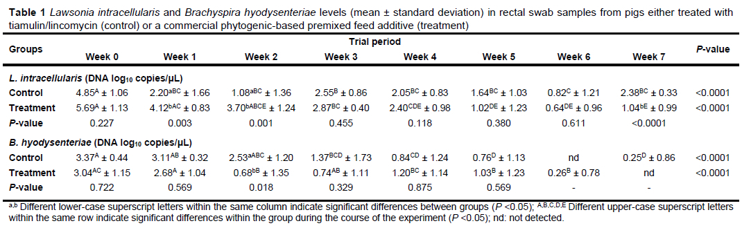
The L. intracellularis and B. hyodysenteriae contents of the rectal swabs from the control and treatment groups are presented in Table 1. Real-time PCR analyses showed that at the beginning of the experiment (week zero), the number of DNA log10 copies per microlitre in the swabs was higher for L. intracellularis than for B. hyodysenteriae, and there was no significant difference between the control and treatment groups for either bacterium. This indicates that the control and treatment groups were uniform at the beginning of the experiment, with the level of co-infection being equally pronounced in both groups of pigs. Furthermore, the finding of a higher level of L. intracellularis than B. hyodysenteriae in both groups of pigs can be partially explained by the hypothesis proposed by Daniel et al. (2023). These authors suggested that L. intracellularis induces lesions in the large intestine in the early stages of infection, and impairs the host intestinal immune response, thereby facilitating the colonisation of B. hyodysenteriae.
There was a prominent reduction in L. intracellularis shedding from week zero to week one (a decrease of 2.65 DNA log10 copies/μL) and from week zero to week two (a decrease of 3.77 DNA log10 copies/μL) in the group treated with tiamulin in their feed. This differed significantly from the treatment group, in which L. intracellularis shedding decreased by only 1.57 DNA log10 copies/μL from week zero to week one, and 1.99 DNA log10 copies/μL from week zero to week two. Corroborating our results, Walter et al. (2001) demonstrated that tiamulin effectively treated pigs with L. intracellularis infection under field-like conditions, eliminating positive faecal PCR results after 14 days of feed-based treatment, initiated at the onset of clinical symptoms. However, by week seven, both groups had had a continuous decrease in the number of L. intracellularis in their rectal swabs, with the exception of the control group in week three, which had a 1.47 DNA log10 copies/μL increase in L. intracellularis at this time.
This isolated increase cannot be attributed to a decreased sensitivity of L. intracellularis to tiamulin, despite the results of Wattanaphansak etal. (2019) and Yeh etal. (2011). Wattanaphansak et al. (2019) indicated that L. intracellularis showed potential resistance to certain antibiotics under in vitro conditions, and Yeh et al. (2011) reported that the effective intracellular and extracellular activities of various antibiotics, including tiamulin, against L. intracellularis strains decreased by two to eight times over an eight-year period. Walter et al. (2001) previously demonstrated that treating PE with tiamulin could affect faecal PCR diagnostic tests by interfering with faecal shedding, making the interpretation of results ambiguous. Therefore, the confounding results of the present study are most likely due to tiamulin's interference with the PCR procedure or an intermittent trend of excretion of L. intracellularis (Schwartz et al., 1999; Jacobson et al., 2010). In accordance with our results, intermittent faecal shedding lasting up to 12 weeks after the first positive PCR result was observed in growing-finishing pigs experimentally challenged with L. intracellularis (Guedes & Gebhart, 2003), and following an acute PE outbreak on a commercial farm (Guedes et al., 2002).
After 14 days of lincomycin supplementation and seven days of antibiotic withdrawal (week seven), significantly higher levels of L. intracellularis were found in the control (2.38 ± 0.03 DNA log10 copies) than in the PFA treatment group (1.04 ± 0.99 DNA log10 copies). The capacity of non-antibiotic additive premixes to be used until the end of fattening, without a withdrawal period, can thus be considered a competitive advantage. In line with these findings, previous studies similarly determined that the addition of a PFA with a similar composition to the one used in the present study significantly decreased L. intracellularis faecal shedding in fattening pigs after two weeks and in weaned piglets after two and four weeks of treatment (Draskovic et al., 2018; Nuntapaitoon et al., 2023). Furthermore, a preparation based on extracts of O. vulgare and A. sativum proved effective in reducing the prevalence of L. intracellularis over a six-week treatment period, with no difference between this group and the control group of piglets, which received tiamulin in their feed throughout the entire experiment (Papatsiros et al., 2009).
A significant decrease in the B. hyodysenteriae DNA load of the rectal swabs between week zero and week seven of the experiment was found in both groups of pigs, with a difference (P = 0.018) between the treatment and the control group only being found in week two (0.68 ± 1.35 and 2.53 ± 1.20 DNA log10 copies/μL, respectively). The B. hyodysenteriae faecal load was below the minimum detection level during week six in the control group and week seven in the treatment group. In contrast with the findings for L. intracellularis, the decline of B. hyodysenteriae DNA log10 copies in the rectal samples was intermittent in the treatment group. However, the sensitivity of B. hyodysenteriae and L. intracellularis detection can be influenced by various factors, such as intermittent excretion patterns, the bacterial load in the faecal samples or rectal swabs, sample pooling, the sensitivity of the PCR protocol, and the DNA extraction procedures (including inhibitory factors) used, all of which must be considered when interpreting the results (Heinonen et al., 2000; Jacobson et al., 2003; Grahofer et al., 2016).
There is little available in vivo data on the antibacterial efficiency of phytogenics for the treatment of pig infections caused by obligatory intracellular agents (Papatsiros et al., 2009; Draskovic et al., 2018; DeliC et al., 2021; Nuntapaitoon et al., 2023), mainly because of the fastidious growth conditions of these agents, and the limited detection methods available (Maele et al., 2015). Additionally, despite the widespread use of phytogenics in animal production, because of their antioxidant and antimicrobial properties and their beneficial impact on growth performance, knowledge about their mode of action is still limited (Silva Júnior et al., 2020). Nonetheless, efforts have been made to demonstrate the in vitro
susceptibility of B. hyodysenteriae and L. intracellularis to active compounds composed of different organic acids and essential oils commonly used as feed additives, as well as to partially explain the mechanisms involved in mitigating the intestinal lesions caused by these bacteria (Álvarez-Ordóñez et al., 2013b; Maele et al., 2015; Gómez-García et al., 2020; Meneguzzi, 2020; de Groot et al., 2022). Álvarez-Ordóñez et al. (2013b) reported the vigorous antibacterial activity of a feed supplement composed of citrus fruit extract against B. hyodysenteriae strains, with minimum inhibitory concentration (MIC) values ranging from 10 ppm to 40 ppm. Similarly, Maele et al. (2015) found that eugenol, carvacrol, thymol, and cinnamaldehyde had low MIC values against three strains of B. hyodysenteriae, with binary essential oil and organic acids combinations having additive effects, but synergism only being observed for a thymol and carvacrol combination. Strong dose-dependent antibacterial activity was reported for thymol and carvacrol, with an approximately 27% higher reduction rate of viable bacterial populations than was found for organic acids (Gómez-García et al., 2020).
It is assumed that multi-component phytogenics based on essential oils, which consist of a wide range of chemical compounds with antimicrobial properties, increase the likelihood of achieving additive or synergistic activities because of their action on different cellular targets (Álvarez-Ordóñez et al., 2013b). Thus, in addition to the bacterial cell membrane, which is the primary cellular target of essential oils because of their lipophilic/hydrophobic nature (Trombetta et al., 2005), it has been established that specific components in essential oils also affect cell proteins embedded in the cytoplasmic membrane by distorting lipid-protein interactions or directly affecting the hydrophobic regions of these proteins (Sikkema et al., 1995). These suggested mechanisms, which exert both physico-chemical effects on bacterial membranes and target specific cellular components, may lower the chances of bacteria developing resistance (Álvarez-Ordóñez et al., 2013b). Considering the results obtained in this study, which suggest the effectiveness of a PFA for treating mixed infections, this represents a significant advantage of natural antimicrobials, particularly in controlling SD. This is especially relevant considering the findings of Yeh et al. (2018) that B. hyodysenteriae isolates from Taiwan have decreased susceptibility to tiamulin and lincomycin, two of the most common antibiotics used in the treatment and control of SD.
It has recently been demonstrated that various non-antimicrobial compounds used as feed additives exhibit beneficial effects in an explant infection model (de Groot et al., 2022). In particular, it was found that phytogenics made from a blend of thymol and carvacrol increased epithelial coverage and downregulated interleukin-1α, interferon-γ, and tumour necrosis factor-α in spiral colon explants infected with B. hyodysenteriae ex vivo. De Groot et al. (2022) suggested that this effect could have been due to the anti-inflammatory properties of carvacrol, thymol, and other phenolic compounds, which are associated with inhibiting the cyclooxygenase-2 cascade. The PFA used in this study, which showed an effect on a combined infection comparable to that of antibiotic treatment, contained a mixture of essential oils from T. vulgaris and O. vulgare, with thymol and carvacrol as the primary active substances. This suggests that similar effects to those described above might be expected in vivo, to a certain extent.
The results for the L. intracellularis and B. hyodysenteriae DNA loads in the floor faecal samples corroborated the levels of L. intracellularis and B. hyodysenteriae shedding found in the rectal swabs (Table 2). At week zero and week one of the study, the PFA-supplemented group had significantly higher numbers of L. intracellularis DNA log10 copies/μL than the control group. However, in week two, a notable decrease in the L. intracellularis contents of the treatment group samples was found, and significant differences between the control group and the PFA-supplemented group were not observed again until week six of the experiment. After the withdrawal of the antibiotic, a significantly lower number of L. intracellularis DNA log10 copies/μL were found in the floor faecal samples from the treatment group (2.58 ± 0.94 DNA log10 copies/μL) than from the control group (3.32 ± 0.58 DNA log10 copies/μL). A significant decline in the L. intracellularis DNA log10 copies/μL in the floor faecal samples from the beginning of the study was found after four weeks of antibiotic supplementation in the control group and two weeks of PFA supplementation in the treatment group. The long-term presence of pathogenic agents in the environment, shed by animals without visible clinical signs, could be the source of infection for susceptible pigs (Guedes & Gebhart, 2003). This was confirmed by the floor faecal sample results from week two to week seven in this study. Furthermore, L. intracellularis could remain viable outside the host for 14 days in faeces at 5-15 °C (Collins et al., 2000).
Contrary to the findings for L. intracellularis, the B. hyodysenteriae DNA log10 copies/μL in weeks zero (P <0.0001), one (P = 0.005), and two (P = 0.030) were higher in the floor faecal samples of the control group than in those of the treatment group. A continuous decline in B. hyodysenteriae bacteria was noted until week six in the control group and week seven in the PFA-supplemented group. Similarly, Delić et al. (2021) showed a positive effect of two essential oil-based phytogenic additives on the degree of infection with B. hyodysenteriae that did not differ from tiamulin treatment.
The significantly lower levels of L. intracellularis and B. hyodysenteriae DNA log10 copies/μL found in the floor faecal samples collected during the last week of the study for the PFA-supplemented group can be attributed to the withdrawal of the antibiotics from the feed of the control group for the last seven days of the experiment, as per the required withdrawal periods for animals intended for slaughter. This reflects one of the main advantages of using antibiotic-alternatives in animal feed for the treatment of bacteria for which the development of resistance has not yet been determined (Yang et al., 2015), while the emergence of antibiotic-resistant strains of L. intracellularis and B. hyodysenteriae suggests an additional advantage (Card et al., 2018; Joerling et al., 2018; Seo et al., 2019).
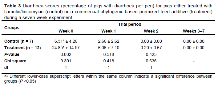
A recent study reported that experimentally infected six-week-old piglets had more pronounced clinical signs, macroscopic changes, and decreased intestinal microbiome diversity in cases of co-infection with L. intracellularis and B. hyodysenteriae than in cases of infection with a single pathogen (Daniel et al., 2023), with co-infection mainly increasing the severity of SD symptoms. Evidence of the beneficial effects of plant-based feed additives on diarrhoeal disorders caused by L. intracellularis or B. hyodysenteriae under field conditions is documented in the literature (Papatsiros et al., 2009; Delic et al., 2018; Draskovic et al., 2018; Nuntapaitoon et al., 2023). However, data on the impact of phytogenics under co-infection conditions remain unavailable. The diarrhoea score results from the present study showed that at the beginning of the experiment, both the control and the treatment groups had pigs with diarrhoea, with a significantly higher incidence in the treatment group (Table 3). After one week of PFA supplementation, the number of pigs with clinical signs had decreased, and there was no significant difference between the control and treatment groups. From week two in the control group and week three in the treatment group, no pigs with diarrhoea were recorded until the end of the experiment. This likely reflects the decrease in the numbers of L. intracellularis and B. hyodysenteriae detected in the rectal swabs through the course of the study, even though these bacteria were present throughout the study period.
In experimentally challenged piglets, differences in microscopic changes in the large intestine were observed between piglets infected with a combination of L. intracellularis and B. hyodysenteriae and piglets infected with only one of the two bacteria, indicating a synergistic effect of these two pathogens (Daniel et al., 2023). In addition to moderate diffuse mucohaemorrhagic colitis and severe diffuse fibrin necrohaemorrhagic catarrhal colitis in the large intestines of co-infected piglets, further histological analysis of the caecum and colon revealed more extensive and intense lesions, including superficial necrosis, haemorrhage, goblet cell hyperplasia, crypt abscesses, and lamina propria neutrophil infiltration, in the co-infected piglets, compared to uninfected piglets and those with PE.
However, co-infected piglets only exhibited more pronounced goblet cell hyperplasia than piglets with SD.
In the control group of co-infected pigs treated with antibiotics in the present study, the architecture of the ileum mucosa was improperly organised into intestinal villi. Increased mucosal thickness, with vanishing villi, was observed, the mucosa was composed of adenomatous and hyperplastic crypts, and goblet cells were reduced in number. Crypts were elongated, dilated, and branched, resulting in the aforementioned thickening of the mucosal layer. Furthermore, dilated crypts were filled with inflammatory cells and cell debris, and the lamina propria was hyperaemic (Figure 1A).
In the group of co-infected pigs treated with the PFA, the architecture of the mucosa was properly organised into intestinal crypts and villi, and the mucosal thickening was reduced compared to the distal ileum of the positive control group of piglets. Goblet cells were slightly proliferated, and the lamina propria mucosa was moderately hyperaemic (Figure 1B). These results suggest that the PFA may have exhibited antibacterial effects on both L. intracellularis and B. hyodysenteriae simultaneously, with a potentially protective effect on the intestinal mucosa. This aligns with the findings of de Groot et al. (2022), where a phytogenic supplement containing thymol and carvacrol exhibited anti-inflammatory effects and increased epithelial coverage in explants infected with an obligatory intracellular agent.
Previous studies have shown that PFAs rich in essential oils improve growth performance in pigs with PE or SD with an efficiency comparable to that of tiamulin (Papatsiros et al., 2009; Delić et al., 2018). The results of this study are in agreement with these findings, as, at the end of the experiment, the average body weight of the pigs that received tiamulin/lincomycin was 116 ± 12.83 kg, and that of the pigs supplemented with the PFA in their feed was 117 ± 15.34 kg, with no significant difference observed between these two groups. It can thus be inferred that the PFA had an effect similar to that of the antibiotics on the performance of the pigs infected with both L. intracellularis and B. hyodysenteriae.
Since this is the first study examining the impact of a PFA on combined L. intracellularis and B. hyodysenteriae infections under field conditions, its significance lies in providing preliminary insights into the efficacy of natural antimicrobials in a practical setting. However, because of the challenging experimental conditions, several limiting factors must be considered, both in interpreting the results and in conducting additional trials. These include using a larger number of animals to monitor prevalence more accurately, as well as optimising factors that influence the detection of pathogens in faecal samples and rectal swabs through the use of specific PCR procedures, in order to minimise variability in results. Additionally, the use of antibiotics in pigs complicates the detection of infection in medicated populations, necessitating larger sample sizes, as indicated by Schwartz et al. (1999).
On farms with concurrent PE and SD infections, it is difficult to separate groups of mono-infected animals. This is another limiting factor of this study that should be considered when elucidating the efficacy of the PFA, as previous studies suggested a synergic effect between these two pathogens. Previous studies have also highlighted the significant role of the intestinal microbiome in disease development (Daniel et al., 2023), underscoring the need to consider the impact of the PFA on dysbiosis. When interpreting production results, the absence of negative controls and the difficulty distinguishing between growth promoting and therapeutic effects complicates the evaluation of the PFA and its comparison to antibiotics. Therefore, monitoring other indicators, such as weight gain, feed intake, and the feed conversion ratio in subclinically infected herds is crucial to determine whether this PFA prevents impaired production.
Conclusions
The present study's findings indicate that a PFA composed of mixtures of different essential oils and plant extracts at a dose of 2 g/kg could effectively control and treat simultaneous L. intracellularis and B. hyodysenteriae infections in finishing pigs. This represents the first report of the efficacy of this PFA as a treatment for L. intracellularis and B. hyodysenteriae co-infection in a clinical trial under field conditions. However, further research is needed to address the limitations of this study, including trials with larger sample sizes and incorporating the monitoring of production performance, to elucidate the extent to which this PFA could replace antibiotics, especially pleuromutilins, in the management of PE and SD in a cost-effective manner.
Acknowledgements
PATENT CO. DOO provided financial support for this trial. The authors also acknowledge the support of the Ministry of Science, Technological Development and Innovation of the Republic of Serbia through a contract signed with the Faculty of Veterinary Medicine, University of Belgrade (No. 451-03-66/2024-03/200143), which funded authors Milica Glišić and Vladimir Draskovic.
Authors' contributions
HCK, MV, and JBN conceived and designed the experiment. CFW and HCK conducted the experiment and performed the analyses. MG analysed the data. CFW, HCK, MG, and VD wrote the manuscript draft. All authors reviewed the manuscript and gave their final approval of the manuscript.
Conflict of interest declaration
PATENT CO. DOO provided financial support for this trial; however, this trial was conducted prospectively and the authors had no preconceptions or biases to influence the results of the study. The authors therefore declare that they have no conflicts of interest.
References
Akase, S., Uchitani, Y., Sohmura, Y., Tatsuta, K., Sadamasu, K., & Adachi, Y., 2009. Application of real time PCR for diagnosis of swine dysentery. J. Vet. Med. Sci., 71, 359-362. doi 10.1292/jvms.71.359 [ Links ]
Álvarez-Ordóñez, A., Carvajal, A., Arguello, H., Martínez-Lobo, F.J., Naharro, G., & Rubio, P., 2013b. Antibacterial activity and mode of action of a commercial citrus fruit extract. J. Appl. Microbiol., 115, 50-60. doi 10.1111/jam.12216 [ Links ]
Alvarez-Ordóñez, A., Martínez-Lobo, F.J., Arguello, H., Carvajal, A., & Rubio, P., 2013a. Swine dysentery: aetiology, pathogenicity, determinants of transmission and the fight against the disease. Int. J. Environ. Res. Public Health, 10, 1927-1947. doi 10.3390/ijerph10051927 [ Links ]
Arnold, M., Crienen, A., Swam, H., von Berg, S., Jolie, R., & Nathues, H., 2019. Prevalence of Lawsonia intracellularis in pig herds in different European countries. Porc. Health Manag., 5, 31. doi 10.1186/s40813-019-0137-6 [ Links ]
Card, R.M., Stubberfield, E., Rogers, J., Nunez-Garcia, J., Ellis, R.J., AbuOun, M., Strugnell, B., Teale, C., Williamson, S., & Anjum, M.F., 2018. Identification of a new antimicrobial resistance gene provides fresh insights into pleuromutilin resistance in Brachyspira hyodysenteriae, aetiological agent of swine dysentery. Front. Microbiol., 9, 1183. doi 10.3389/fmicb.2018.01183 [ Links ]
Carranza, A., Parada, J., Tamiozzo, P., León, M.F., Camacho, P., Di Cola, G., Corona-Barrera, E., Ambrogi, A., & Zielinski, G., 2021. Identification and distribution of Brachyspira species in feces from finishing pigs in Argentina. Vet. World, 14, 607-613. doi 10.14202/vetworld.2021.607-613 [ Links ]
Collins, A., Love, R.J., Pozo, J., Smith, S.H., & McOrist, S., 2000. Studies on the ex vivo survival of Lawsonia intracellularis. J. Swine Health Prod., 8, 211-215. [ Links ]
Daniel, A.G., Pereira, C.E., Dorella, F., Pereira, F.L., Laub, R.P., Andrade, M.R., Barrera-Zarate, J.A., Gabardo, M.P., Otoni, L.V.A., Macedo, N.R., Correia, P.A., Costa, C.M., Vasconcellos, A.O., Wagatsuma, M.M., Marostica, T.P., Figueiredo, H.C.P., & Guedes, R.M., 2023. Synergic effect of Brachyspira hyodysenteriae and Lawsonia intracellularis coinfection: Anatomopathological and microbiome evaluation. Animals, 13, 2611. doi 10.3390/ani13162611 [ Links ]
de Groot, N., Meneguzzi, M., de Souza, B., & de O. Costa, M., 2022. In vitro screening of non-antibiotic components to mitigate intestinal lesions caused by Brachyspira hyodysenteriae, Lawsonia intracellularis and Salmonella enterica Serovar Typhimurium. Animals, 12, 2356. doi 10.3390/ani12182356 [ Links ]
Delić, N., Drašković, V., Stevanović, J., Savić, B., Lakić, N., Bošnjak-Neumüller, J., & Stanimirović, Z., 2018. The efficacy of two phytogenic feed additives in the control of swine dysentery. Acta Vet.-Beograd, 68, 178-189. doi 10.2478/acve-2018-0016 [ Links ]
Delić, N., Nikšić, D., Petričević, M., Stanojković, A., Živković, V., Lazarević, M., & Maksimović, N., 2021. The effect of phytogenic additives on the degree of bacterial infection B. hyodysenteriae in weaned piglets. In: Proceedings of the 13th International Symposium Modern Trends in Livestock Production, Serbia, pp. 217226. [ Links ]
Dors, A., Czyzewska-Dors, E., & Woźniakowski, G., 2019. A survey on the occurrence of Brachyspira pilosicoli and Brachyspira hyodysenteriae in growing-finishing pigs. F1000Res., 8, 1702. doi 10.12688/f1000research.20639.3 [ Links ]
Dors, A., Pomorska-Mól, M., CzyZewska, E., Wasyl, D., & Pejsak, Z., 2015. Prevalence and risk factors for Lawsonia intracellularis, Brachyspira hyodysenteriae and Salmonella spp. in finishing pigs in Polish farrow-to-finish swine herds. Pol. J. Vet. Sci., 18, 825-831. doi 10.1515/pjvs-2015-0107 [ Links ]
Drašković, V., Bosnjak-Neumuller, J., Vasiljevic, M., Petrujkic, B., Aleksic, N., Kukolj, V., & Stanimirovic, Z., 2018. Influence of phytogenic feed additive on Lawsonia intracellularis infection in pigs. Prev. Vet. Med., 151, 4651. doi 10.1016/j.prevetmed.2018.01.002 [ Links ]
Gómez-García, M., Argüello, H., Puente, H., Mencía-Ares, Ó., González, S., Miranda, R., Rubio, P., & Carvajal, A., 2020. In-depth in vitro evaluation of the activity and mechanisms of action of organic acids and essential oils against swine enteropathogenic bacteria. Front. Vet. Sci., 7, 963. doi 10.3389/fvets.2020.572947 [ Links ]
Grahofer, A., Overesch, G., Nathues, H., & Zeeh, F., 2016. Effect of soy on faecal dry matter content and excretion of Brachyspira hyodysenteriae in pigs. Vet. Rec. Open, 3, e000159. doi 10.1136/vetreco-2015- 000159 [ Links ]
Guedes, R.M. & Gebhart, C.J., 2003. Onset and duration of fecal shedding, cell-mediated and humoral immune responses in pigs after challenge with a pathogenic isolate or attenuated vaccine strain of Lawsonia intracellularis. Vet. Microbiol., 91, 135-145. doi 10.1016/S0378-1135(02)00301-2 [ Links ]
Guedes, R.M., Gebhart, C.J., Armbruster, G.A., & Roggow, B.D., 2002. Serologic follow-up of a repopulated swine herd after an outbreak of proliferative hemorrhagic enteropathy. Can. J. Vet. Res., 66, 258-263. [ Links ]
Hampson, D.J., Fellström, C., & Thomson, J.R., 2006. Swine dysentery. In: Diseases of swine. Ed: Straw, B.E., Zimmerman, J.J., D'Allaire, S., & Taylor, D.J., Blackwell Publishing, Oxford, UK, pp. 785-805. [ Links ]
Hansen, C.F., Hernández, A., Mansfield, J., Hidalgo, A., La, T., Phillips, N.D., Hampson, D.J., & Pluske, J.R., 2011. A high dietary concentration of inulin is necessary to reduce the incidence of swine dysentery in pigs experimentally challenged with Brachyspira hyodysenteriae. Br J Nutr., 106, 1506-1513. doi 10.1017/S000711451100208X [ Links ]
Heinonen, M., Fossi, M., Jall, J.P., Saloniemi, H., & Tuovinen, V., 2000. Detectability and prevalence of Brachyspira species in herds rearing health class feeder pigs in Finland. Vet. Rec., 146, 343-347. doi 10.1136/vr.146.12.343 [ Links ]
Hidalgo, Á., Carvajal, A., Vester, B., Pringle, M., Naharro, G., & Rubio, P., 2011. Trends towards lower antimicrobial susceptibility and characterization of acquired resistance among clinical isolates of Brachyspira hyodysenteriae in Spain. Antimicrob. Agents Chemother., 55, 3330-3337. doi 10.1128/AAC.01749-10 [ Links ]
Jacobson, M., Fellström, C., & Jensen-Waern, M., 2010. Porcine proliferative enteropathy: an important disease with questions remaining to be solved. Vet J., 184, 264-268. doi 10.1016/j.tvjl.2009.05.010 [ Links ]
Jacobson, M., Fellström, C., Lindberg, R., Wallgren, P., & Jensen-Waern, M., 2003. Experimental swine dysentery: comparison between infection models. J. Med. Microbiol., 53, 273-280. doi 10.1099/jmm.0.05323-0 [ Links ]
Joerling, J., Barth, S.A., Schlez, K., Willems, H., Herbst, W., & Ewers, C., 2018. Phylogenetic diversity, antimicrobial susceptibility and virulence gene profiles of Brachyspira hyodysenteriae isolates from pigs in Germany. PloS One, 13, e0190928. doi 10.1371/journal.pone.0190928 [ Links ]
Karlsson, M., Aspán, A., Landén, A., & Franklin, A., 2003. Further characterization of porcine Brachyspira hyodysenteriae isolates with decreased susceptibility to tiamulin. J. Med. Microbiol., 53, 281-285. doi 10.1099/jmm.0.05395-0 [ Links ]
Karuppannan, A.K. & Opriessnig, T., 2018. Lawsonia intracellularis: Revisiting the disease ecology and control of this fastidious pathogen in pigs. Front Vet Sci., 5, 181. doi 10.3389/fvets.2018.00181 [ Links ]
Katedangsakulwut, S., Tantilertcharoen, R., Bunpapong, N., Lampraphat, N., Therarachatamongkol, S., & Nuntapaitoon, M., 2021. Efficacy of phytogenic feed additive in control of Lawsonia intracellularis in fattening pigs. Thai J. Vet. Med., 51, 261-262. [ Links ]
Komine, M., Cunha, T.O., Mullaney, T.P., Smedley, R.C., & Langohr, I.M., 2016. Pathology in Practice. J. Am. Vet. Med. Assoc., 248, 897-899. doi 10.2460/javma.252.8.937 [ Links ]
Lekagul, A., Tangcharoensathien, V., & Yeung, S., 2019. Patterns of antibiotic use in global pig production: a systematic review. Vet. Anim. Sci., 7, 100058. doi 10.1016/j.vas.2019.100058 [ Links ]
Lindecrona, R.H., Jensen, T.K., Andersen, P.H., & M0ller, K., 2002. Application of a 5' nuclease assay for detection of Lawsonia intracellularis in fecal samples from pigs. J. Clin. Microbiol., 40, 984-987. doi 10.1128/JCM.40.3.984-987.2002 [ Links ]
Luo, W., Qin, H., Chen, D., Wu, M., Meng, K., Zhang, A., Pan, Y., Qu, W., & Xie, S., 2020. The dose regimen formulation of tilmicosin against Lawsonia intracellularis in pigs by pharmacokinetic-pharmacodynamic (PK-PD) model. Microb. Pathog., 147, 104389. doi 10.1016/j.micpath.2020.104389 [ Links ]
Maele, L.V., Heyndrickx, M., Maes, D., De Pauw, N., Mahu, M., Verlinden, M., Haesebrouck, F., Martel, A., Pasmans, F., & Boyen, F., 2015. In vitro susceptibility of Brachyspira hyodysenteriae to organic acids and essential oil components. J. Vet. Med. Sci., 78, 325-328. doi 10.1292/jvms.15-0341 [ Links ]
Massacci, F.R., De Luca, S., Cucco, L., Tentellini, M., Perreten, V., Pezzotti, G., & Magistrali, C.F., 2018. Multiresistant Brachyspira hyodysenteriae shedding by pigs during the fattening period. Vet. Rec., 183, 264264. doi 10.1136/vr.104886 [ Links ]
McOrist, S. & Gebhart, C.J., 2006. Proliferative enteropathies. In: Diseases of swine. Ed: Straw, B.E., Zimmerman, J.J., D'Allaire, S., & Taylor, D.J., Blackwell Publishing, Oxford, UK, pp. 727-738. [ Links ]
Meneguzzi, M. 2020. Investigation of the effectiveness of non-antimicrobial compounds against Brachyspira hyodysenteriae, Lawsonia intracellularis, and Salmonella enterica serovar Typhimurium. MSc thesis, University of Minnesota, USA. [ Links ]
Mirajkar, N.S., Davies, P.R., & Gebhart, C.J., 2016. Antimicrobial susceptibility patterns of Brachyspira species isolated from swine herds in the United States. J. Clin. Microbiol., 54, 2109-2119. doi 10.1128/JCM.00834-16 [ Links ]
Mølbak, L., Johnsen, K., Boye, M., Jensen, T.K., Johansen, M., Møller, K., & Leser, T.D., 2008. The microbiota of pigs influenced by diet texture and severity of Lawsonia intracellularis infection. Vet. Microbiol., 128, 96107. doi 10.1016/j.vetmic.2007.09.012 [ Links ]
Nathues, H., Holthaus, K., & Grosse Beilage, E., 2009. Quantification of Lawsonia intracellularis in porcine faeces by real-time PCR. J. Appl. Microbiol., 107, 2009-2016. doi 10.1111/j.1365-2672.2009.04389.x [ Links ]
Nathues, H., Oliveira, C.J.B., Wurm, M., Grosse Beilage, E., & Givisiez, P.E.N., 2007. Simultaneous detection of Brachyspira hyodysenteriae, Brachyspira pilosicoli and Lawsonia intracellularis in porcine faeces and tissue samples by multiplex-PCR. J. Vet. Med. A, 54, 532-538. doi 10.1111/j.1439-0442.2007.00995.x [ Links ]
Neirynck, W., Boyen, F., Chantziaras, I., Vandersmissen, T., Vyt, P., Haesebrouck, F., Dewulf, J., & Maes, D., 2020. Implementation and evaluation of different eradication strategies for Brachyspira hyodysenteriae. Porc. Health Manag., 6, 27. doi 10.1186/s40813-020-00162-2 [ Links ]
Nuntapaitoon, M., Katedangsakulwut, S., Tantilertcharoen, R., Bunpapong, N., Iampraphat, N., Therarachatamongkol, S., & Gatine, J., 2021. Potential risk factors for Brachyspira hyodysenteriae, Lawsonia intracellularis and Salmonella spp. infection and their prevalence in commercial swine farms in Thailand. Thai J. Vet. Med., 51, 715-722. doi 10.14456/tjvm.2021.86 [ Links ]
Nuntapaitoon, M., Katedangsakulwut, S., Tantilertcharoen, R., Bunpapong, N., Iampraphat, N., Therarachatamongkol, S., & Gatine, J., 2023. Declined Lawsonia intracellularis in feces by phytogenic feed additive supplementation in fattening pigs in different herds system. Thai J. Vet. Med., 53, 445-452. [ Links ]
World Organisation for Animal Health (OIE), 2024. Enhanced surveillance systems to support responsible antimicrobial use in animals. Available at: https://www.woah.org/en/document/enhanced-surveillance-systems-to-support-responsible-antimicrobial-use-in-animals/ (accessed 17 May 2024). [ Links ]
Papatsiros, V.G., Tzika, E.D., Papaioannou, D.S., Kyriakis, S.C., Tassis, P.D., & Kyriakis, C.S., 2009. Effect of Origanum vulgaris and Allium sativum extracts for the control of proliferative enteropathy in weaning pigs. Pol. J. Vet. Sci., 12, 407-414. [ Links ]
Phillips, N.D., La, T., Adams, P.J., Harland, B.L., Fenwick, S.G., & Hampson, D.J., 2009. Detection of Brachyspira hyodysenteriae, Lawsonia intracellularis and Brachyspira pilosicoli in feral pigs. Vet. Microbiol., 134, 294299. doi 10.1016/j.vetmic.2008.08.006 [ Links ]
Pringle, M., Landén, A., Unnerstad, H.E., Molander, B., & Bengtsson, B., 2012. Antimicrobial susceptibility of porcine Brachyspira hyodysenteriae and Brachyspira pilosicoli isolated in Sweden between 1990 and 2010. Acta Vet. Scand., 54, 54. doi 10.1186/1751-0147-54-54 [ Links ]
Reiner, G., Winkelmann, M., & Willems, H., 2011. Prevalence of Lawsonia intracellularis, Brachyspira hyodysenteriae, and Brachyspira pilosicoli infection in hunted wild boars (Sus scrofa) in Germany. Eur. J. Wildl. Res., 57, 443-448. doi 10.1007/s10344-010-0451-4 [ Links ]
Schwartz, K., Knittel, J., Walter, D., Roof, M., & Anderson, M., 1999. Effect of oral tiamulin on the development of porcine proliferative enteropathy in a pure-culture challenge model. Swine Health Prod., 7, 5-11. [ Links ]
Seo, B.J., Moon, S.H., Lee, S.M., Lee, S.Y., Jung, B.Y., Kim, W.I., Lee, C.S., Oh, Y., & Cho, H.S., 2019. Recent antimicrobial susceptibility of Lawsonia intracellularis field isolates from pigs with proliferative hemorrhagic enteropathy in Korea. Thai J. Vet. Med., 49, 81-85. [ Links ]
Sikkema, J.A.N., de Bont, J.A., & Poolman, B., 1995. Mechanisms of membrane toxicity of hydrocarbons. Microbiol. Rev., 59, 201-222. doi 10.1128/mr.59.2.201-222.1995 [ Links ]
Silva Júnior, C.D., Martins, C.C., Dias, F.T., Sitanaka, N.Y., Ferracioli, L.B., Moraes, J.E., Pizzolante, C.C., Budiño, F.E.L., Pereira, R., Tizioto, P., Paula, V.R.C., Coutinho, L.L., & Ruiz, U.S., 2020. The use of an alternative feed additive, containing benzoic acid, thymol, eugenol, and piperine, improved growth performance, nutrient and energy digestibility, and gut health in weaned piglets. J. Anim. Sci., 98, skaa119. doi 10.1093/jas/skaa119 [ Links ]
Šperling, D., Smola, J., & Čízek, A., 2011. Characterisation of multiresistant Brachyspira hyodysenteriae isolates from Czech pig farms. Vet. Rec., 168, 215-215. doi 10.1136/vr.c4247 [ Links ]
Stege, H., Jensen, T.K., Møller, K., Baekbo, P., & Jorsal, S.E., 2001. Risk factors for intestinal pathogens in Danish finishing pig herds. Prev. Vet. Med., 50, 153-164. doi 10.1016/s0167-5877(01 )00194-5 [ Links ]
Stevanović, Z.D., Bosnjak-Neumüller, J., Pajić-Lijaković, I., Raj, J., & Vasiljević, M., 2018. Essential oils as feed additives-Future perspectives. Molecules, 23, 1717. doi 10.3390/molecules23071717 [ Links ]
Suh, D.K. & Song, J.C., 2005. Prevalence of Lawsonia intracellularis, Brachyspira hyodysenteriae and Salmonella in swine herds. J. Vet. Sci., 6, 289-293. doi 10.4142/jvs.2005.6.4.289 [ Links ]
Trombetta, D., Castelli, F., Sarpietro, M.G., Venuti, V., Cristani, M., Daniele, C., Saija, A., Mazzanti, G., & Bisignano, G., 2005. Mechanisms of antibacterial action of three monoterpenes. Antimicrob. Agents Chemother., 49, 2474-2478. doi 10.1128/AAC.49.6.2474-2478.2005 [ Links ]
Walter, D., Knittel, J., Schwartz, K., Kroll, J., & Roof, M., 2001. Treatment and control of porcine proliferative enteropathy using different tiamulin delivery methods. J. Swine Health Prod., 9, 109-113. [ Links ]
Wattanaphansak, S., Pereira, C.E.R., Kaenson, W., Assavacheep, P., Tantilertcharoen, R., Resende, T.P., Barrera-Zarate, J.A., de Oliveira-Lee, J.S.V., Klein, U., Gebhart, C.J., & Guedes, R.M.C., 2019. Isolation and in vitro antimicrobial susceptibility of porcine Lawsonia intracellularis from Brazil and Thailand. BMC microbiology, 19, 1-7. doi 10.1186/s12866-019-1397-7 [ Links ]
Whitney, M.H., Shurson, G.C., & Guedes, R.C., 2006. Effect of dietary inclusion of distillers dried grains with solubles on the ability of growing pigs to resist a Lawsonia intracellularis challenge. J. Anim. Sci., 84, 18601869. doi 10.2527/jas.2004-574 [ Links ]
Xu, T., Guo, Y., Zhang, Y., Cao, K., Zhou, X., Qian, M., & Han, X., 2023. Alleviative effect of probiotic ferment on Lawsonia intracellularis infection in piglets. Biology, 12, 879. doi 10.3390/biology12060879 [ Links ]
Yang, C., Chowdhury, M.A., Huo, Y., & Gong, J., 2015. Phytogenic compounds as alternatives to in-feed antibiotics: potentials and challenges in application. Pathogens, 4, 137-156. doi 10.3390/pathogens4010137 [ Links ]
Yeh, J.C., Lo, D.Y., Chang, S.K., & Kuo, H.C., 2018. Antimicrobial susceptibility patterns of Brachyspira species isolated in Taiwan. Microb. Drug Resist., 24, 685-692. doi 10.1089/mdr.2017.0188 [ Links ]
Yeh, J.Y., Lee, J.H., Yeh, H.R., Kim, A., Lee, J.Y., Hwang, J.M., Kang, B.K., Kim, J.M., Choi, I.S., & Lee, J.B., 2011. Antimicrobial susceptibility testing of two Lawsonia intracellularis isolates associated with proliferative hemorrhagic enteropathy and porcine intestinal adenomatosis in South Korea. Antimicrob. Agents Chemother., 55, 4451-4453. doi 10.1128/AAC.00408-11 [ Links ]
Submitted 11 July 2023
Accepted 9 January 2024
Published February 2025
# Corresponding author: jasna.bosnjak@patent-co.com
