




Services on Demand
Journal

Article
 English (pdf)
 Article in xml format
 Article references
 Automatic translation
 Send this article by e-mail
Indicators
Related links
-
 Cited by Google
 -
 Similars in Google
Share

 Permalink
South African Journal of Surgery
On-line version ISSN 2078-5151Print version ISSN 0038-2361
S. Afr. j. surg. vol.64 n.2 Cape Town 2026
https://doi.org/10.36303/sajs.04106
CASE SERIES
Acuphagia: Insights into a rare surgical emergency
R VallabhI; S VallabhII; S GovenderIII; M NaidooIV, V, VI; DL ClarkeIV, V, VII; N MakiwaneVI; SJ MalatjiVI
IVictoria Mxenge Hospital, South Africa
IINelson R Mandela School of Medicine, University of KwaZulu-Natal, South Africa
IIIPietermaritzburg Metropolitan Hospital Complex, South Africa
IVDepartment of Surgery, University of KwaZulu-Natal, South Africa
VDepartment of Surgery, University of the Witwatersrand, South Africa
VIDepartment of Surgery, Ngwelezane Hospital, South Africa
VIIDepartment of Surgery, Greys Hospital, South Africa
SUMMARY
Acuphagia, the intentional ingestion of sharp metallic objects, is a high-risk form of pica associated with significant morbidity, including perforation, bleeding, and sepsis. This case series highlights key clinical lessons: a high index of suspicion is required due to unreliable histories, and imaging is essential for diagnosis and monitoring of multiple or migrating foreign bodies. Management must be individualised, ranging from conservative observation to endoscopic or surgical intervention depending on presentation. Recurrent ingestion and underlying psychiatric disease are common, necessitating early multidisciplinary involvement. In resource-limited settings, delays in presentation further increase the risk of complications, underscoring the need for structured diagnostic pathways and coordinated care.
Keywords: acuphagia, foreign body ingestion, psychiatric comorbidity, laparotomy, pica
Case presentations
Case 1: Patient A
Case 1 is a 36-year-old man who presented with a three-month history of a painful swelling on the right side of his lower chest and abdomen, occasionally draining pus. He was being treated for pulmonary tuberculosis and had a previous left arm fracture. He is a heavy smoker and had no known drug allergies. During the examination, he was stable and did not have a fever. Doctors noticed a draining abscess on the right side of his chest with swelling, dark skin, and tenderness. Breath sounds were decreased in the lower right lung area. There were clinically no abdominal abnormalities, but inflammation from the chest wall had extended to the right upper quadrant abdominal wall. An initial chest X-ray from another facility raised concerns about metallic foreign bodies. A follow-up scan confirmed multiple metallic objects in the stomach and upper part of the small intestine (Figure 1a-c). Cultures from the pus grew Escherichia coli and Aeromonas caviae, which were sensitive to ciprofloxacin.
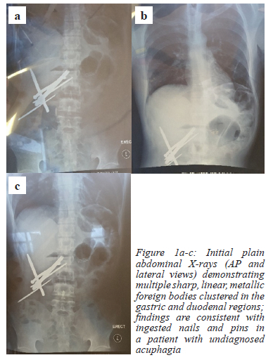
The patient underwent surgery to drain the abscess. During the procedure, doctors found multiple sharp metallic objects in the stomach and duodenum with fluoroscopy, resulting in the decision to perform an upper midline laparotomy. A gastrotomy was performed, and several nails were removed from the stomach and duodenum. No perforation was found. The stomach was closed in two layers, and the abscess cavity was cleaned and packed with jelonet and gauze. After surgery, the patient developed a persistent wound infection with fluid build-up in the right upper quadrant that required extended hospital care. A subsequent gastroscopy revealed a duodenal ulcer and a retained coin. This coin was missed during the gastrotomy and was removed during the procedure. Fluoroscopy was used intraoperatively and the coin was not noted. We suspect that the patient may have swallowed the coin while in hospital after the operation but before the endoscopy. A psychiatric evaluation was requested. Although the patient denied swallowing nails, he admitted to swallowing a coin, attributing the incident to witchcraft. He was eventually discharged, to be followed up for psychiatric care and ongoing wound treatment.
Case 2: Patient B
Case 2 was a 21-year-old man who intentionally swallowed a sewing needle. He had a history of swallowing foreign objects and had previously had a needle removed through a proctoscopy. Upon admission, he was stable and asymptomatic. There were clinically no abdominal abnormalities. An abdominal X-ray showed the sewing needle near the pylorus, with no signs of obstruction or perforation. Since he had no clinical or X-ray complications, he was treated conservatively with regular abdominal examinations and daily imaging. Stool inspection confirmed the needle passed out through the rectum several days later, and he was discharged with a referral for psychiatric evaluation.
Case 3: Patient C
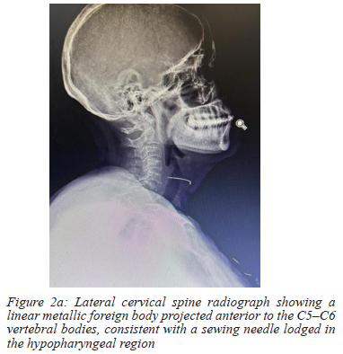
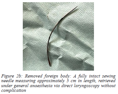
Case 3 was a 61-year-old woman known with schizophrenia and on treatment for HIV and high blood pressure. She presented with a four-day history of dysphagia and a feeling of something stuck in her throat, especially when lying down. She was unsure if she swallowed the object accidentally or on purpose but mentioned a prior incident of swallowing a needle. Upon admission, she was stable with no difficulty in breathing or stomach complaints. Laboratory tests showed metabolic alkalosis, low sodium, low potassium, and low calcium. A lateral X-ray of her cervical spine showed a metallic object anterior to the C5-C6 vertebrae, consistent with a foreign body lodged in the hypopharyngeal region (Figure 2a). She began receiving intravenous antibiotics and treatment for her electrolyte levels. On day three, the needle was successfully removed via exploration of the pharynx under general anaesthesia without any complications (Figure 2b). She remained stable after the procedure and was discharged with a plan for psychiatric follow-up.
Discussion
Acuphagia is particularly dangerous within the spectrum of pica because it involves the intentional swallowing of sharp metallic objects, leading to potentially severe surgical complications.1,2 Unlike other types of non-nutritive ingestion, acuphagia has a higher risk of gastrointestinal perforation, bleeding, sepsis, and airway injury.2 The presented cases demonstrate the variety of this condition, ranging from slow progression to abscess formation, asymptomatic migration, and urgent hypopharyngeal obstruction. Together, they highlight the need for careful monitoring and prompt diagnostic action.
Delays in diagnosis are a significant challenge. Patients often deny swallowing anything, give inconsistent accounts, or attribute their symptoms to other causes, complicating timely identification.3,4 In the first case, delayed diagnosis led to a chronic infection that created spontaneous skin openings, mimicking other serious health issues. Such unusual presentations can result in repeated ineffective treatments and prolonged suffering until the diagnosis is determined. This diagnostic uncertainty emphasises the importance of maintaining suspicion, especially in patients with unexplained infections, recurrent abscesses, or known mental health issues.3
Standard X-rays remain a key diagnostic tool for suspected metallic object ingestion, especially in areas with limited resources.5 In this series, X-rays were vital for confirming diagnoses and shaping treatment decisions. However, the limitations of X-rays must be recognised, including their inability to detect non-metallic materials or accurately show complications.5,6 For stable patients, computed tomography (CT) scans provide better detail, allowing for precise location of foreign bodies and early detection of perforations or abscesses, with reported sensitivities above 90%.6 Careful use of advanced imaging is important for balancing diagnostic accuracy and resource availability.
Endoscopic removal is the preferred method for foreign bodies that can be reached in the upper gastrointestinal tract.7,8 However, acuphagia has unique technical and safety challenges. The sharpness, number, and irregular shapes of the swallowed objects significantly increase the risk of injury or bleeding during removal.7 In this series, endoscopy was beneficial as a supplementary method, helping to remove a retained coin and inspecting the condition of the stomach and duodenum. Protective measures, like overtubes and caps, can reduce injury risks during retrieval, but their effectiveness decreases when objects move beyond the reach of flexible endoscopes.7,8
Surgical intervention is the only option in cases complicated by perforation, abscesses, or the presence of multiple sharp objects in different areas.8 The first case illustrates the issues caused by delayed diagnosis, leading to the need for open surgery and extensive recovery. While less invasive techniques have had good results in some cases, open surgery is crucial, especially for patients in unstable conditions or with infections and complex contamination.10
Conservative treatment may be suitable for selected patients who are asymptomatic, stable and demonstrate progression of the foreign body, as seen in the second case. This method aligns with established guideline recommendations, provided there is thorough clinical observation and ongoing imaging.5,8 However, conservatively managing such cases should not be seen as completely safe; the risk of delayed perforation necessitates clear escalation protocols and diligent supervision.
The third case highlights the serious risks of foreign body impaction in the throat or upper oesophagus. These situations pose an immediate risk to breathing and can lead to aspiration, mediastinitis, or injury to the vasculature if not immediately addressed.8 X-rays of the cervical spine were crucial in pinpointing the foreign body, allowing for timely surgical removal under general anaesthesia. The severe metabolic imbalances in this patient further underline the need for careful management in the perioperative period.
Mental health issues were a common thread in all cases and played a key role in both initial presentations and recurrences. Acuphagia is closely linked to psychiatric disorders like schizophrenia and obsessive-compulsive spectrum conditions, with recurrence rates approaching one-third in some reports.1,9,10 Despite this, psychiatric evaluations are often delayed or insufficient, particularly in public healthcare settings where mental health resources are scarce. Not addressing the underlying mental health problems leads to repeated ingestion, increased morbidity, and rising healthcare costs.3
Effective management of acuphagia requires a coordinated approach involving the surgeon, gastroenterologist, radiologist, anaesthetist and psychiatrist. Psychiatric evaluation should be an essential part of the process rather than an optional adjunct, helping to guide risk assessment, behavioural intervention, and long-term prevention strategies.9,10 Without this integrated approach, surgical treatment addresses only the immediate complication while leaving the underlying pathology unresolved.
In summary, acuphagia is a significant surgical challenge marked by diagnostic ambiguity, psychological complexity, and the potential for serious complications. Early identification, appropriate imaging, and decisive treatment are critical for favourable outcomes. This case series highlights the need for increased awareness and structured multidisciplinary pathways to reduce both the immediate surgical dangers and the long-term burden associated with this rare but serious condition.1,2,8
Conflict of interest
The authors declare no conflict of interest.
Funding source
No funding was required.
Ethical approval
This case series was conducted in accordance with the ethical standards as per the South African Journal of Surgery Policy. Formal ethical approval was not required as this was a retrospective review of anonymised case data; an institutional case series exemption was granted by the Department of Surgery at Ngwelezane Hospital. Informed consent for publication was obtained from all patients included in the series.
ORCID
R Vallabh https://orcid.org/0009-0001-3618-8629
S Vallabh https://orcid.org/0009-0007-6303-0943
S Govender https://orcid.org/0000-0003-0825-2387
M Naidoo https://orcid.org/0000-0002-1581-8871
DL Clarke https://orcid.org/0000-0002-8467-1455
N Makiwane https://orcid.org/0009-0003-1157-152X
SJ Malatji https://orcid.org/0009-0007-8018-0759
REFERENCES
1. Mishori R, McHale C. Pica: An age-old eating disorder that's often missed. J Fam Pract. 2014;63(7):E1-4. PMID: 25198212. [ Links ]
2. Hong KH, Kim YJ, Kim JH, et al. Risk factors for complications associated with upper gastrointestinal foreign bodies. World J Gastroenterol. 2015 14;21(26):8125-31. https://doi.org/10.3748/wjg.v21.i26.8125. [ Links ]
3. Robertson AR. Self‐harm by sharp foreign body ingestion. Suicide Life Threat Behav. 2018;49(3):735-8. https://doi.org/10.1111/sltb.12474. [ Links ]
4. Jaini PA, Haliburton J, Rush AJ. Management challenges of recurrent foreign body ingestions in a psychiatric patient: A case report. J Psychiatr Pract. 2023;29(2):167-73. https://doi.org/10.1097/PRA.0000000000000694. [ Links ]
5. ASGE Standards of Practice Committee; Ikenberry SO, Jue TL, Anderson MA, et al. Management of ingested foreign bodies and food impactions. Gastrointest Endosc. 2011;73(6):1085-91. https://doi.org/10.1016/j.gie.2010.11.010. [ Links ]
6. Gmeiner D, von Rahden BHA, Meco C, et al. Flexible versus rigid endoscopy for treatment of foreign body impaction in the oesophagus. Surg Endosc. 2007;21(11):2026-9. https://doi.org/10.1007/s00464-007-9252-6. [ Links ]
7. Bekkerman M, Sachdev AH, Andrade J, Twersky Y, Iqbal S. Endoscopic management of foreign bodies in the gastrointestinal tract: A review of the literature. Gastroenterol Res Pract. 2016;2016:8520767. https://doi.org/10.1155/2016/8520767. [ Links ]
8. Birk M, Bauerfeind P, Deprez PH, et al. Removal of foreign bodies in the upper gastrointestinal tract in adults: ESGE clinical guideline. Endoscopy. 2016;48(5):489-96. https://doi.org/10.1055/s-0042-100456. [ Links ]
9. Butler W, Allen L. Acuphagia on the obsessive-compulsive spectrum in an adolescent male. Case Rep Psychiatry. 2020;2020:8885503. https://doi.org/10.1155/2020/8885503. [ Links ]
10. Kariholu PL, Jakareddy R, Hemanthkumar M, Paramesh KN, Pavankumar NP. Pica - a case of acuphagia or hyalophagia? Indian J Surg. 2008;70(3):144-6. https://doi.org/10.1007/s12262-008-0040-x. [ Links ]
 Correspondence:
R Vallabh
Email: rashae27vallabh@gmail.com
