


Services on Demand
Article
 English (pdf)
  Article in xml format
 Article references
Indicators

Related links
 Cited by Google
 Similars in Google
Share

 Permalink
Journal of the South African Veterinary Association
On-line version ISSN 2224-9435
Print version ISSN 1019-9128
J. S. Afr. Vet. Assoc. vol.82 n.4 Pretoria Dec. 2011
ARTICLE ARTIKEL
Radiographic changes in Thoroughbred yearlings in South Africa
C FurnissI,*; A CarstensII; S S van den BergIII
IDepartment of Companion Animal Clinical Studies, Faculty of Veterinary Science, University of Pretoria, Private Bag X04, Onderstepoort, 0110 South Africa
IIDepartment of Companion Animal Clinical Studies, Section Diagnostic Imaging, Faculty of Veterinary Science, University of Pretoria Private Bag X04, Onderstepoort, 0110 South Africa
IIIc/o Department of Companion Animal Clinical Studies, Equine Specialist Services, PO Box 1412, Suider-Paarl, 7624 South Africa
ABSTRACT
This study involves the evaluation of pre-purchase radiographic studies of South African Thoroughbred yearlings. Radiographic changes were recorded and compared with similar international studies. The study differs from other studies in that a lower prevalence of pedal osteitis (1.26 %), dorsal osteochondral fragmentation of the metatarsophalangeal joint (1.60 %), distal metacarpal sagittal ridge changes (15.7 %), ulnar carpal bone lucencies (8.33 %), carpal osteophytes (1.19 %), distal intertarsal and tarsometatarsal joint radiographic changes (9.92 %), tarsal osteochondrosis lesions (4.40 %) and stifle osteochondrosis lesions (0.4 %) was found. The prevalence of dorsal osteochondral fragments in the metacarpophalangeal joint was similar to other studies (1.60 %). A higher prevalence of vascular channels as well as irregular borders and lucencies was evident in the proximal sesamoid bones. There was a higher prevalence of palmar metacarpophalangeal and plantar metatarsophalangeal osteochondral fragments (2 % and 7.10 % respectively). Palmar metacarpal disease, metacarpal supracondylar lysis, proximal sesamoid bone fractures and carpal osteochondral fragmentation were absent in the current study. Additional findings recorded in the current study were proximal interphalangeal joint hyperextension (left front 15.13 %, right front 18.91 %), the solar angle (right front 2.38º, left front 2.79º), the prevalence of carpal bone 1 (30.95 %) and carpal bone 5 (1.59 %). Management, nutrition and genetics in the various groups of Thoroughbred yearlings should be further investigated in order to explain the reasons for the differences recorded in the current study.
Keywords: equine, pre-purchase radiographic studies, radiographic changes.
INTRODUCTION
Radiographic studies as part of a pre-purchase examination of yearlings at national yearling sales was initiated in Kentucky, USA, in 1993 with the repository system, a facility which allows the viewing of radiographs, coming into effect in 199614. In New Zealand the repository system was 1st established in January 2003 at Karaka, Auckland17.InSouth Africa the 1st repository was opened at the 2007 National Yearling Sale in Germiston where over 300 sets of radiographs were lodged, while in the same year at the Keeneland National Yearlings sale in Kentucky, USA, 4700 sets of radiographs were lodged14.
The South African National Yearling Sale is held annually in Germiston, Gauteng, where between 550 and 600 yearlings are sold annually26. This is the largest Thoroughbred sale in South Africa and in 2008 showed a gross income of R201 050 000 with the highest priced yearling sold at R3 million26. In 2008, 269 of 600 yearlings were radiographed (44.83 %).
Radiographic changes recorded from pre-purchase radiographs of Thoroughbred yearlings have been investigated in Thoroughbred populations around the world, not only in terms of the prevalence of specific radiographic changes but also the impact these changes have on the sale price and future racing career of the Thoroughbred3,10,11,13,14,17,24. The changes identified can either be classified as developmental or traumatic in origin. Osteochondritis dissecans (OCD) is a developmental orthopaedic disease and most commonly manifests as sagittal ridge defects of the distal metacarpus, distal intermediate ridge defects of the tibia, trochlear ridge lesions in the tarsus and lesions in the stifle joint in the Thoroughbred yearling 10,11,13,14,17,24. The most common traumatic radiographic lesions identified in the same studies are within the distal joints of the tarsus with the prevalence varying from 6.1 % to 31 %9,14,17. Evidence has been found linking these degenerative changes of the distal joints of the tarsus to osteochondrosis in juvenile horses1,27. In a study of Thoroughbred yearlings palmar metacarpal disease (41.3 %) and supracondylar lysis (4.8 %) were identified in the metacarpophalangeal joint (MCP) and is likely linked to repetitive trauma sustained during exercise2,14.
The purpose of this study is to describe the prevalence of radiographic changes in pre-purchase radiographs of Thoroughbred yearlings presented at the 2008 National Yearling Sale in Germiston, South Africa. This will enable the veterinarian involved in the pre-purchase examination to inform his/her client of the normal and abnormal radiographic findings. Our hypothesis was that the prevalence of radiographic changes is similar to studies done in other Thoroughbred populations around the world.
MATERIALS AND METHODS
Sampling and radiographic studies
Radiographic studies of 269 Thoroughbred yearlings were obtained from the repository of the 2008 National Yearling Sale in Germiston, South Africa. These radiographs form part of a pre-purchase examination that the owners/buyers request and are taken between 1 month and 1 day before the sale. The owner/ buyer chooses whether or not to lodge the radiographic studies at the repository.
A radiographic examination of a yearling comprised a minimum of 36 radiographs evaluating the front digit (LM), MCP joint (D15ºPrPaDO, D30ºMPaLO, D30ºLPaMO, LM flexed), metatarsophalangeal (MTP) joint (D15ºPrPlDO, D15ºPr30ºMPlDiLO, D15ºPr30ºLPlDiMO, LM), carpus (D 30º-40º) LPaMO, D(20º- 30º) MPaLO, LM flexed), tarsus (D65º MPlLO, D10ºLPlMO, LM) and stifle joint (Cd20ºLCrMO, CdCr, LM). The Thoroughbred Breeders Association of South Africa requires the views described. Poor quality radiographs were excluded from the study.
Each radiograph was evaluated by the primary researcher (CF). Approximately 10 % of all radiographs were scrutinised by the co-author (AC). All differences pertaining to the radiographic changes were discussed and CF re-evaluated all areas where differences in opinion were found and adapted the reports accordingly.
Categorisation of radiographic changes
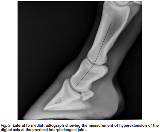
The digit: radiographic changes recorded included the angle of phalanx 3 (P3) in relation to the solar surface (solar angle), the inclination of the digital axis and enthesophytes/osteophytes/periosteal reactions on the phalanges (Fig. 1). The digital axis was evaluated by observing the angle of the proximal interphalangeal (PIP) joint and the distal interphalangeal (DIP) joint and classifying this angle as normal, hyperextended or flexed (Fig. 2). A normal axis was represented by a line drawn bisecting phalanx 2 (P2) into equal dorsal and palmar halves being parallel to the dorsal cortex of P327. Enthesophytes/ osteophytes were identified as new bone formation exceeding 2 mm in height and present in specific areas such as the extensor process of P3 and the distal and proximal aspects of P2.

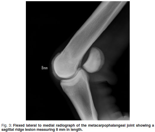
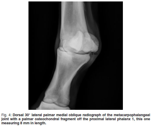
Metacarpophalangeal and metatarsophalangeal joints and proximal sesamoid bones: dorsal and palmar/plantar osteochondral fragments were classified as articular or non-articular. The longest diameters of the osteochondral fragments were measured. Osteochondral fragments were considered to originate from phalanx 1 (P1) when they were found in close proximity to P1 with a fracture bed identified in the parent bone. The flexed LM view of the MCP joint highlighted the dorsal aspect of the sagittal ridge where the presence of a semicircular notch or flattening of the visible sagittal ridge was recorded (Fig. 4). Subchondral cyst-like lesions were defined as an area of lucency in the subchondral bone of which the longest diameter was measured.
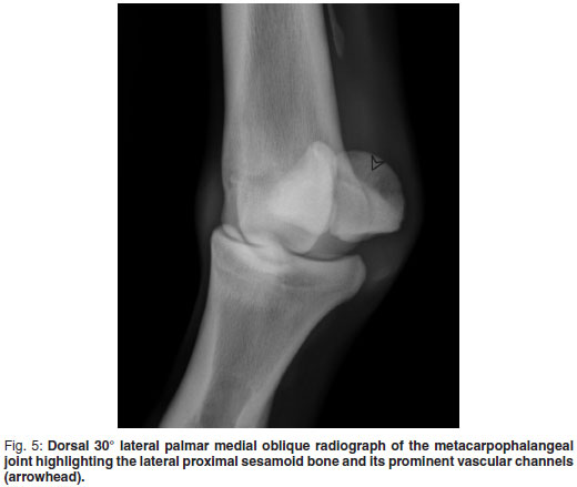
Linear lucencies on the abaxial surface of the proximal sesamoid bones (PSB) were recorded as vascular channels, counted and classified into regular or irregular vascular channels. Regular vascular channels were defined as linear lucencies that had parallel sides for the entire length and were less than or equal to 2 mm in width. Irregular vascular channels were defined as linear lucencies that had non-parallel sides for any portion of their length or were more than 2 mm in width14. Lucent areas on the abaxial surface of the PSBs were measured. Osteophytes, which were defined as new bone formation on the articular apices and bases of the PSBs, were recorded14.
Enthesophytes differed from osteophytes as they were defined as new bone formation at the insertion of the interrosseus medius or origin of the distal sesamoidean ligaments14.
The carpus: the presence of osteophytes, osteochondral fragments, ulnar carpal bone (Cu) lucencies, the presence of carpal bone 1 (C1) and carpal bone 5 (C5) were recorded.
The tarsus: all osteochondral fragments were measured and recorded. Variations in appearance of the distal aspect of the trochlear ridges were ignored as they were seen as normal variations9. Radiographic changes involving the distal intertarsal (DIT) and tarsometatarsal (TMT) joints which included new bone formation, irregularity of the subchondral joint surfaces and/or lucencies were recorded and these changes were grouped together.
The stifle: subchondral cyst-like lesions and osteochondral fragments were measured and recorded.
Data analysis
Radiographic changes were described and the percentages of the total radiographic changes noted. The mean as well as the largest and smallest solar angle for each P3 was calculated. All findings and measurements were entered into an Excel spreadsheet (Microsoft Excel 2007, Microsoft Corp, Redmond, WA, USA). The radiographic changes identified in the South African yearlings were compared with other yearling populations (Tables 9-11).
RESULTS
The digit
The average solar angle of the RF (right front) and LF (left front) P3 was 2.38º and 2.79º respectively. Hyperextension of the PIP joint was found in 36 of 238 cases in the LF digit (15.13 %) and 45 of 238 cases in the RF digit (18.91 %). Hyperextension in the DIP joint was identified in 18 of 238 cases in the LF digit (7.56 %) and in 23 of 238 cases in the RF digit (9.66 %) (Fig. 3). Few of the DIP joints were in a flexed position (1 of 238 in the left digit, 0.42 % and 2 of 238 in the right digit, 0.84 %). Table 1 shows the radiographic changes of the digits.
The metacarpophalangeal joint and proximal sesamoid bones
The most common radiographic change recorded was the change on the sagittal ridge with 30 of 255 LF MCP joints affected (11.80 %) and 29 of 255 RF MCP joints affected ( 11.40 %) (Table 2). The change was bilateral in 19 (47.50 %) of the 40 yearlings affected. Proximopalmar osteochondral fragments were more common than proximodorsal osteochondral fragments with a total of 5 yearlings affected (1.96 %). The largest proximopalmar osteochondral fragment measured 7.4 mm in length while the smallest measured 2.3 mm in length. Four yearlings had proximodorsal osteochondral fragments in the MCP joint (1.57 %) measuring 2.0 mm, 2.1 mm, 3.5 mm and 1 mm in length. All osteochondral fragments in the MCP joints were found to be articular.
Most MCP joint PSBs had 2 or 3 vascular channels present on their abaxial surface with a combination of regular and irregular vascular channels (Fig. 5). The medial PSB in both LF and RF had an irregular abaxial border in 57 of 255 (22.4 %), and 32 of 255 MCP joints (12.50 %), more often than the lateral PSB, which was affected in 15 of 255 (5.90 %) and 17 of 255 (6.70 %) MCP joints. Only a few PSB osteophytes were seen with 1 of 255 (0.40 %) recorded in the medial PSB of the LF MCP joint and 3 of 255 (1.20 %) in the medial PSB of the RF MCP joint.
The metatarsophalangeal joint and proximal sesamoid bones
Articular proximoplantar fragments of P1 were more prevalent in the LH MTP joint with 12 of 255 (4.71 %) MTP joints affected in comparison with the RH MTP joint, where 5 of 255 (1.96 %) joints were affected. The length of these osteochondral fragments ranged from 22-85 mm in the LH MTP joint and 20-69 mm in the RH MTP joint. Proximodorsal osteochondral fragments of P1 were recorded in the same frequency in the LH and RH MTP joints with a total of 4 yearlings affected (1.57 %). The dorsal osteochondral fragments in the LH MTP joint were 2.0 and 4.7 mm in length on 2 separate joints while the in the RH MTP joint these osteochondral fragments were 3.2 and 0.5 mm in length. Subchondral cyst-like lesions were identified in the LH P1 in 2 yearlings. Changes on the sagittal ridge of the distal metatarsus were identified in only 1 yearling (0.40 %).
An irregular border of the PSBs of the MTP joint was seen more frequently in the medial PSB in both the LH MTP joint (8.98 %) and the RH MTP joint (8.59 %). Lucencies showed a similar pattern of distribution. Osteophytes were identified on the PSBs with a prevalence 5.47 % and 8.59 %. Visible vascular channels were less common in the MTP joint than in the MCP joint. All radiographic changes of the MTP joint are listed in Table 4.
The carpus
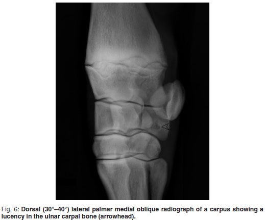
Carpal bone 1 was present in 78 yearlings (30.95 %). Exactly half of these yearlings had C1 bilaterally. Carpal bone 5 was found less commonly (4 of 252, 1.49 %). A circular lucency in the Cu was identified in 21 Thoroughbred yearlings (8.33 %) with only 5 being bilateral (23.81 %). These circular lucencies varied in size ranging from 2.2-7.0 mm in length (Fig. 6). Osteophytic new bone formation on the carpal bones was recorded in a total of 3 yearlings (1.19 %).
The tarsus
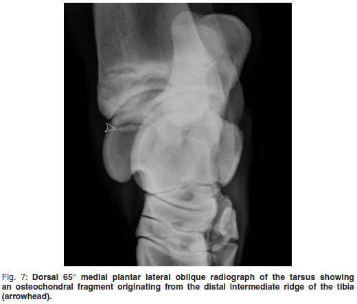
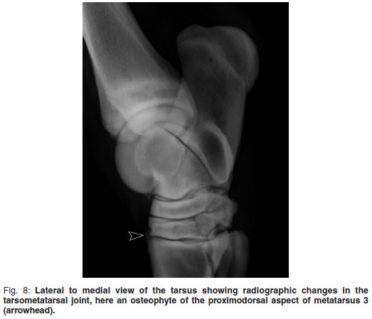
Radiographic changes in the DIT and TMT joints were present in 25 of 252 yearlings (9.92 %) (Fig. 8). Forty-eight per cent of these radiographic changes were bilateral. Concavity and/or fragmentation of the distal intermediate ridge of the tibia was present in 5 of 252 left tarsal joints (1.98 %) and 6 of 252 right tarsal joints (2.38 %) and 33 % of these were bilateral (Fig. 7). The largest osteochondral fragment recorded was 19 mm × 6 mm while the smallest recorded was 23 mm in length. Four tarsal joints affected had multiple fragments. Other manifestations of osteochondrosis were found such as lucencies in the medial malleolus in 1 of 252 yearlings (0.4 %) and changes in the lateral and medial trochlear ridges of the talus in 8 of 252 yearlings (3.17 %). Wedging or collapse of the central and 3rd tarsal bones was identified in 4 of 252 yearlings (1.59 %), 50 % being bilateral.
The stifle
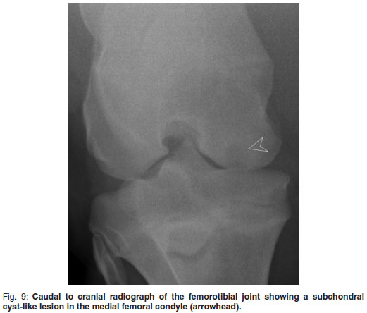
One subchondral cyst-like lesion was recorded in the right stifle of a yearling (0.4 %). Lucency and fragmentation of the patella was identified in the right stifle of another yearling (0.4 %).
Comparison of radiographic changes with other similar studies
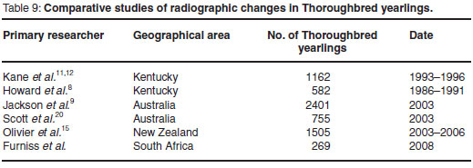
Similar studies on pre-purchase radiographs in Thoroughbred yearlings were compared and are listed in Table 9 . The radiographic findings of each study were compared to the current study and the prevalences are represented in Tables 10 and 11.
DISCUSSION
This study provides prevalence data on the radiographic changes identified in pre-purchase radiographs of a select group of Thoroughbred yearlings at the 2008 National Yearling Sale, Germiston, South Africa. The hypothesis that radiographic changes are similar to studies done in other Thoroughbred yearling populations was rejected, and it was concluded that the radiographic findings in South African Thoroughbreds are not similar to studies of Thoroughbreds elsewhere in the world. These differences are discussed below.
In the current study there was a lower prevalence of pedal osteitis (1.26 %) than in other studies where the prevalence recorded was between 2 % and 11 %10,14. The only radiographic change identified in the current study, in terms of pedal osteitis, was new bone formation on P3. This radiographic change was recorded in the left P3 in 2 yearlings and in the right P3 in 1 yearling. In 1 study a D65ºPr-PaLDiO view or upright pedal view of P3 was also included14. The addition of this view would increase the likelihood of identifying changes of pedal osteitis5,27. In the latter view, P3 is seen dorsopalmarly in its entirety and signs of bone resorption, marginal fractures or widening of the vascular channels along the margin of P3 would be more readily identified5. The digit radiographic sets in the current study were limited to a LM view only, since this was the only view required by the TBA.
The most common radiographic finding in the digits was deviation in the digital axis, which was not recorded in selected studies used for comparison. This may be significant as it may have an effect on the future athletic career of the yearling. Malalignment of the digital bones is seen in 72.8 % of horses with front limb lameness15,18. With hyper-extension of the proximal and distal interphalangeal joints, greater stress is placed on the deep digital flexor tendon (DDFT) and the navicular bone and has been associated with heel bruising, lamellar tearing at the toe, pedal osteitis, navicular disease, distal sesamoidean ligament injury, DDFT tenopathy as well as injuries to tendons proximal to the digit7. The disruption of the digital axis may progress to the long-toe low heel syndrome commonly seen in Thoroughbred racehorses. This condition delays break-over and contributes to increased compression of the dorsal joint margins of the carpus as well as the MCP, PIP and DIP joints7. Without early correction of the digital axis in long-toe low heel syndrome, chronic changes may develop that may shorten the athletic career of the race horse. As no exact angles were measured, these joints were only described as flexed, normal or hyperextended.
The solar angle of P3 was found to be lower than normal, with the average solar angle in the RF and LF digit being 2.38º and 2.79º, respectively. This angle in normal horses has been recorded to be between 5 and 10º 5,6,27. This discrepancy in the angle of P3 has been suggested to be breed-related as low angles have been recorded in Thoroughbred populations6. The other studies of radiographic changes in Thoroughbred yearlings did not measure the solar angle, therefore the results cannot be compared. The solar angle has been found to be influenced by trimming of the hoof with the P3 to heel distance reduced more than the P3 to toe distance, thus decreasing the angle of the P315. The horn of the hoof is softer at the heel area and thus more easily rasped than the horn in the toe area. This may result in more horn removed contributing to the low solar angle.
The prevalence of proximodorsal osteochondral fragments in the MCP joints of South African Thoroughbred yearlings was recorded as being 1.57 % in Thoroughbred yearlings evaluated, which is similar to other studies that recorded prevalences between 1.2 and 1.6 %10,14,24. Prevalence of proximodorsal osteochondral fragments was found to be similar in both MCP and MTP joints. In other studies these fragments were found to be twice as common in the MTP joint10,14. This difference between the population of Thoroughbred yearlings may be attributed to stud farm management, as these fragments have been linked to trauma7.
There is a higher prevalence of proximopalmar and proximoplantar osteochondral fragments in the MCP and MTP joint in the current study. Similar studies recorded prevalence of between 0.3 % and 0.5 %10,14. Proximoplantar osteochondral fragments were found to be more common in the MTP joints, which was similar to other studies. As these osteochondral fragments have been attributed to osteochondrosis and trauma, it is difficult to explain the relatively higher prevalence of palmar/plantar fragments in this study population7,21. Palmar/plantar osteochondral fragments have been shown to have a genetic predisposition in Standardbreds7. As the genetic contribution is unknown, it is difficult to speculate whether or not this factor plays a role in the prevalence of palmar/plantar osteochondral fragments in the current study. In this foal crop of 2006 there are no other radiographic changes in other joints that suggest that trauma plays a role.
The prevalence of changes on the sagittal ridge of the MCP joint was lower (15.69 %). In other studies of Thoroughbred yearlings the prevalence of sagittal ridge lesions varied between 7.5 and 60.8 % in the MCP joint10,11,14,24. In 1 study the prevalence of sagittal ridge radiographic changes was nearly 4 times the number recorded in this study (60.6 %)14. Sagittal ridge lesions have been linked to joint effusion and lameness and thus are likely to be a significant finding28. A limitation of the current study is the fact that sagittal ridge lesions were not classified into types and thus it is not possible to accurately prognosticate on the sagittal ridge findings. As discussed above, these sagittal ridge lesions have been classified under osteochondrosis although it is difficult to distinguish between osteochondrosis and trauma7. In this study, 47.50 % of sagittal ridge changes identified were found to be bilateral and this characteristic may suggest a developmental aetiology. Factors that influence the development of osteochondrosis such as nutrition, biomechanics and genetics may play less of a role in South African Thoroughbreds, thus resulting in a lower incidence.
The most common change seen in the MCP joints in a previous study was flattening of the distal palmar 3rd of MC3 condyles with a prevalence of 41.3 %, with half of these changes being bilateral14. These changes have been seen in working Thoroughbred racehorses2. Palmar supracondylar lysis was identified in MCP joints of Thoroughbred yearlings in the same study with a total of 54 of 1127 Thoroughbred yearlings affected (4.8 %)14. Signs of flattening of the distal palmar 3rd of MC3 and supracondylar lysis were not recorded in the population of South African Thoroughbred yearlings. Both of these conditions have been linked to exercise and the exercise programmes of the different Thoroughbred populations could explain the variation in the prevalence of these conditions.
There was a slightly higher prevalence of vascular channels, irregular abaxial borders and lucencies in the PSBs of the MCP joint in the current study. Vascular channels are a common finding in Thoroughbred yearling radiographs3,14. Radiographic changes in the PSBs have been described as a grade III sesamoiditis with 9.5 % and 10.9 % of front and hind fetlocks affected, respectively10. This change could be present due to an increase in exercise prior to the sale but may also be associated with pathology in the nterosseus medius7.
No PSB fractures were present in the current study, in contrast to a prevalence of fractures in the PSBs of the MTP joint in 27 of 1102 yearlings (2.5 %) in Kentucky14. This prevalence of PSB fractures may reflect the management practices at the studs in the Kentucky region. There may be an increased risk of injury during rearing of the horse or in the preparation for the sales.
The prevalence of carpal bone 1 and 5 was 30.95 % and 1.59 % respectively. This radiographic finding was not recorded in the other studies of Thoroughbred yearlings used as a comparison. The South African Thoroughbreds fall into the normal reference range for these radiographic features5.
There was a lower prevalence of lucencies in the Cu bone in the current study. It has been found that ulnar carpal bone lucencies and associated fragments can be an incidental finding in horses from all disciplines23. It is difficult to explain this lower prevalence in the South African Thoroughbred yearling population. It may be speculated that these lucencies could represent osteochondrosis lesions.
There was a lower prevalence of osteophytes in the carpus with similar studies showing a prevalence of between 1.7 % and 2.6 %14,24. This radiographic change, indicative of a degenerative process in the joint, may reflect the differences in the exercise programmes of the different Thoroughbred populations. Poor conformation may also play a role7.
No osteochondral fragments associated with the joints in the carpus were present in South African Thoroughbred yearlings. Other studies of Thoroughbred yearlings found a prevalence for osteochondral fragments of 0.3 to 2.2 % of carpi examined10,14,24. Again the presence of this radiographic change in other groups of Thoroughbred yearlings may reflect the level of exercise to which they are exposed to prior to the Yearling Sales.
There was a lower prevalence of radiographic changes in the DIT and TMT joints (9.92 %). Other studies had a recorded prevalence of 17.5 % and 30 %, respectively14,17,25. It has been found that osseous spurs in the DIT and TMT joints could not be linked to hind limb lameness25. Further research is needed to explain the low incidence of tarsal radiographic changes in the current study.
The prevalence of osteochondrosis lesions in the tarsus (4.4 %) and stifle (0.8 %) is generally lower compared with other studies10,14,17,24. Concavity/fragmentation of the distal intermediate ridge of the tibia was the most common osteochondrosis lesion and was present in 4.4 % of yearlings in a previous study compared with 3.17 % in the current study14. The lower prevalence of lesions indicative of osteochondrosis may be attributed to a difference in management, genetics or nutrition or a combination of these factors in the South African population of Thoroughbred yearlings. Other reasons for this low prevalence recorded could be the low sample population in the current study or the fact that yearlings with tarsal or stifle lesions may not have had their radiographs lodged in the repository.
The aetiopathogenesis of osteochondrosis is often multifactorial and poorly understood; genetics, nutrition and biomechanics are factors that have been implicated in the development of this condition16,19,20. The South African Thoroughbred population is not an isolated genetic pool. Seventy per cent of the top 100 yearlings at the 2008 National yearling sale were sired by stallions from the USA. This statistic suggests a strong link to the USA gene pool. There is a great deal of movement of Thoroughbred racehorses in and out of South Africa for breeding and racing purposes. In 2008, a total of 295 Thoroughbreds were imported into South Africa for these purposes26. This suggests that the local Thoroughbred racehorses do not have an isolated gene pool and thus this factor is unlikely to play a role in the low prevalence of OCD lesions present. Another factor influencing the development of osteochondrosis is nutrition. Further investigation of this factor in the different Thoroughbred populations is warranted. Individual management practices may also play a role in the development of osteochondrosis lesions. The differing management practices may be reflected in the amount of time foals spend on pasture or confined in a stable, as well as the exercise programme of the yearling prior to the sale16. Management practices in the various study populations should be further investigated to try to account for the low prevalence of osteochondrosis lesions seen in the current study.
The prevalence of flattening of the distal palmar 3rd MC3, supracondylar lysis, sesamoid fractures, osteophytes and fragmentation of the carpus and radiographic changes in the distal joints of the tarsus is higher in other populations of Thoroughbreds and these changes are all related to exercise7. The exercise programme of yearlings in preparation for the sales may account for the traumatic and degenerative radiographic changes. As discussed above, the prevalence of palmar metacarpal disease and supracondylar lysis in the fetlocks of the USA Thoroughbreds may be due to a more intensive training programme prior to the sale. Joint injury rates and exercise programmes have been shown to differ between trainers3,8.In South Africa the yearling's pre-sale training programme is restricted to walking and trotting. In South Africa there is a large amount of affordable labour and thus studs usually employ 1 handler per yearling. This may decrease the risk of injury during the pre-sale exercise programme. Yearling sales preparation has been studied using information from 18 farms in New Zealand. This research showed a significant difference in training and incidence of lameness between farms8. This may apply to Thoroughbred yearling populations worldwide.
Disease of the suspensory apparatus or interosseus medius pathology may account for the mild increase in vascular channels seen as well as the irregular borders of the PSBs noted. It may be speculated that due to the high percentage of Thoroughbred yearlings with hyperextension of the PIP joint in the current study (15.13 % in the LF digit and 18.9 % in the RF digit) there may be a greater strain on the interosseus tendon in this population of Thoroughbred yearlings. This may lead to an increase in the number of vascular channels seen in the PSBs and or result in an irregular border at the insertion of the interosseus tendon on the PSB. This is speculation as no attempt was made to link the radiographic changes in the PSBs to hyperextension in the PIP joint. This may be investigated in future studies. One may further speculate that the long-toe low-heel syndrome may also play a role in increasing the number of vascular channels in the PSBs by the same principles discussed above.
Farriery done on the stud farms may play a role in the radiographic changes seen affecting the digital axis, solar angle and sesamoid changes. Incorrect hoof trimming or an excessively long interval between trims could lead to mediolateral and dorsopalmar hoof imbalance and may lead to radiographic changes seen7.
Similar studies in Thoroughbred yearlings involved study populations of between 582 and 1505 yearlings10,14,17,24. The South African study population consisted of only 269 Thoroughbred yearlings. This is a relatively small study population and this may also play a role in the low prevalence of radiographic changes recorded.
One of the previous studies was done from radiographs that were taken over 4 years from 1993 to 199614. Another study was done over 6 years from 1986 to 1991 with other studies done later in the new millenium10,14. The current study is limited to radiographic studies taken in a single year, 2008. This may have an influence on the prevalence of radiographic changes seen in the South African population of Thoroughbred yearlings, as seasonal weather conditions vary from year to year, which may influence the quality and quantity of nutrition of the Thoroughbred yearling. A more representative study population would consist of radiographs of Thoroughbred yearlings taken over a number of years in order to eliminate any effects due to variation of seasonal weather conditions.
The history of the population of Thoroughbred yearlings under investigation was not obtained. The practice of radiographing weanlings has recently gained popularity in South Africa, resulting in some horses undergoing arthroscopic surgery before the sale. One study showed the incidence of a history of arthroscopy prior to a yearling sale to be as high as 13 %22. This would result in a decrease in the prevalence of radiographic changes present in pre-purchase radiographic examinations performed.
The owner of the radiographs has a choice whether to lodge the radiographs at the repository or not. This principle also applies to other repositories around the world4. Radiographic studies of yearlings with many radiographic changes may not have been submitted to the repository as radiographic findings affect the price of the yearling to be sold. This principle applies to other studies whose study population depends on the lodging of radiographs at a repository. Therefore, studies which base their study population on Thoroughbred yearlings whose radiographs appear in the sales repository are biased studies as they do not represent the entire Thoroughbred yearling population. Whether South Africans are less likely to lodge all radiographic studies at the repository is unknown. The repository system has only recently been introduced at South African sales and therefore it may be speculated that South Africans may be more apprehensive over radiographic changes and their effect on the sale of their yearling. This may account for the lower prevalence of radiographic changes seen in the current study.
Many studies have researched the effect of radiographic changes on the athletic ability of the Thoroughbred racehorse by following the career of the racehorse after the yearling sales13. The prevalence and distribution of radiographic changes has been found to have little effect on the future of racing performance of the Thoroughbred racehorse12,14,17. A reason for this finding could be the short athletic career of the Thoroughbred racehorse. A racehorse is 1st raced as a 2 year old as the races with the highest stakes are often in the 1st 2 years of its athletic career. It may be speculated that by the time these lesions start causing degenerative changes in the joint, the career of the animal is over. For this reason many Thoroughbred racehorse owners have questioned the role of pre-purchase radiographic examinations. However, the information gained is important to establish what lesions are present in the yearling and to use this to attempt to minimise or prevent the condition in the future.
The current study provides prevalence data of radiographic changes in the South African Thoroughbred yearlings, which show some differences when compared with similar studies done in Thoroughbred yearlings around the world.
ACKNOWLEDGEMENTS
The Thoroughbred Breeders Association is thanked for providing the radiographic studies.
REFERENCES
1. Bjornsdöttir S, Ekamn S, Eksell P, Lord P 2004 High detail radiography and distology of the centrodistal tarsal joint of Icelandic horses age 6 months to 6 years. Equine Veterinary Journal 36:5-11 [ Links ]
2. Blevins B E, Widmer W R 1990 Radiology in racetrack practise. Veterinary Clinics of North America: Equine Practice 6:31-60 [ Links ]
3. Bolwell C F, Rogers C W, French N P, Firth E C 2011 Exercise in Thoroughbred yearlings during sales preparation: A cohort study. Equine Veterinary Journal June 8. doi: 10.1111/j.2042-3306.2011.00370.x.( in press) [ Links ]
4. Bramlage, L R 1993 Osteochondrosis and the sale horse. Proceedings of the Thirty-Ninth Annual Convention of the American Association of Equine Practitioners, San Antonio, Texas, USA, December 5-8,1993:87-89 [ Links ]
5. Butler J A, Colles C M, Dyson S J, Kold S E, Poulos P W 2000 Clinical radiology of the horse (2nd edn). Blackwell Scientific Publications, London [ Links ]
6. Cripps P J, Eustace R A 1999 Radiological measurements from the feet of normal horses with relevance to laminitis. Equine Veterinary Journal 31:427-432 [ Links ]
7. Dyson S J, Ross M W 2003 Diagnosis and management of lameness in the horse. Saunders, St Louis, Missouri [ Links ]
8. Jackson F, M, Whitton C, Vizard A, Anderson G, Clarke A 2003 A prospective study of presale radiographs of Thoroughbred yearlings. Online at: http://www.rirdc.infoservices.com.au (accessed 5 November 2009) [ Links ]
10. Howard B A, Embertson R M, Rantanen N W, Bramlage L R 1993 Survey radiographic findings in Thoroughbred sale yearlings. Proceedings of the Annual Convention of the American Association of Equine Practitioners 38:397-402 [ Links ]
11. Jackson M, Whitton C, Vizard A, Anderson G, Clarke A 2003 A prospective study of presale radiographs of Thoroughbred yearlings. Online at: http://www. rirdc.infoservices.com.au (accessed 5 November 2009) [ Links ]
12. Jorgensen H S, Proschowsky H, Flak-Ronne J, Willeberg P, Hesselholt M 1997 The significance of routine radiographic findings with respect to subsequent racing performance and longevity in Standardbred trotters. Equine Veterinary Journal 29:55-59 [ Links ]
13. Kane A J, McIlwraith C W, Park R D, Rantanen N W, Morehead J P, Bramlage L R 2003 Radiographic changes in Thoroughbred yearlings. Part 2: Associations with racing performance. Equine Veterinary Journal 35:66-374 [ Links ]
14. Kane A J, Park R D, McIlwraith C W, Rantanen N W, Morehead J P, Bramlage L R 2003 Radiographic changes in Thoroughbred yearlings. Part 1: Prevalence at the time of the yearling sales. Equine Veterinary Journal 35:354-365 [ Links ]
15 Kummer M, Geyer H, Imboden I, Auer J, Lischer C 2006 The effect of hoof trimming on radiographic measurements of the front feet of normal Warmblood horses. Veterinary Journal 172:58-66 [ Links ]
16. McIlwraith C W, Trotter G W 1996 Joint disease in the horse. WB Saunders Company, Philadelphia [ Links ]
17. Olivier L J, Baird D K, Baird, A N, Moore, G E 2008 Prevalence and distribution of radiographically evident lesions on repository films in the hock and stifle joints of yearling thoroughbred horses in New Zealand. New Zealand Veterinary Journal 26:202-209 [ Links ]
18. Page B T, Hagen T L 2002 Breakover of the hoof and its effects on structures and forces within the hoof. Journal of Equine Veterinary Science 22:258-264 [ Links ]
19. Pearce S G, Firth E C, Grace N D, Fennessy P F 1998 Effect of copper supplementation on the evidence of developmental orthopaedic disease in pasture-fed New Zealand Thoroughbreds. Equine Veterinary Journal 30:211-218 [ Links ]
20. Pool R R 1993 Difficulties in definition of equine osteochondrosis; differentiation of developmental and acquired lesions. Equine Veterinary Journal, Supplement 16:5-12 [ Links ]
21. Pool R R, Meagher D M 1990 Pathologic findings and pathogenesis of racetrack injuries. Veterinary Clinics of North America, Equine Practice 6:1-30 [ Links ]
22. Preston S A, Zimmel D N, Chmielewski T L, Trumble T N, Brown M P, Boneau J C, Hernandez J A 2010 Prevalence of various presale radiographic findings an association of findings with sales price in Thoroughbred yearlings sold in Kentucky. Journal of the American Veterinary Medical Association 236: 440-445 [ Links ]
23. Reed S R, Jacksone B F, McLlwraith C W, Pilsworth R, Knapp S, Wood J L, Price J S, Verheyen K L 2011 Discriptive epidemiology of joint injuries in Thoroughbred racehorses in training. Equine Veterinary Journal Mar 15. doi: 10.1111/j.2042-3306.2010.00352.x. (in press)
24. Scott N J, Hance S, Todhunter P, Adams P, Adkins A R 2005 Incidence of radiographic changes in Thoroughbred yearlings. 755 cases. Advances in Equine Nutrition III: 347 [ Links ]
25. Simon V, Dyson S J 2010 Radiographic anatomic variation of the carpus in horses with carpal lameness and control horses. Veterinary Radiology and Ultrasound 51(6):601-606 [ Links ]
26. Thoroughbred Breeders Association SA 2008 Sales results Germiston national yearling sales. Online at: http://www.tba.co.za (accessed 26 August 2009) [ Links ]
27. Thrall D E 2007 Textbook of veterinary diagnostic radiology. Saunders Elsevier, St Louis, Missouri [ Links ]
28. Yovich J V, Mcllwraith C W, Stashak T S 1985 Osteochondritis dissecans of the sagittal ridge of the third metacarpal and metatarsal bones in horses. Journal of the American Veterinary Medical Association 186:1186-1191 [ Links ]
Received: November 2010
Accepted: September 2011
* Author for correspondence. E-mail: cknox@seaworld.org.za

