Services on Demand
Article
Indicators
Related links
-
 Cited by Google
 -
 Similars in Google
Share
Journal of the Southern African Institute of Mining and Metallurgy
On-line version ISSN 2411-9717
Print version ISSN 2225-6253
J. S. Afr. Inst. Min. Metall. vol.116 n.3 Johannesburg Mar. 2016
http://dx.doi.org/10.17159/2411-9717/2016/v116n3a6
PAPERS - MINE PLANNING AND EQUIPAMENT SELECTION (MPES)
Microscopic analyses of Bushveld Complex rocks under the influence of high temperatures
G.O. Oniyide; H. Yilmaz
School of Mining Engineering, The University of the Witwatersrand, Johannesburg, South Africa
SYNOPSIS
The South African platinum mines in the Bushveld Complex (BC) have unique features that distinguish them from the gold mines. They have a higher horizontal stress closer to the surface, lower occurrence of seismic activity (caused mainly by pillar failure), and are sited in an area of high geothermal gradient. One of the future challenges of platinum mining is the increasing temperatures as the mining depth increases. High temperatures invariably bring about higher ventilation costs and require different approaches to human factors and ergonomics. Excavation stability would also be another area of concern due to increasing stresses. In this investigation, the effect of temperature on the physical and chemical properties of selected Bushveld rocks (chromitite, pyroxenite, norite, leuconorite, gabbronorite, mottled anorthosite, varitextured anorthosite, granite, and granofels) was studied by means of optical microscopy and scanning electron microscopy using energy-dispersive X-ray spectrometry (SEM-EDX). The rock specimens were heated in a temperature-controlled oven a rate of 2°C /min to 50°C, 100°C, and 140°C and kept at temperature for five consecutive days. Samples were allowed to cool to ambient temperature (approximately 20°C) before image capturing. Micrographs of the specimens were taken before and after heat treatment. All the SEM samples were also subjected to heating and cooling on alternate days for ten days in order to observe the effect of repeated heating and cooling on the rocks. The results of the optical microscopy analyses showed minor physical changes in the rocks. The SEM images revealed that cracks initiating at lower temperatures extend with increasing temperature. The chemical analyses showed that the temperature range considered in this research is not high enough to induce changes in the chemical composition of rock samples.
Keywords: Bushveld Complex, microscopic analyses, heat treatment of rock.
Introduction
Clauser (2011) observed that the thermal regime of the Earth is governed by its heat sources and sinks, transport processes and heat storage, and their equivalent physical properties. Clauser and Huenges (1995) explained that the interior heat of the Earth is transmitted to its surface by conduction, radiation, and advection. They further stated that the thermal conductivity of igneous rocks is isotropic, while the thermal conductivity of sedimentary and metamorphic rocks displays anisotropy.
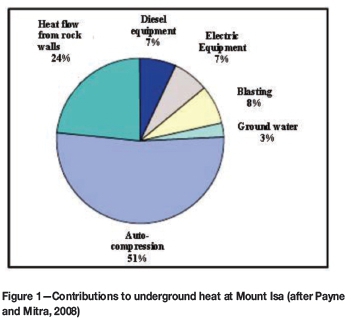
Geothermal heat and heat from autocompression, which are sources of underground heat, exist in all mining situations to varying degrees, depending on the geothermal gradient and surface temperature. Other sources of underground heat are blasting, mining equipment, and groundwater, as shown in Figure 1. From the figure, it can be seen that autocompression -that is, compression of air by its own weight -makes the highest heat contribution, followed by heat flow from the rock mass (Brake and Bates, 2000; Payne and Mitra, 2008).
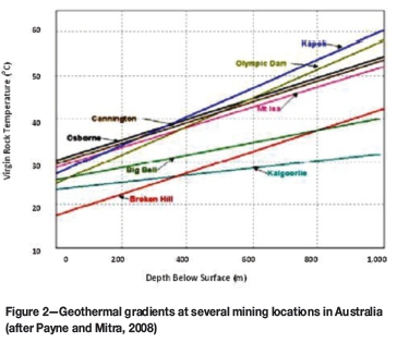
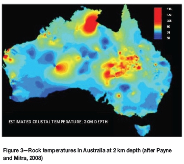
Payne and Mitra (2008) also stated that the geothermal gradient of the upper crust is between about 15°C/km and 40°C/km. A comparison is made between the geothermal gradient of Australia and South Africa. Brake and Bates (2000) asserted that Australia is the world's hottest and driest continent. They explained that the geothermal gradient of Australia is between 23°C/km and 30°C/km. Biffi et al. (2007) reported the average geothermal gradient for the South African gold and platinum mines as 13°C/km and 23°C/km respectively. Figures 2 to 5 show the heat flow pattern in Australia and South Africa.
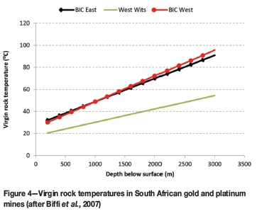
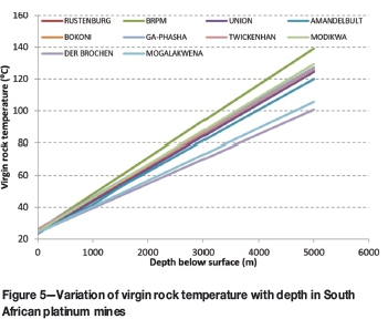
Biffi et al. (2007) reported that the geothermal gradient of the South African platinum mines is about twice that of the gold mines. They also pointed out that the rock mass in the platinum mines, mainly norite, cools faster than quartzite, which is the major host rock in the Witwatersrand Basin. However, Witwatersrand quartzite has a higher thermal conductivity and lower heat capacity than Bushveld Complex rocks (Jones, 2003, 2013, pers. Com.). The thermal conductivity and heat capacity of quartzite and norite are 6.37 and 2.32 Wm-1K-1 and 810 and 840 Jkg-1K-1 respectively. This implies that quartzite will absorb and release heat faster than norite. The heating and cooling of rocks may lead to micro-factures in rocks. This is one of the subjects of this investigation.
Comparing Figures 2 and 5, it can be seen that apart from Broken Hill, Kalgoorille, and Big Bell mines, which have similar a geothermal gradient to the platinum mines, all the other Australian mines shown in Figure 2 have higher gradients. Mt Isa is Australia's deepest mine, which started operations in 1924 and is now mining at 1900 m below surface. At the current depth of mining, the virgin rock temperature at Mt Isa mine is approximately 80°C. This temperature will be reached at a depth of 2200 m below surface at Bafokeg Rasimone platinum mine. This means that the investigation into the effect of temperature on the behaviour of rocks in mines will be beneficial not only to South African mines, but also to all other mines across the globe that are located in high geothermal gradient areas. Most of the previous researchers, such as Biffi et al. (2007), Brake and Bates (2000), and Payne and Mitra (2008), focused on strategies of improving ventilation or cooling of the mines and safeguarding the health of the underground workers.
The aim of the present research is to examine the effect of heat on rock texture and composition, in terms of initiation or extension of micro-cracks and changes in chemical composition. In the mining context, an attempt was made understand the effect of temperature drop on the surfaces of underground openings due to ventilation by the help of microscopic analysis. Planar cracks, which are perpendicular to the mineral grain boundaries, have been reported to form during the cooling of igneous rocks. These cracks were attributed to a mismatch of physical properties across grain boundaries during cooling (Richter and Simmons, 1974; Fonseka et al., 1985). The micro-cracks affect the compressive and tensile properties, sonic velocity, and thermal conductivity of the rocks.
Microscopy
Image were captured by means of two types of microscope:
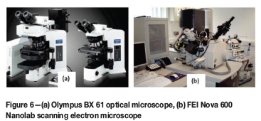
> Optical microscope (OM). The OM used is an Olympus BX61 (Figure 6a). The advantage of the OM is that the colour of the minerals that make up the rock samples can be seen, since surface coating of the specimens is not required. The OM uses visible light and a system of lenses to magnify images of small samples. Using the Olympus BX61, the images were captured in both bright field (sample contrast is due to absorbance of light in the sample) and dark field illumination (sample contrast is due to light scattered by the sample)
> Scanning electron microscope (SEM). The SEM used is FEI Nova 600 Nanolab, (Figure 6b). The SEM has higher resolution than the OM, but the specimens were gold-palladium coated in order to allow for good quality image. The SEM produces image by scanning the sample with a focused beam of electrons. The interaction of the electrons from the beam and the atoms in the samples produces signals that contain information about the surface topography and composition.
Rock samples and location
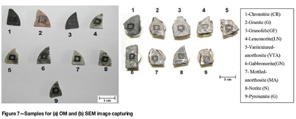
The rock samples were prepared from geological core samples obtained from mines in the western and northern Bushveld Complex (BC). Chromitite, pyroxenite, norite, and leuconorite were prepared from cores samples from Bafokeng Rasimone Platinum Mine (BRPM) in the western BC; gabbronorite, mottled anorthosite, and varitextured anorthosite from Khomanani Mine (a subsidiary of Anglo American Platinum) in the western BC; while granite and granofels were from Mogalakwena Mine (also a subsidiary of Anglo American Platinum) in the northern BC.
The rock specimens were cut to a thickness of 2.5 mm and the surfaces to be examined were smoothed with the aid of a lapping machine. The surfaces of the SEM specimens were coated with a thin layer of gold-palladium alloy. The layer, which is typically 10-20 nm thick, is necessary to make the specimens conductive. Figures 7 (a) and (b) show the samples prepared for the optical and scanning electron microscope respectively.
Heat treatment
The thermal treatment of the rock specimens was done in a temperature-regulated oven. The samples were heated to 50°C, 100°C, and 140°C at a rate of 2°C /min and kept at constant temperature for five consecutive days. These temperatures are approximately the same as the temperatures in the mines at depths of 1073 m, 3276 m, and 5038 m below surface respectively. Samples were allowed to cool to ambient temperature (approximately 20°C) before image capturing. Micrographs of specimens were taken before and after heat treatment. For SEM analyses, a single specimen from each rock type was subjected the specified temperature range, that is 50°C, 100°C, and 140°C, while different specimens were heat-treated for OM analyses. All the SEM samples were also subjected to heating and cooling on alternate days for ten days in order to observe the effect of repeated heating and cooling of the rocks in the mine face.
Result of optical image capturing
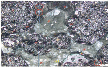
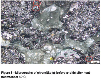
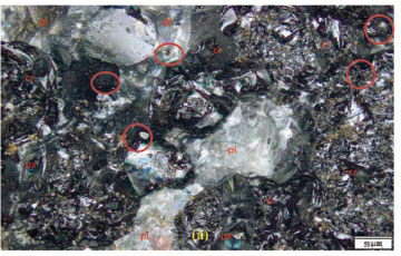
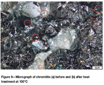
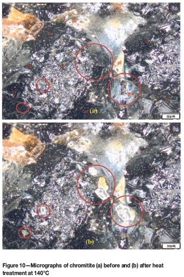
The essence of the optical image capturing was to examine whether heat treatment of the rock sample will have a significant effect of the physical properties in terms of the colours of the minerals. All the samples were imaged using the BX 61 Olympus optical microscope under bright field illumination. Figures 8 to 10 show the micrographs of chromitite before and after heat treatment at different temperatures.
Voordouw, Gutzmer, and Beukes (2009) reported the average modal mineral abundance for the UG1 and UG2 seams (Table I). Schouwstra, Kinloch, and Lee (2000) stated that UG2 consists predominantly of chromite (60 to 90 vol.%), with lesser silicate minerals (5 to 30% pyroxene and 1 to 10% plagioclase), while other minerals are present in minor concentrations.
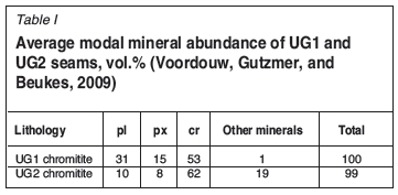
The colour of plagioclase (pl) ranges from white to colorless, cream, gray, yellow, orange, and pink; chromite (chr) is mostly black, while pyroxene (px) ranges from dark green to black. Plagioclase is a silicate of sodium, calcium, and aluminium; pyroxene is a silicate of magnesium and iron; while chromite is an oxide of chromium, iron, and magnesium (Schouwstra, Kinloch, and Lee, 2000).
The major chromitite minerals are indicated in Figures 8 in red. Figures 8(a) and (b) show the micrographs of chromitite before heat treatment and when cooled to ambient temperature after heat treatment at 50°C. The physical changes observed (red circles) are in the chromite-rich areas. In Figure 8(b), heat treatment exposed yellow-looking minerals (plagioclase, rutile or serpentine) and whitish minerals (quartz, talc, or plagioclase) - the upper two circles. The lower circle shows the disappearance of a whitish mineral, which may be talc or plagioclase, that flaked off from its original position due to heating and cooling of the specimen.
In Figure 9 (a) and (b), it can be seen that heat treatment at 100°C does not cause significant physical change in the specimen. However, some spots of a white mineral appeared after the heat treatment (red circles in Figure 9). A major difference is observed after heat treatment at 140°C (Figure 10). Some of the dark minerals, such as chromite and pyroxene, were displaced by quartz or plagioclase.
In general, the heating and cooling of rocks is seen to cause changes in the physical characteristics of the minerals.
Result of scanning electron microscopy
The SEM was used to examine the effects of heat treatment on crack extension/widening. The changes in the elemental composition of the rock samples were also determined using energy-dispersive X-ray spectrometry (EDX) equipment (known as INCA) attached to the SEM. Figures 11 and 12 show the SEM micrographs of chromitite before and after heat treatment and repeated heating and cooling.
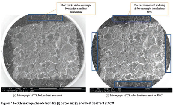
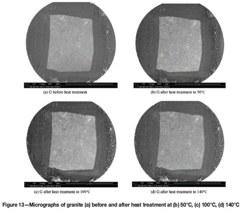
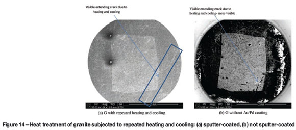
Of all the samples heat-treated, chromitite was the most affected by the heat treatment, followed by pyroxenite. Only few cracks were observed in mottled anorthosite, leuconorite, norite, gabbronorite, granofels, and varitextured anorthosite. No visible cracks were observed in granite (Figure 13) when heated from ambient temperature through 140°C, but when subjected to repeated heating and cooling, a clearly visible macro-crack extending from the lower to the upper end of the sample was observed, as shown in Figure 14.
Result of EDX
The elemental composition of all the samples were determine for each temperature range, that is at ambient, 50°C, 100°C, and 140°C. The results of the chemical characterization of the samples are presented in in Figure 15 and Table II.
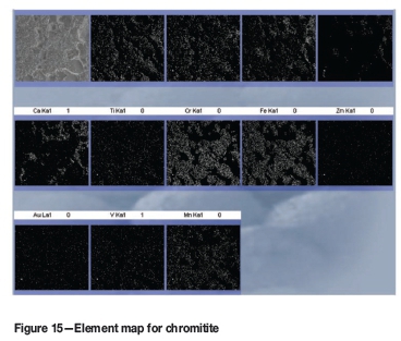
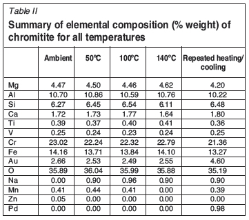
An element map is an image showing the spatial distribution of elements in a sample. In Figure 15, the letters 'K' and 'L' following the chemical symbols refer to the first and second electron shells respectively, while 'a' represents alpha X-rays. The bright portions in the maps indicate the dominance of elements in the group, and the maps that look similar indicate a strong correlation between the elements. It should be noted that in Table II and Figure 15, oxygen is the most abundant element. The reason is that oxygen reacts with almost all the other elements to form silicates and oxides. A careful examination of all the elemental compositions in Table II shows that there is not much difference in the mineral compositions of the specimen when heat treated from ambient temperature up to 140°C. However, temperature caused some elements to be displaced while others resurface. For example, the element maps and Table II reveal that zinc appears only in ambient conditions. The sudden increase in the values for gold and palladium in the sample that underwent repeated heating and cooling results from sputter coating.
Conclusion
The results of the optical microscope analyses show that physical changes take place in the rocks subjected to heat treatment. However, the observed changes are not so significant as to affect the physical behaviour of the rocks.
The scanning electron microscope images revealed that crack initiation starts at lower temperature and the cracks extend with increasing temperature. This is an indication that temperature would influence the texture and strength of rocks in underground mines with high geothermal gradients.
The chemical analyses of the samples subjected to different temperatures show that the temperature range used in this research is not high enough to induce noteworthy chemical changes in the samples.
References
Biffi, M., Stanton, D., Rose H., and Pienaar, D. 2007. Ventilation strategies to meet future needs of the South African platinum industry. Journal of the Southern African Institute of Mining and Metallurgy, vol. 107, no.1. pp. 59-66. [ Links ]
Brake, D.J. and Bates, G.P. 2000. An interventional study. Proceedings of the International Conference on Physiological and Cognitive Performance in Extreme Environments. Lau, W.M. (ed.). Defence Scientific and Technology Organisation, Australian Department of Defence, Canberra. pp. 170-172. [ Links ]
Clauser, C. 2011. Thermal storage and transport properties of rocks, I: Heat capacity and latent heat. Encyclopedia of Solid Earth Geophysics. Gupta, H. (ed.). 2nd edn. Springer. [ Links ]
Clauser, C. and Huenges, E. 1995. Thermal conductivity of rocks and minerals. Rock Physics and Phase Relations: A Handbook of Physical Constants. Ahrens, T.J. (ed.). AGU Ref. Shelf, vol. 3. AGU, Washington DC. pp. 105-126. [ Links ]
Fonseka, G.M., Murrell, S.A.F., and Barnes, P. 1985. Scanning electron microscope and acoustic emission studies of crack development in rocks. International Journal of Rock Mechanics and Mining Sciences and Geomechanics Abstracts, vol. 22, no. 5. pp. 273-289. [ Links ]
Jones, M.Q.W. 2003. Thermal properties of stratified rocks from Witwatersrand gold mining areas. Journal of the South African Institute of Mining and Metallurgy, vol. 103, no. 3. pp. 173-185. [ Links ]
Richter, D. and Simmons, G. 1974. Thermal expansion behaviour of igneous rocks. International Journal of Rock Mechanics and Mining Sciences and Geomechanics Abstracts, vol. 11. pp. 403-411. [ Links ]
Schouwstra, R.P., Kinloch, E.D., and Lee, C.A. 2000. A short geological review of the Bushveld Complex. Platinum Metals Review, vol. 44, no. 1. pp. 33-39. [ Links ]
Payne, T. an Mitra, R 2008. A review of heat issue in underground metalliferous mines. Proceedings of the 12th US/ North American Mine Ventilation Symposium, Reno, Nevada, June 2000. Wallace, K.G. Jr (ed.). pp. 197-201. [ Links ]
Voordouw, R., Gutzmer, J., and Beukes N.J. 2009. Intrusive origin for Upper Group (UG1, UG2) stratiform chromitite seams in the Dwars River area, Bushveld Complex, South Africa. Mineralogy and Petrology, vol. 97. pp.75-94. [ Links ]


