


Services on Demand
Article
 English (pdf)
 Article in xml format
 Article references
Indicators

Related links
-
 Cited by Google
 -
 Similars in Google
Share

 Permalink
African Entomology
On-line version ISSN 2224-8854
Print version ISSN 1021-3589
AE vol.30 Pretoria 2022
http://dx.doi.org/10.17159/2254-8854/2022/a13589
RESEARCH ARTICLE
Larvicidal potential of copper sulphide nano aqua dispersions against Aedes aegypti (Linnaeus)
KK SandhuI, II; N VashishatI; A SidhuIII
IDepartment of Zoology, Punjab Agricultural University, Ludhiana, India
IIDepartment of Zoology, Akal University, Talwandi Sabo, Punjab, India
IIIDepartment of Chemistry, Punjab Agricultural University, Ludhiana, India
ABSTRACT
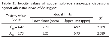
Nanotechnology has emerged as promising field in insect pest management. Aedes aegypti (Linnaeus) a well-known vector of dengue, chikungunya, and dengue haemorrhagic fever has no commercial management practice for their eradication at the larval stage. In the present study, copper sulphide one of the most detoxified form of copper with biopotential properties was synthesised by standard methodology using sonochemical irradiation method and was evaluated for their larvicidal potential against Ae. aegypti. Treated larvae were observed for various morphological changes as compared to control. Larvae were most susceptible to CuSNPs at 7 ppm showing 100% mortality within 24 h. LC50 and LC90 values calculated with the help of POLO software were 4.42 and 5.73 ppm. The epithelium layer of treated larvae was damaged as compared to control. Remarkable results of copper sulphide nanoformulations at low dosage against Ae. aegypti larvae advocates their further exploration for vector control programmes.
Keywords: aqueous nanoformulations, metal nanoparticles, mortality, mosquito, vector control
INTRODUCTION
Mosquitoes are key vectors for dissemination of various parasites and pathogens as they are haem feeders, thus drawing attention due to their medical importance. Aedes aegypti (Linnaeus) is one of the most significant species in the world. This mosquito can be identified by the lyre form marking on the upper surface of its thorax and white colouration on its legs. It originated in Africa and extensively dispersed in tropical and subtropical zones (Powell et al. 2018). This species is a carrier of Dengue Fever Virus (DENV) causing dengue fever, chikungunya fever, and dengue haemorrhagic fever (Yang et al. 2009). Dengue is an emerging disease which infects millions of people in the world (Goncalvez et al. 2007; WHO 2015). In over 128 nations, more than 3.9 billion people are at risk of dengue, with about 96 million cases annually (WHO 2017). This disease is endemic in all the states of India and major cause of hospitalisation (Kumar et al. 2018).
Therefore, the control of vector mosquito is an important public health concern (Magro et al. 2019) for ending epidemics in the tropical areas. Generally, synthetic insecticides like malathion and permethrin are sprayed to control the adult mosquitoes (Nwabor 2019). However, these chemicals have adverse effects on the non-target species, environment and human health (Margulis-Goshen et al. 2013). Further, a number of mosquito species have become resistant towards them (Wattanachai & Tintanon 1999). Control strategies at the larval stages are the most pertinent concept as far as their practical eradication is concerned. The control of larval stages is a strategy to tackle the resurgence in vectors, in more effective way (Wilke &Marrelli. 2012). Moreover, environmental safety is considered most important. In order to be acceptable, an insecticide should also show its potential not to affect non-target species (WHO 2018) rather than only showing mortality of target species (Ghosh et al. 2012).
Nanotechnology has developed remarkably in the last decade and many innovative materials were created with a wide range of applications of nanoparticles in medical, pharmaceutical, industrial, biotechnological and scientific fields. In recent years, there has been emphasis on the application of nanotechnology in insect pest management (Prabhakar et al. 2017). Nanoparticles were considered as a useful tool loaded with herbicides, fungicides, larvicides and targeting specific tissues in desired plants or insects to release their charge to desired part of the target animal to achieve desired results (Duhan et al. 2017). Furthermore, nanopesticide-based formulations have a potential and bright future for the development of more effective and safer biopesticide/pesticides (Deka et al. 2021).
On the nanotechnological front, studies on copper nanoparticles (Cu NPs) and copper oxide nanoparticles (CuO NPs) have been reported for their larvicidal, antibacterial, antifungal and antimicrobial properties. Valodkar et al. (2011) synthesised nanoparticles of 5-10 nm of silver and copper using latex from Euphorbiaceae. These nanoparticles have excellent antibacterial activity towards Gram positive and Gram negative bacteria. Ramyadevi et al. (2011) conducted a study on synthesis of copper nanoparticles by polyol process and showed their anti-larval and anti-parasitic properties.
Sulphide analogues of transition metals are hypothesised as one of the most detoxified forms of these metals (Wang et al. 2013; Sidhu et al 2017). Various transition metals applied in various forms were detoxified in the environment by sulphidation. The eminent bio efficacy and detoxified nature of metal sulphide nanoparticles are drawing the attention of scientists for their use against a variety of environmental menaces. Copper sulphide (CuS) is one of the most detoxified forms of copper (Baek 2017; Baek & Kim 2017; Guo et al. 2013; Wang et al. 2013), natural, thermally stable (Rao et al. 2016), water insoluble (Guo et al. 2013) still retaining the biopotential of copper (Chakraborty et al. 2016; Prabhavathi et al. 2015). It is quite safe and non-toxic to humans (Guo et al. 2013) and therefore may be used in the environment against vector mosquitoes without causing harm to non-target species. There is limited literature on synthesis and biopotential of water-dispersed CuS nanoparticles which prompted us to prepare the novel surface protected aqueous nano formulations of copper sulphide for the evaluation of larvicidal potential against Ae. aegypti. The possible morphological alterations of larvae by NCuS are also given in the paper.
MATERIALS AND METHODS
Synthesis of copper sulphide nano-aqua dispersions (NCuS)
Copper nitrate trihydrate (Cu(NO3)2.3H2O) solution (0.001 M, 30 ml), prepared in ethylene glycol, was added dropwise to 30 ml of sodium sulphide aqueous solution (0.001 M), during sonication, along with a pinch of cetyltrimethyl ammonium bromide (CTAB) (surfactant) (Sidhu et al. 2017). This reaction mixture was subjected to ultrasonic irradiation at temperature 40 | 25 °C having pulse of 05 on and 01 off and amplitude of 60%. The solution was irradiated with microwaves for 30 sec and allowed to cool down at room temperature. An amount of 0.3 g of capping agent, polyvinyl pyrollidone (PVP) dissolved in 20 ml distilled water was added to the above prepared solution during sonication, which was continued for another 15 min, to get surface stabilised copper sulphide nanoparticles (NCuS). The prepared NCuS (0.0005 M, 35.85 μg/ml) were retained as such as stock solution, which was further diluted by adding autoclaved water, as and when required.
Morphological characterisation of prepared NCuS solution
The morphology and size of nanoparticles were recorded in a Hitachi Transmission Electron Microscope Hi-7650 at an accelerated voltage of 200 kV by casting a drop of particle solution onto a 200-mesh carbon coated copper grid from EMN Laboratory, Punjab Agricultural University, Ludhiana.
Mosquito larvae collection
Water samples were collected in the monsoon season from different peri-domestic fresh water collections like desert coolers, earthen pots, fire buckets, water tanks, plates under pots and roadside ditches in different zones of Punjab Agricultural University Ludhiana district (30.9010 °N and 75.8573 °E) using plastic dippers from June to August 2019. From the various types of mosquito larvae present in the collected water samples, Ae. aegypti larvae were recognised from the other types of mosquito larvae (if present) on the basis of their morphological features by following the standard keys given by Becker et al. (2010) and Bar & Andrew (2013). All mosquito larvae were adequately fed with a mixture of dog biscuits and yeast ground in the ratio of 3:1 (2 mg/100 ml) as used by Mavundza et al.
(2013).
Larvicidal bioassay
Varying concentrations of the NCuS (0.0005 M, 35.85 μg/ml) at 5, 6, 7 and 8 ppm were prepared by diluting it with de-chlorinated water. Twenty 4th-instar Ae. aegypti larvae were exposed to these concentrations in triplicate. In addition, a vehicle-control and a control set with PVP and only dechlorinated water respectively were run in triplicate. The experimental sets were kept in a Bio-Oxygen Demand incub ator at 26 ± 2 °C. Mortality of larvae was recorded in copper sulphide nano-aqua dispersions treated, vehicle control and control sets after 3, 6, 12and 24 h. Treated larvae were observed under a microscope for various outer morphological changes.
Statistical analysis
Statistical analysis was performed by comparing the mortality data recorded from NCuS treated sets with control and vehicle control sets by using ANOVA (Duncan multiple range test) with the help of SPSS software version 16.0. LC50 and LC90 values after 24 h post-exposure, were worked out by log probit technique (Finney 1952) employing the POLO programme (Robertson et al. 1980).
RESULTS
Surface protected copper sulphide nanoparticles (NCuS) in aqua-dispersed form were synthesised by sonochemical irradiation method. Cu2+ ions from Cu(NO3)2 in ethylene glycol (HOCH2CH2OH) solution were made to combine with S2- from sodium sulphide (Na2S) solution, while sonication, forming copper sulphide nanoparticles, which were stabilised using stabilising agent PVP.
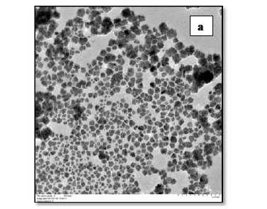
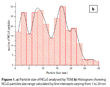
Characterisation of copper sulphide nano-aqua dispersions The topo-morphological characters and particles size of synthesised surface protected copper sulphide nanoparticles (CuS NPs) in aqua-dispersed form by Transmission Electron Microscopy (TEM). Shape analogy indicated from TEM morphograph showed distorted spherical to oval shapes of CuS nanoparticles with slight aggregations. The average particle size was 16 nm (Figure 1a) and range of CuS nanoparticles size is from 1 to 29 nm (Figure 1b).
Larvicidal activity
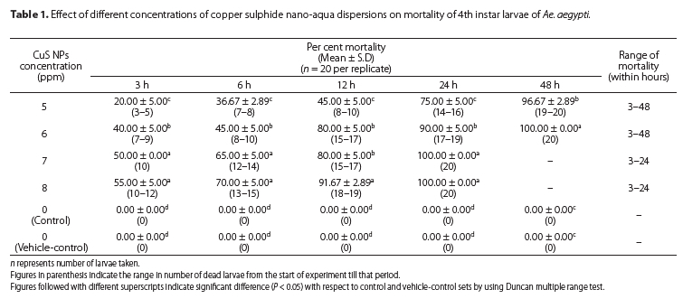
Copper sulphide nanoparticles were tested for their larvicidal potential against Ae. aegypti at 5, 6, 7 and 8 ppm to assess their larvicidal potential. With the exposure of 5 ppm of copper sulphide nanoparticles (NCuS), the percent mortality was observed as 20.00 ± 5.00 after 3 h and 36.67 ± 2.89, 45.00 ± 5.00, 75.00 ± 5.00, 96.67 ± 2.89 after 6, 12, 24 and 48 h, respectively, whereas at 6 ppm percent mortality increased, i.e. 40.00 ± 5.00, 45.00 ± 5.00, 80.00 ± 5.00, 90.00 ± 5.00 and 100.00 ± 0.00 after 3, 6, 12, 24 and 48 h respectively (Table 1) which is statistically significant. With the treatment of 7 and 8 ppm of NCuS 100% mortality was observed within 24 h while 50.00 ± 0.00, 65.00 ± 5.00 and 80.00 ± 5.00 and55.00 ± 5.00, 70.00 ± 5.00 and 91.67 ± 2.89% mortality was observed after 3, 6 and 12 h respectively. Statistically there is no relationship betweenpercentmortality at 7 and 8 ppm of NCuS. No larval mortality was recorded in the untreated control andvehick control sete. Duringthe present study, larvae were m st susceptible to CuS NPs at 7 ppm and it was found to be the most effective concentration out of the four tested concentrations, as it resulted into 100% larval mortality within 24 h or before their conversion to the next developmental stage, i.e. pupae. No mortality was recorded in control and vehicle-control sets.
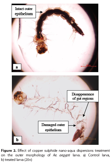
The values for LC50 and LC90 of copper sulphide nanoparticles after 24 h of post-exposure against fourth instar larvae of Ae. aegypti was calculated to be 4.42 ppm and 5.73 ppm (Table 2) respectively by the log probit technique. Exposure to effective copper sulphide nanoparticles resulted in destruction of the outer epithelial layer of a larva while an untreated larva was intact and normal (Figure 2). There was disappearance of the gut in treated larvae while it is present in the control larva.
DISCUSSION
Copper nanoaprticles (CuNPs) are considered to have antifungal, antibacterial and antiplasmodial potential; so we intend to evaluate if they coul Fbe appiiedaemesquitolarvieiOaleganti. Previous studies demonirated that bulk copper particles were less efficient as compared to CuNPs against fungal and bacterial infections (Rai et al. 2018). To the best of the authors' knowledge, it is the first report on the use of copper sulphide nanoparticles as larvicidal agents against Ae. aegypti. No previous report has been documented about copper sulphide nano aqua dispersions as larvicidal agents against any of the mosquito species, but several nanoparticles such as silver (Yadav & Prakash 2016), gold (Soni & Prakash 2012), palladium (Minal & Prakash 2018), copper (Ramyadevi et al. 2011), zinc (Vijayakumar et al. 2015), silica and carbon(Murugan et al. 2016) nanoparticles, were exemplifiedtobe thebest candidatesformosquitocontrol.
The appreciable effective larvicidal concentration is 7 ppm in our results which is much lower than the previously reported studies like silver doped copper oxide nanosheets had mortality effects at 300 ppm concentration against larvae of Spodoptera littoralis (Atwa et al. 2017). In another study, Shaker et al (2016) synthesised copper oxide nanoparticles used in control and management of cotton leafworm (S. littorals) as an example for the pest which attacks large scale of crops and vegetables by applying six different concentrations 1000, 500, 250, 125, 62.5 and 31.25 mg/l. Treated larvae showed 100% mortality with LC50 of 62.5 mg/l which is different from our results and they reported significant impact on the biological features of the insect as well. Minal & Prakash (2019) tested the larvicidal efficacy of Azadirachta indica leaf extract mediated bimetallic nanoparticles. They concluded that monometallic forms of nanoparticles, i.e. copper and zinc alone showed less larvicidal efficacy as compared to copper-zinc bimetallic nanoparticles against Culex quinquefasciatus. Copper oxide nanoparticles were synthesised using seed of Artocarpus heterophyllus and proved them as an anti-larval application (Mishra et al. 2015). Selvan et al. (2018) also synthesised copper oxide nanoparticles using a leaf extract of Tridax procumbens and proved them to have larvicidal properties against Ae. aegypti. Suresh et al. (2020) demonstrated that metal oxide nanoparticles act as potential mosquitocidal agents and also reported by Gayathri et al. (2018) that magnesium oxide (MgO) nanoparticles had efficient larvicidal potential against Ae. aegypti, Anopheles stephensi and Culex quinquefasciatus.
Sidhu et al. (2017) reported in vitro anti-fungal evaluation at 7 μg/ml of copper sulphide nanoformulations against Alternaria alternata, Drechslera oryzae and Curvularia lunata. They found the average particle size was 12 nm and range of size of CuS NPs was 10-30 nm which was at par of with our results. Analogous studies were carried out by Naik et al. (2014) in which copper oxide nanoparticles were synthesised using a leaf extract of Gloriosa superba. Scanning electron microscopic (SEM) images indicate that they have particle size in the range of 5-10 nm which is smaller than observed in the present study. In another study, Lee et al. (2013) synthesised Cu nanoparticles of 37-110 nm using Magnolia kobus leaf extract which was greater in size as compared to present study and have shown antibacterial activity against Escherichia coli.
The nanoparticles entered the larval body through the tracheal system. In this study, various morphological alterations such as damaged epithelium and gut disappearance were observed. These were analogous to the findings of Kaur et al (2019) in which nanoemulsion of E. globulus affected the cuticle layer of the abdomen of larvae of Ae. aegypti. Such morphological changes in the form of distortion and damage in various body parts of larvae treated with copper sulphide nano-formulations results in larval mortality and could be used as part of a mosquito control programme.
Copper sulphide is natural as well as non-toxic and has been reported to be metabolised in the human body to cure various ailments like cancer and atherosclerosis (Guo et al. 2013). Moreover, copper and sulphur belong to essential micro- and macro-nutrients for the living kingdom which presents that these copper sulphide nanoformulations are somewhat non-toxic. The remarkable mortality of Ae. aegypti larvae at a low dosage of CuS NPs in the present study, advocates their possible use in the vector control strategies and further research of CuS NPs in the field along with biosafety tests could lead to a very promising avenue for vector control.
ACKNOWLEDGEMENTS
The authors are grateful to the Head, Department of Zoology, for providing all the necessary facilities, and Department of Science and Technology, Government of India, New Delhi, for providing infrastructural facilities under FIST grant.
ORCID IDs
KK Sandhu - https://orcid.org/0000-0003-3129-3670
N Vashishat - https://orcid.org/0000-0003-0071-8955
A Sidhu - https://orcid.org/0000-0003-0013-9558
REFERENCES
Atwa AA, Salah NA, Khafagi WE, Al-Ghamdi A. 2017. Insecticidal effects of pure and silver-doped copper oxide nanosheets on Spodoptera littoralis (Lepidoptera: noctuidae). Canadian Entomologist 149(5): 677-690. https://doi.org/10.4039/tce.2017.36 [ Links ]
Baek SW, Kim MS. 2017. Antibacterial filter comprising copper based sulfur compound. US Patent 2016/0332104A1. Baek SW. 2017. Artificial biomaterial comprising copper based compound. US Patent 2017/0035933A1.
Bar A, Andrew J. 2013. Morphology and morphometry of Aedes aegypti adult mosquito. Annual Research & Review in Biology 3: 52-69. [ Links ]
Becker N, Petric D, Zgomba M, Boase C, Dahl C, Madon M, Kaiser A. 2010. Mosquitoes and their Control. 2nd ed. New York, U.S.A.: Springer Publications. p. 9-40. https://doi.org/10.1007/978-3-540-92874-4_2 [ Links ]
Chakraborty P, Adhikar J, Chatterjee S, Biswas B, Chattopadhyay T. 2016. Facile synthesis of copper sulfide nanoparticles: antibacterial and antifungal activity study. Rasayan Journal of Chemistry 9: 77-83. [ Links ]
Deka B, Babu A, Baruah C, Barthakur M. 2021. Nanopesticides: A systematic review of their prospects with special reference to tea pest management. Frontiers in Nutrition 8: 686131. https://doi.org/10.3389/fnut.2021.686131 [ Links ]
Duhan JS, Kumar R, Kumar N, Kaur P, Nehra K, Duhan S. 2017. Nanotechnology: the new perspective in precision agriculture. Biotechnology Reports 15: 11-23. https://doi.org/10.1016/j.btre.2017.03.002 [ Links ]
Finney DJ. 1971. Probit Analysis. New York, U.S.A.: Cambridge University Press p. 68-72. [ Links ]
Gayathri B, Muthukumarasamy N, Velauthapillai D, Santhosh SB, Asokan V. 2018. Magnesium incorporated hydroxyapatite nanoparticles: preparation, characterisation, antibacterial and larvicidal activity. Arabian Journal of Chemistry 11(5): 645-654. https://doi.org/10.1016/j.arabjc.2016.05.010 [ Links ]
Ghosh A, Chowdhury N, Chandra G. 2012. Plant extracts as potential mosquito larvicides. Indian Journal of Medical Research 135: 581-598. [ Links ]
Goncalvez AP, Engle RE, St. Claire M, Purcell RH, Lai C-J. 2007. Monoclonal antibody-mediated enhancement of dengue virus infection in vitro and in vivo and strategies for prevention. Proceedings of National Academy of Sciences of USA 04: 94229427. https://doi.org/10.1073/pnas.0703498104
Guo L, Panderi I, Yan DD, Szulak K, Li Y, Chen Y, Ma H, Niesen DB, Seeram N, Ahmed A, et al. 2013, A comparative study of hollow copper sulfide nanoparticles and hollow gold nanosphere on degradability and toxicity. ACS Nano 7(10): 8780-8793. https://doi.org/10.1021/nn403202w. [ Links ]
Kaur N, Kocher DK, Sidhu A. 2019. Synthesis and testing of Eucalyptus globulosa oil-based nanoemulsion for its larvicidal potential against Aedes aegypti. African Entomology 27(2): 433-438. https://doi.org/10.4001/003.027.0433 [ Links ]
Kumar S, Arjun MC, Gupta SK, Nongkynrih B. 2018. Malaria, dengue and chikungunya in India - an update. Indian Journal of Medical Specialities 9: 25-30. https://doi.org/10.1016/j.injms.2017.12.001 [ Links ]
Lee H, Song YJ, Kim BS. 2013.Biological synthesis of copper nanoparticles using Magnolia kobus leaf extract and their antibacterial activity. Journal of Chemical Technology and Biotechnology 88: 1971-1977. [ Links ]
Magro M, Bramuzzo S, Baratella D, Ugolotti J, Zoppellaro G, Chemello G, Olivotto I, Ballarin C, Radaelli G, Arcaro B, et al. 2019. Self- assembly of chlorin-e6 on y-Fe2O3 nanoparticles: application for larvicidal activity against Aedes aegypti. Journal of Photochemistry and Photobiology B: Biology 194: 21-31. https://doi.org/10.1016/j.jphotobiol.2019.03.004 [ Links ]
Margulis-Goshen K, Magdassi S, Ishaaya I, Palli S, Horowitz A. Advanced Technologies for Managing Insect Pests. Dordrecht, The Netherlands: Springer; 2013. p. 295-314. https://doi.org/10.1007/978-94-007-4497-4_15 [ Links ]
Mavundza EJ, Maharaj R, Chukwujekwu JC, Finnie JF, van Staden J. 2013. Larvicidal activity against Anopheles arabiensis of 10 South African plants that are traditionally used as mosquito repellents. South African Journal of Botany 88: 86-89. https://doi.org/10.1016/j.sajb.2013.05.007 [ Links ]
EMinal PS, Prakash S. 2019. Efficacy of bimetallic copper-zinc nanoparticles against larvae of microfilariae vector in laboratory. International Journal of Scientific Research 8: 72-75. [ Links ]
Minal SP, Prakash S. 2018.Characterization and nano-efficacy study of palladium nanoparticles against larvae of Anopheles stephensi (Liston). International Journal of Advanced Engineering and Nanotechnology 3(10): 1-5. [ Links ]
Mishra PM, Sahoo SK, Naik GK, Parida K. 2015. Biomimetic synthesis, characterization and mechanism of formation of stable silver nanoparticles using Averrhoa carambola L. leaf extract. Materials Letters 160: 566-571. https://doi.org/10.1016/j.matlet.2015.08.048 [ Links ]
Murugan K, Nataraj D, Madhiyazhagan P, Sujitha V, Chandramohan B, Panneerselvam C, Dinesh D, Chandirasekar R, Kovendan K, Suresh U, et al. 2016. Carbon and silver nanoparticles in the fight against the filariasis vector Culex quinquefasciatus: genotoxicity and impact on behavioral traits of non-target aquatic organisms. Parasitology Research 115(3): 1071-1083. https://doi.org/10.1007/s00436-015-4837-9 [ Links ]
Naik R, Prashantha SC, Nagabhushana H, Sharma SC, Nagabhushana BM, Nagaswarupa HP, Premkumar HB. 2014. Low temperature synthesis and photoluminescence properties of red emitting Mg2 SiO4: Eu3+nanophosphor for near UV light emitting diodes. Sensors and Actuators B Chemical 195: 140-149. https://doi.org/10.1016/j.snb.2014.01.018 [ Links ]
Nwabor FO. 2019. Synthetic insecticides, phytochemicals and mosquito resistance. Academia Journal of Biotechnology 5: 118-125. [ Links ]
Powell RJ, Gloria-Soria A, Kotsakiozi P. 2018. Recent history of Aedes aegypti: vector genomics and epidemiology records. Bioscience 68(11): 854-860. https://doi.org/10.1093/biosci/biy119 [ Links ]
Prabhakar M, Tyagi BK, Chandrasekaran N, Mukherjee A. 2017. Biological nanopesticides: a greener approach towards the mosquito vector control. Environmental Science and Pollution Research 25: 10151-10163. [ Links ]
Prabhavathi SP, Ranjith Ranjam SR, Mauthamuthu Johnson T. 2015. Microwave synthesis, characterization and biological activities of CuS and CdS nanoparticles. World Journal of Pharmaceutical Research 4: 710-720. [ Links ]
Rai M, Ingle AP, Pandit R, Paralikar P, Shende S, Gupta I, Biswas JK, da Silva SS. 2018. Copper and copper nanoparticles: role in management of insect pests and pathogenic microbes. Nanotechnology Reviews 7(4): 303-315. https://doi.org/10.1515/ntrev-2018-0031 [ Links ]
Ramyadevi J, Jeyasubramanian K, Marikani A, Rajakumar G, Rahuman AA, Santhoshkumar T, Kirthi AV, Jayaseelan C, Marimuthu S. 2011. Copper nanoparticles synthesized by polyol process used to control hematophagous parasites. Parasitology Research 109(5): 1403-1415. https://doi.org/10.1007/s00436-011-2387-3 [ Links ]
Rao H, Sun W, Ye S, Yan W, Li Y, Peng H, Liu Z, Bian Z, Huang C. 2016. Solution-processed CuS NPs as an inorganic holeselective contact material for inverted planar perovskite solar cells. ACS Applied Materials & Interfaces 8(12): 7800-7805. https://doi.org/10.1021/acsami.5b12776 [ Links ]
Robertson JL, Russell RM, Savin NE. 1980. POLO: A User's Guide to Probit or Logit Analysis. Berkeley, CA, U.S.A.: Pacific South-West Forest and Range Experiment Station. https://doi.org/10.2737/PSW-GTR-38 [ Links ]
Selvan SM, Anand VK, Govindaraju K, Tamilselvan S, Kumar VG, Subramanian KS, Kannan M, Raja K. 2018. Green synthesis of copper oxide nanoparticles and mosquito larvicidal activity against dengue, zika and chikungunya causing vector Aedes aegypti. IET Nanobiotechnology 12(8): 1042-1046. https://doi.org/10.1049/iet-nbt.2018.5083 [ Links ]
Shaker AM, Zaki AH, Rahim EFA, Khader MH. 2016. Novel CuO nanoparticles for pest management and pesticides degradation. Advances in Environment Biology 10: 274-283. [ Links ]
Sidhu A, Barmota H, Bala A. 2017. Antifungal evaluation studies of copper sulfide aqua-nanoformulations and its impact on seed quality of rice (Oryza sativa). Applied Nanotechnology 7: 681-689. [ Links ]
Soni N, Prakash S. 2012. Efficacy of fungus mediated silver and gold nanoparticles against Aedes aegypti larvae. Parasitology Research 110(1): 175-184. https://doi.org/10.1007/s00436-011-2467-4 [ Links ]
Suresh M, Jeevanandam J, Chan YS, Danquah MK, Kalaiarasi JMV. 2020. Opportunities for metal oxide nanoparticles as a potential mosquitocide. Bionanoscience 10(1): 292-310. https://doi.org/10.1007/s12668-019-00703-2 [ Links ]
Valodkar M, Jadeja RN, Thounaojam MC, Devkar RV, Thakore S. 2011. In vitro toxicity study of plant latex capped silver nanoparticles in human lung carcinoma cells. Materials Science and Engineering 31(8): 1723-1728. https://doi.org/10.1016/j.msec.2011.08.001. [ Links ]
Vijayakumar S, Vinoj G, Malaikozhundan B, Shanthi S, Vaseeharan B. 2015. Plectranthus amboinicus leaf extract mediated synthesis of zinc oxide nanoparticles and its control of methicillin resistant Staphylococcus aureus biofilm and blood sucking mosquito larvae. Spectrochimica Acta Part A: Molecular and Biomolecular Spectroscopy 137: 886-891. https://doi.org/10.1016/j.saa.2014.08.064 [ Links ]
Wang Z, von dem Bussche A, Kabadi PK, Kane AB, Hurt RH. 2013. Biological and environmental transformation of copper-based nanomaterials. ACS Nano 7(10): 8715-8727. https://doi.org/10.1021/nn403080y. [ Links ]
Wattanachai P, Tintanon B. 1999. Resistance of Aedes aegypti to chemical compounds in aerosol insecticide products in different areas of Bangkok, Thailand. Journal of Communicable Disease 25: 188-191 [ Links ]
WHO [World Health Organisation]. 2015. Dengue and severe dengue. Fact sheet No. 117. Geneva, Switzerland: World Health Organization [ Links ]
WHO [World Health Organisation] 2017. https://www.who.int/en/news-room/fact-sheets/detail/dengue-and-severe-dengue
WHO [World Health Organisation]. 2018. Biosafety and Biosecurity. Fact sheet 1. Geneva, Switzerland: World Health Organization. [ Links ]
Wilke BB, Marrelli MT. 2012. Genetic control of mosquitoes: population suppression strategies. Journal of the São Paulo Institute of Tropical Medicine 54(5): 287-292. https://doi.org/10.1590/S0036-46652012000500009. [ Links ]
Yadav S, Prakash S. 2016. Control of filariasis by silver nanoparticles: A green method for health care. In: Humanitarian Technology Conference (R10-HTC), IEEE Region 10 (pp. 1-7). IEEE. https://doi.org/10.1109/R10-HTC.2016.7906826
 Correspondence:
KK Sandhu
Email: komalpreetkaur903@gmail.com
Received:8 August 2020
Accepted: 24 November 2021

