


Servicios Personalizados
Articulo
 Inglés (pdf)
 Articulo en XML
 Referencias del artículo
Indicadores

Links relacionados
-
 Citado por Google
 -
 Similares en Google
Compartir

 Permalink
South African Journal of Childhood Education
versión On-line ISSN 2223-7682
versión impresa ISSN 2223-7674
SAJCE vol.12 no.1 Johannesburg 2022
http://dx.doi.org/10.4102/sajce.v12i1.1138
REVIEW ARTICLE
Functional near-infrared spectroscopy as a tool to assess brain activity in educational settings: An introduction for educational researchers
Candida BarretoI; Mojtaba SoltanlouII, III
IDepartment of Integrated Studies of Learning Language, Science, and Mathematics in the Primary School, Faculty of Education, University of Johannesburg, Johannesburg, South Africa
IISchool of Psychology, University of Surrey, Guildford, United Kingdom
IIINRF SARChI Chair: Department of Integrated Studies of Learning Language, Science, and Mathematics in the Primary School, Faculty of Education, University of Johannesburg, Johannesburg, South Africa
ABSTRACT
BACKGROUND: Educational research has been conducted mainly by using behavioural approaches. Whilst such methods provide invaluable insights into the field, several important questions such as 'how do we learn?' and 'what mechanisms cause individual differences?' cannot be answered thoroughly by using only behavioural approaches. In the last three decades, the advances of neuroimaging technologies and computational power have allowed researchers to investigate these questions beyond behavioural measures that provide complementary knowledge about human brain.
AIM: One of the most recent neuroimaging techniques that holds much promise for use in educational settings is functional near-infrared spectroscopy (fNIRS). This article aims to introduce the fNIRS technique to educational researchers interested in neurocognitive mechanisms of academic learning and achievements to further promote the growing field of Educational Neuroscience.
METHOD: We present the properties of the fNIRS device, its basic principles and important considerations when planning an fNIRS study.
RESULTS: Functional near-infrared spectroscopy is a portable, cost-effective and easy-to-handle neuroimaging device that allows experimentation in naturalistic settings such as in the school.
CONCLUSION: Even though several articles describe different applications and technical features of the fNIRS technique, there is still a need for materials with a more accessible language for those unfamiliar with neuroscientific and technical terms.
Keywords: functional near-infrared spectroscopy (fNIRS); educational neuroscience; neuroimaging; academic education; reading; mathematics.
Introduction
Although the global literacy and numeracy rates have improved in the last decades, there are still inequalities around the world (Roser & Ortiz-Ospina 2016). This discrepancy displays an urgent need to equate these numbers across the world (Tilak 1987). Educational research is one of the fields that provide evidence-based insights to address those global issues (Bruer 2016). However, most of this research relies on theoretical and behavioural approaches and do not necessarily provide insights about underlying cognitive mechanisms of children's academic achievements as one of the crucial factors in individual differences, in addition to several others, such as socio-economic status and educational facilities (McCandliss 2010; Thomas, Ansari & Knowland 2019). As a growing field in the last decade, Educational Neuroscience is a multidisciplinary field that tries to bring cognitive neuroscientists and educational scientists together to further disclose those neurocognitive mechanisms of academic achievements in ecologically valid settings such as schools (Goswami 2006). The aim of this article is to introduce one of the promising neuroimaging methods to educational researchers who are interested in further investigation of individuals' learning. Outcomes of studies with functional near-infrared spectroscopy (fNIRS) might, ultimately, contribute to diminishing the global inequities in education.
The advancement of neuroimaging techniques has provided opportunities to investigate the neurocognitive mechanisms underlying the complex cognitive processes of learning, such as in reading and mathematics competence (Ansari & Coch 2006; Szűcs & Goswami 2007). Functional magnetic resonance imaging (fMRI) is a neuroimaging modality that has been frequently used for studying the process of reading and learning mathematics (Ansari 2008; Jasińska et al. 2021). It is a non-invasive technique that measures brain activity, using strong magnetic fields. Although application of fMRI in educational studies has helped us to have a better understanding of individuals' learning, its requirements restrict its appropriateness in educational investigations (Gervain et al. 2011). For instance, it requires the individuals to be laid inside the scanner with no movement, except some button presses; it is very noisy, which may intimidate children; it is incompatible with metal implants because the fMRI measurement relies on a strong magnetic field; and it is an expensive technique (Pinti et al. 2018; Soltanlou et al. 2018b). Specifically, with regard to educational research, fMRI can only be applied in a very controlled in-lab setting. Such an environment cannot simulate the everyday world of children's written or oral production of responses.
Another commonly used method to investigate the brain mechanisms in educational studies of reading and mathematics is electroencephalography (EEG). It measures the electrical signals of the brain by placing electrodes on the participant's head (Xu & Zhong 2018). Electroencephalography is also sensitive to movement artefacts such as fMRI (Gervain et al. 2011). It has a lower spatial resolution, which means that it is more difficult to infer the original sources of the brain signal (Mehta & Parasuraman 2013). Also, the preparation and the placement of the electrodes in the individual's scalp may be tiring in case of a dense coverage of the head, especially for children (Curtin & Ayaz 2018; Ferrari & Quaresima 2012). Although fMRI and EEG have significantly contributed to the field of education and are quite appropriate for some conditions, a more flexible approach for naturalistic studies, in which participants may need to move freely in a known environment, would benefit the field (Da Silva Ferreira Barreto et al. 2020).
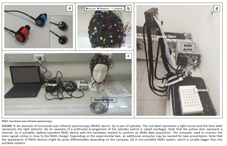
A fast-growing neuroimaging method that increasingly appears to be appropriate for educational settings is fNIRS. It is a non-invasive technique that indirectly measures brain activity (similar to fMRI) by placing sources and detectors (see Figure 3a) of near-infrared (NIR) light on the participant's head (Jobsis 1977). This technique has many advantages over other neuroimaging methods, such as fMRI and EEG (Curtin & Ayaz 2018). For instance, it has an easy and fast setup, is comparatively of low cost (Gervain et al. 2011; Lloyd-Fox, Blasi & Elwell 2010) and less susceptible to movement artefacts, and the portable models allow the participants to move (Balardin et al. 2017; Pinti et al. 2018). These characteristics of fNIRS devices have made it a promising tool to investigate the neurocognitive mechanisms of academic skills in naturalistic situations that simulate everyday life or are actual everyday settings, such as classrooms (Jasińska et al. 2021; Soltanlou et al. 2018b).
All these advantages have made its use in studying reading, numerical and mathematical cognition, problem-solving and interaction between teacher and student in the field of Educational Neuroscience (Jasińska et al. 2021; Soltanlou et al. 2018b). As an example, in so-called 'hyperscanning' studies, the brain activations of the teacher and the students are simultaneously monitored, using fNIRS, whilst they were interacting in a teaching-learning task (Barreto et al. 2021). In another study, the fNIRS tool was used to track longitudinal changes in brain responses during arithmetic problem-solving after 1 year of schooling (Artemenko, Soltanlou, et al., 2018), and changes related to arithmetic training in typically developing children (Soltanlou et al. 2018b), as well as with children with developmental dyscalculia (Soltanlou et al. 2018b).
Considering that fNIRS has been a promising tool for studies of educational neuroscience, this article aims to introduce fNIRS in non-technical and neuroscientific language to educational researchers who might be interested in combining neuroimaging methods with their behavioural educational studies.
Principles of functional near-infrared spectroscopy measurements
Neurovascular coupling
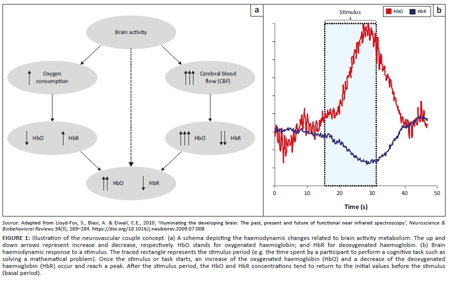
Functional near-infrared spectroscopy indirectly measures the brain activity by estimating the changes in blood supplies in specific brain regions. Neurovascular coupling (NVC) is the mechanism that links neuronal activity to haemodynamic changes (changes in blood supplies) in the brain (Hendrikx et al. 2019). Amongst all substances involved in such NVC, the oxygenated and deoxygenated haemoglobin (HbO and HbR, respectively) is most relevant to understanding the fNIRS measurements (Ferrari & Quaresima 2012). When a stimulus, such as pictures, sounds or questions, is presented to a participant, the brain regions involved in processing that stimulation are activated. This activation causes an increase in oxygen consumption in the corresponding brain regions (Len-Carrion & Len-Dominguez 2012). The cerebral blood flow (CBF) increases in those regions to regulate oxygen consumption. It leads to more oxygenated blood and, consequently, deoxygenated blood decay (Lloyd-Fox et al. 2010). After some seconds of stimulus presentation, the brain activation of the related region is characterised by an increase of HbO and a decrease of HbR (Figure 1). Once the stimulus ends, the concentration changes in HbO and HbR tend to return for the called basal period (the period without stimulation). These metabolic variations of HbO and HbR are called haemodynamic response function (HRF), which takes around 15 s from the stimuli start to return to the basal period (Figure 1b).
The Beer-Lambert law and computation of oxygenated haemoglobin and deoxygenated haemoglobin variations
The NIR light wavelengths vary from 700 nm to 900 nm. This range is considered an optical window for the human tissue, skin and bone, making them almost transparent to the NIR light (Jobsis 1977). Therefore, when an NIR light is emitted into the participants' head, it penetrates the skull and reaches the brain. Some portions of this light scatter and can be detected by the light detectors placed on the head, some portions are absorbed by some molecules called chromophores (Jobsis 1977; Villringer 1997) and some portions are lost in the medium. The HbO and HbR are the main chromophores related to brain activity and absorb most of NIR light (Len-Carrion & Len-Dominguez 2012). The inference about brain activity is made by computing the changes of HbO and HbR concentrations and applying the NVC concept. This calculation is made with the Beer-Lambert law (Equation 1) (Baker et al. 2014; Gervain et al. 2011):
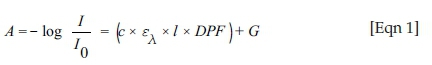
The left side of this equation is related to the light emitted in the scalp and the light scattered and detected by the detector (I and I0, respectively). The right side is related to the features of the medium where the light is propagated such as the tissue so that C is related to concentration of the light-absorbing molecules. The extinction coefficient (ελ) represents the characteristic of the light-absorbing molecule for a specific type of light such as NIR. The path through which light travels is represented by the distance between source and detector (called the light path denoted by l in Eqn. 1). Also, the differential path length (DPF) denotes the non-linear trajectory of light in the biological tissue and G is called the scattering term. We will not present details about the computation with Beer-Lambert law because it is out of the scope of this article.
Types of functional near-infrared spectroscopy techniques
Three types of fNIRS instrumentation are currently available: the continuous wave (CW), the time domain (TD) and the frequency domain (FD). They vary according to the emitted and transmitted light features considered in the measurements (Figure 2).
In the CW systems, the intensity of the light emitted is constant, and the detectors measure only the light intensity that passes through the tissue and is scattered back. Other parameters of the light as a signal, such as frequency, phase and time spent for the light to cross the tissues, are not considered. Therefore, CW systems only provide information of HbO and HbR concentration changes. In the TD systems, an extremely short pulse of light is emitted into the tissue. In this sophisticated and complex model, the time of arrival of the photons (tiny particles of light) to detectors is also measured.
In the FD systems, the intensity of the light emitted is not constant in time, but rather changes. When passing through the tissue not only intensity but also the phase of the light signal changes (i.e. the time that a signal goes up and down). In this case, both the intensity of the transmitted light and the phase shift are measured. Although the time and FDs provide information about the absolute values of HbO and HbR, these systems are complex and not so cost-effective (Boas et al. 2014; Scholkmann et al. 2014). The CW is the most affordable and most used system in the fNIRS literature, which is the focus of this article.
The functional near-infrared spectroscopy device
The fNIRS device consists of NIR light sources and detectors called optodes that are usually attached into caps or bands to be placed on the participant's head (Figures 3a and 3b). The optodes are connected to the light source amplifier and a computer unit (Figure 3c). A pairwise source-detector with the distance of 3 cm is known as a channel, which is the brain region between those two optodes. The number of channels varies according to the available system, the research question and the sample, the brain region of interest (ROI) and some other factors within each study. The portable systems are small, normally battery operated (Figure 3c) and more suitable for naturalistic experimentation in everyday life, such as schools (Balardin et al. 2017; Pinti et al. 2018). However, these systems usually have a fewer optodes, limiting the brain region coverage as compared with stationary lab-based systems. The lab-based systems (Figure 3d) have more optodes, which usually cover the whole head. However, they are less transportable and more suitable for experiments performed inside a laboratory.
Planning a functional near-infrared spectroscopy study
As a common research practice, the first step when planning an fNIRS study is to define an appropriate research question that needs the application of the fNIRS method to be answered. It is essential to have clear hypotheses to create an efficient experimental 'paradigm'. Educational studies with fNIRS are typically designed to answer questions about which brain regions show activations when individuals perform a cognitive/behavioural task such as solving arithmetic problems or reading a sentence (Soltanlou et al. 2018b). In the following section, we discuss planning an fNIRS study and highlight the specificities related to this technique.
The sample size, inclusion and exclusion criteria
Like any educational or psychological study, it is recommended to calculate the sample size at the beginning of the study by using a program such as G*Power. This approach strengthens the reliability of statistical analysis and the experiment's outcomes. In an fNIRS study, this calculation further depends on the number of fNIRS measurement channels (Faul et al. 2007, 2009). It is also important to consider the number of 'discarded' participants because of noisy data. 'Noisy data' means data with meaningless information that comes from irrelevant activities to the brain such as head movements or optodes weekly attached to the head. Those data are called artefacts and contaminate the brain signals measured with fNIRS signal. If the fNIRS data have high level of artefacts, it should not be included in the analysis. Furthermore, anything that may confound the fNIRS results should be considered as exclusion criteria. For instance, participants who suffer from blood pressure problems or diabetes or participants who use medications that may change the blood pressure or individuals with a history of caffeine, drug and alcohol abuse should not be considered in an fNIRS study. Another critical point is the participants' age. Whilst it is usually considered in any educational or psychological study, it needs further caution in fNIRS studies because the haemodynamics responses change with age (Wu et al. 2016).
Experimental design
Typically, the experimental paradigms employed in fNIRS studies are the (1) block design, (2) event-related or (3) resting state (Tak & Ye 2014). The block design has been frequently used in fNIRS studies in an educational context (Jasińska et al. 2021; Soltanlou et al. 2018). In this design, the stimuli are present in groups called blocks (Amaro & Barker 2006). For instance, in 30 s of a block, the participants should solve as many multiplication problems as they can, and then they will have a short rest time and again start a new block (Artemenko et al. 2018b; Jasińska et al. 2021). The blocks must last long enough to allow the complete haemodynamic response (usually longer than 15 s) but not too long (usually shorter than 60 s) to avoid signals saturation. Also, the blocks should be repeated multiple times (usually between 4 and 10 blocks in educational studies) to provide a robust statistical inference (Bonomini et al. 2015) or, in case one needs to discard some noisy blocks with high level of artefacts. The block design studies are usually shorter, and therefore suitable for special populations and statistically provide very strong signals. It has some limitations as well. For example, one may need to decide whether to include the blocks that contain both correct and incorrect responses or whether the number of presented problems within each block should be fixed (then, because of individual differences, there will have to be different length of blocks within and between participants) or the duration of the block should be fixed (then, because of individual differences, there will be a different number of solved problems within each block across participants).
In the event-related design, the tasks or stimuli are presented for a very short period and with adequate time for response from each other (Plichta et al. 2007b). Different conditions or problems (e.g. subtraction and multiplication problems) are presented in a mixed order. The event-related studies allow the analysis of every single problem and random order of presented problems (to avoid order effect) (Plichta et al. 2007a). A drawback of this design is different duration of the problems across participants (Amaro & Barker 2006). They also usually last longer than block designs.
The resting-state studies are usually performed to investigate the brain mechanism not related to a specific cognitive task or stimuli (Wang, Dong & Niu 2017). In this design, participants are asked to stay quiet and not engage in any mental effort (Geng et al. 2017). Sometimes, the participants are requested to keep their eyes closed or open their eyes, looking at a fixation cross. Keeping children still is difficult; therefore, a practice that has been used with this population is to watch a video designed to provide minimum cognitive load but keep them interested in the experiment (Vanderwal, Eilbott & Castellanos 2019). This paradigm has been extensively used in fMRI and has been applied in fNIRS studies, revealing important brain networks that are not necessarily task related.
The appropriate design is chosen based on several factors, such as the sample and the research questions (Gervain et al. 2011). For example, a block design might work better in young children because it is usually shorter than an event-related design, or if a researcher is interested in the brain response to each individual problem, event-related designs would provide more precise information (Amaro & Barker 2006). Resting-state designs are not so common in educational settings, which usually ask participants to engage in mental tasks.
Placement of the optodes
As a result of technical limitations, the fNIRS measures only activity related to the human brain cortex, which is the outer brain layer and with the depth of about 1 cm - 1.5 cm. The brain cortex is divided into lobes, frontal, parietal, temporal and occipital (see Figure 4).
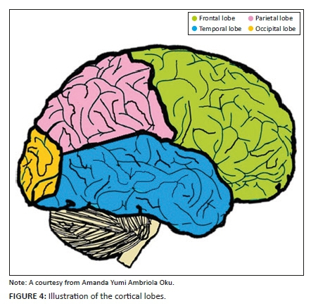
Although the brain works as a network system with regions connected functionally and anatomically, the activity of each lobe is related to cognitive functions that are necessary for educational achievements. The frontal lobe is related to cognitive functions such as working memory, attention and emotional regulation (McKendrick et al. 2014; Takeuchi et al. 2017), the parietal lobe is associated with numerical cognition and manipulation of numbers as magnitudes (Hyde et al. 2010; Soltanlou et al. 2018a) and the temporal lobes are activated when participants perform reading tasks (Joyal et al. 2017; Ojemann et al. 1988).
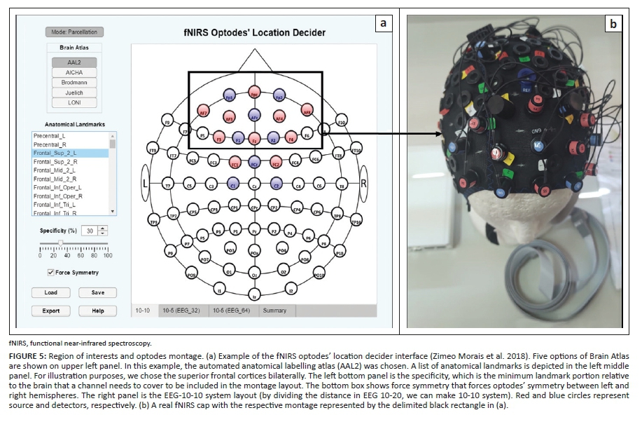
The 'region of interest' (ROI) is a term used in neuroimage studies referring to the brain region that is being investigated in a study. The ROI is defined based on the research hypothesis and information retrieved from the relevant literature. Once the ROI is defined, it is necessary to decide the optodes' position (the 'montage') on the participant's head so that the fNIRS light can achieve the cortical ROI. To guide this choice, researchers use the EEG 10-20 international system. It is a standardised method, which defines the position of the sensors in the scalp (Figure 5a) (Malmivuo & Plonsey 1995; Tatum 2013). In this system, letters (FP = frontal pole, F = frontal, C = central, T = temporal, P = parietal, Z = midline and O = occipital) and numbers (1, 3, 5, 7, 9; 2, 4, 6, 8, 10) are used to represent the electrodes' position in relation to the cortical lobes and hemisphere lateralisation. Even and odd numbers represent the right and left cortical hemisphere, respectively. The fNIRS channel is a pairwise source-detector, instead of a single electrode detector such as in the EEG system. Therefore, researchers use some software to simulate the light propagation and to give more accurate anatomical landmarks so as to have an anatomical approximation about where the light is propagating into the cortex. Softwares such as fNIRS Optodes' Location Decider (fOLD) or PHOBE define the optodes' placement in the scalp by considering anatomical projections into MRI atlases (Pollonini, Bortfeld & Oghalai 2016; Zimeo Morais, Balardin & Sato 2018). Those projections are transformed into the EEG 10-20 system to guide the placement of the optodes on the participant's head.
Pre-processing of the functional near-infrared spectroscopy signals
The artefacts may influence the results by adding signals that do not necessarily originate from the brain (Huppert 2016). Some artefacts are related to the participant's head movement. Moving the head may detach the optodes and influence the signals (Brigadoi et al. 2014). Also, thick or dark hair may cause poor attachment between the optodes and the scalp, interfering in the quality of the signals (Di Lorenzo et al. 2019). Physiological signals related to heart beats and respiratory cycles oscillate in specific frequencies (1 Hz for a heartbeat and 0.25 Hz for respiration) that may interfere with the fNIRS signals (Huppert 2016; Pinti et al. 2019). Head layers other than the brain such as skin, skull and the fluid around the cortex may also cause additional artefacts (Zhou et al. 2020).
A pre-processing process is performed to remove or to diminish the artefacts before computing any statistical analysis with the fNIRS data (Behrendt et al. 2018; Di Lorenzo et al. 2019). The noise related to physiological artefacts can be attenuated by applying frequency filters (Figure 6) as proposed by Nguyen et al. (2018) and Pinti et al. (2019). The short-distance channels are those with source-detector distances usually less than 1 cm, and they measure non-brain signals from the skull and scalp. Computational and mathematical manipulations of these values have been used to remove the artefacts related to the non-brain sources (Brigadoi & Cooper 2015; Zhou et al. 2020). Thus, the approach to remove or to diminish the noise effects varies according to the artefacts' type and source (Tak & Ye 2014).
Statistical analysis of functional near-infrared spectroscopy signals
The analysis of fNIRS data varies with the design used in the experiment. The methods of block average, the area under the curve and general linear model (GLM) have been frequently used to analyse data from this experimental paradigm (Luke et al. 2021; Tak & Ye 2014). The block average consists of averaging the fNIRS signals over the same block condition so that, for each condition, a mean value of HbO and HbR is computed (Artemenko et al. 2018b; Soltanlou et al. 2017). The mean values of different conditions are compared via statistical tests such as t-test or analysis of variance. The brain-behaviour relationship is computed by correlation measurements, such as Pearson's or Spearman's correlation coefficient, between the mean values of the fNIRS signals The brain-behaviour relationship is computed by correlation measurements, such as Pearson's or Spearman's correlation coefficient, between the mean values of the fNIRS signals and the task performance (Pfeifer, Scholkmann & Labruyère 2018). The area under the curve is a method that has been applied to analyse fNIRS data from experiments with different sizes of blocks. It consists of computing the area delimited by the HbO and HbR curves and comparing these values across conditions (Dresler et al. 2009; Obersteiner et al. 2010). Finally, the GLM, which is also called statistical parametric maps (SPM) (Ye et al. 2009), applies statistical concepts of regression models to create activation maps denoting the brain activations related to the task (Huppert et al. 2009; Tak & Ye 2014). As it is out of the scope of this article, we will not go through the mathematical aspects of these methods.
Conclusion and perspectives
In this article, we presented an introduction to fNIRS as a research tool for educational researchers in the framework of Educational Neuroscience. This article is an effort to familiarise the readers with the fNIRS technical vocabulary, ranging from preparing an fNIRS study to the data analysis processes.
A few cautions should be considered when planning an fNIRS study: (1) the fNIRS measures only cortical activity; therefore, it is not suitable to investigate deeper brain layers (Len-Carrin & Len-Domnguez 2012). (2) fNIRS studies provide insights about the association and not causation between the brain and behavioural measurements. For instance, it is possible to infer that the activation of a particular cortical region is associated with some task performance but it does not imply that one causes another. (3) In terms of an experimental design, it must be discussed and established well before data collection takes place. A poor experimental design may waste time and resources, as a neuroimaging study is usually much more effortful than an equivalent behavioural study. The best approach would be to perform a pilot study to make the necessary adjustments before the official data collection. (4) It is also important to remind users that fNIRS is approved for research only and has not been approved for clinical interventions.
The fNIRS has been applied to a variety of studies within educational neuroscience field. Functional near-infrared spectroscopy has been used to investigate the neural mechanisms underlying mathematical cognition (Artemenko et al. 2018a, 2018b; Soltanlou et al. 2018a). Also, fNIRS was used to measure brain activity related to the use of game-based approach to teaching and learning mathematics (Cakir et al. 2015, 2016). Recent and cutting-edge studies have applied the modern machine learning algorithms to fNIRS signals to predict the students' performance after watching online classes, and the student's brain signals using the teacher's during a hyperscanning paradigm (Barreto et al. 2021; Oku & Sato 2021). Neural mechanisms of language development and literacy have also been frequently investigated using fNIRS (Jasińska et al. 2021; Joyal et al. 2017).
Even though fNIRS has been applied to educational experiments to investigate the neural correlates associated with mathematics and literacy (Jasińska et al. 2021; Soltanlou et al. 2018), a rich and complex field is still available to be explored. This technique has the potential of measuring cortical activation of subjects under realistic situations, bringing previous static laboratories to the dynamic settings in schools and classrooms (Balardin et al. 2017; Brockington et al. 2018; Dresler et al. 2009). Open questions related to population such as pre-school children, infants and babies that could hardly be addressed previously because of technical limitations can be explored with the fNIRS. In addition, fNIRS is suitable to perform multimodal studies in which additional neurophysiological measurements such as heart rate, skin conductance and eye tracking and EEG can be measured simultaneously (e.g. Katus et al. 2019; Soltanlou et al. 2018a). It provides a robust set of measurements to support the understanding of the teaching and learning mechanisms.
To conclude, it is worth mentioning that most of the research in the field of Educational Neuroscience has been performed in developed countries because of financial support. Compared with other neuroimaging modalities, the affordable cost of the fNIRS device has provided the opportunity for researchers from developing countries to participate in the field (Katus et al. 2019). It reinforces the global and diverse features of science, which hopefully contributes to diminishing educational inequities across the world.
Acknowledgements
The authors would like to thank Amanda Yumi Ambriola Oku for providing the image of the cortex illustration.
Competing interests
The authors declare that they have no financial or personal relationships that may have inappropriately influenced them in writing this article.
Authors' contributions
The first author wrote the article whilst the second author proofread and effected the necessary corrections.
Ethical considerations
This article followed all ethical standards for a research without direct contact with human or animal subjects.
Funding information
This research received no specific grant from any funding agency in the public, commercial or not-for-profit sectors.
Data availability
Data sharing is not applicable to this article as no new data were created or analysed in this study.
Disclaimer
The views and opinions expressed in this article are those of the authors and do not necessarily reflect the official policy or position of any affiliated agency of the authors.
References
Amaro, E. & Barker, G.J., 2006, 'Study design in fMRI: Basic principles', Brain and Cognition 60(3), 220-232. https://doi.org/10.1016/j.bandc.2005.11.009 [ Links ]
Ansari, D., 2008, 'Effects of development and enculturation on number representation in the brain', Nature Reviews Neuroscience 9(4), 278-291. https://doi.org/10.1038/nrn2334 [ Links ]
Ansari, D. & Coch, D., 2006, 'Bridges over troubled waters: Education and cognitive neuroscience', Trends in Cognitive Sciences 10(4), 146-151. https://doi.org/10.1016/j.tics.2006.02.007 [ Links ]
Artemenko, C., Soltanlou, M., Dresler, T., Ehlis, A.C. & Neurk, H.C., 2018a, 'The neural correlates of arithmetic difficulty depend on mathematical ability: Evidence from combined fNIRS and ERP', Brain Structure and Function 223(6), 2561-2574. https://doi.org/10.1007/s00429-018-1618-0 [ Links ]
Artemenko, C., Soltanlou, M., Ehlis, A.-C., Neurk, H.C. & Dresler, T., 2018b, 'The neural correlates of mental arithmetic in adolescents: A longitudinal fNIRS study', Behavioral and Brain Functions 14(1), 5. https://doi.org/10.1186/s12993-018-0137-8 [ Links ]
Baker, W.B., Parthasarathy, A.B., Busch, D.R., Mesquita, R.C., Greenberg, J.H. & Yodh, A.G., 2014, 'Modified Beer-Lambert law for blood flow', Biomedical Optics Express 5(11), 4053. https://doi.org/10.1364/BOE.5.004053 [ Links ]
Balardin, J.B., Zimeo Morais, G.A., Furucho, R.A., Trambaiolli, L., Vanzella, P., Biazoli, C. et al., 2017, 'Imaging brain function with functional near-infrared spectroscopy in unconstrained environments', Frontiers in Human Neuroscience 11, 258. https://doi.org/10.3389/fnhum.2017.00258 [ Links ]
Barreto, C., Bruneri, G.d.A., Brockington, G., Ayaz, H. & Sato, J.R., 2021, 'A new statistical approach for fNIRS hyperscanning to predict brain activity of preschoolers' using teacher's', Frontiers in Human Neuroscience 15, 622146. https://doi.org/10.3389/fnhum.2021.622146 [ Links ]
Behrendt, H.F., Firk, C., Nelson III, C.A. & Perdue, K.L., 2018, 'Motion correction for infant functional near-infrared spectroscopy with an application to live interaction data', Neurophotonics 5(1), 015004. https://doi.org/10.1117/1.NPh.5.1.015004 [ Links ]
Boas, D.A., Elwel, C.E., Ferraric, M. & Tagad, G., 2014, 'Twenty years of functional near-infrared spectroscopy: Introduction for the special issue', NeuroImage 85, 1-5. https://doi.org/10.1016/j.neuroimage.2013.11.033 [ Links ]
Bonomini, V., Zucchelli, L., Re, R., Ieva, F., Spinelli, L., Contini, D. et al., 2015, 'Linear regression models and k-means clustering for statistical analysis of fNIRS data', Biomedical Optics Express 6(2), 615-630. https://doi.org/10.1364/BOE.6.000615 [ Links ]
Brigadoi, S., Ceccherini, L., Cutini, S., Scarpaa, F., Scatturina, P., Selb, J. et al., 2014, 'Motion artifacts in functional near-infrared spectroscopy: A comparison of motion correction techniques applied to real cognitive data', NeuroImage 85, 181-191. https://doi.org/10.1016/j.neuroimage.2013.04.082 [ Links ]
Brigadoi, S. & Cooper, R.J., 2015, 'How short is short? Optimum source-detector distance for short-separation channels in functional near-infrared spectroscopy', Neurophotonics 2(2), 025005. https://doi.org/10.1117/1.NPh.2.2.025005 [ Links ]
Brockington, G., Balardin, J.B., Zimeo Morais, G.A., Malheiros, A., Lent, R., Moura, L.M. et al., 2018, 'From the laboratory to the classroom: The potential of functional near-infrared spectroscopy in educational neuroscience', Frontiers in Psychology 9, 1840. https://doi.org/10.3389/fpsyg.2018.01840 [ Links ]
Bruer, J.T., 2016, 'Where is educational neuroscience?', Educational Neuroscience 1, 2377616115618036. https://doi.org/10.1177/2377616115618036 [ Links ]
Cakir, M.P., Çakir, N.K., Ayaz, H. & Lee, F.J., 2015, 'An optical brain imaging study on the improvements in mathematical fluency from game-based learning', in Proceedings of the 2015 Annual Symposium on Computer-Human Interaction in Play, Association for Computing Machinery (CHI PLAY '15), New York, NY, pp. 209-219.
Cakir, M.P., Çakir, N.K., Ayaz, H. & Lee, F.J., 2016, Neural correlates of computational fluency training with a mobile game: An optical brain imaging study, AERA Online Paper Repository.
Curtin, A. & Ayaz, H., 2018, 'The age of neuroergonomics: Towards ubiquitous and continuous measurement of brain function with fNIRS', Japanese Psychological Research 60(4), 374-386. https://doi.org/10.1111/jpr.12227 [ Links ]
Da Silva Ferreira Barreto, C., Morais, G.A.Z., Vanzella, P. & Sato, J.R., 2020, 'Combining the intersubject correlation analysis and the multivariate distance matrix regression to evaluate associations between fNIRS signals and behavioral data from ecological experiments', Experimental Brain Research 238, 2399-2408. https://doi.org/10.1007/s00221-020-05895-8 [ Links ]
Di Lorenzo, R., Pirazzoli, L., Blasi, A., Bulgarelli, C., Hakuno, Y. & Minagawa, Y. et al., 2019, 'Recommendations for motion correction of infant fNIRS data applicable to multiple data sets and acquisition systems', NeuroImage 200, 511-527. https://doi.org/10.1016/j.neuroimage.2019.06.056 [ Links ]
Dresler, T., Obersteiner, A., Schecklmann, M., Vogel, A.C.M., Ehlis, A.C., Richter, M.M. et al., 2009, 'Arithmetic tasks in different formats and their influence on behavior and brain oxygenation as assessed with near-infrared spectroscopy (NIRS): A study involving primary and secondary school children', Journal of Neural Transmission (Vienna, Austria: 1996) 116, 1689-1700. https://doi.org/10.1007/s00702-009-0307-9 [ Links ]
Faul, F., Erdfelder, E., Lang, A.G. & Buchner, A., 2007, 'G*Power 3: A flexible statistical power analysis program for the social, behavioral, and biomedical sciences', Behavior Research Methods 39(2), 175-191. https://doi.org/10.3758/BF03193146 [ Links ]
Faul, F., Erdfelder, E., Lang, A.G. & Buchner, A., 2009, 'Statistical power analyses using G*Power 3.1: Tests for correlation and regression analyses', Behavior Research Methods 41(4), 1149-1160. https://doi.org/10.3758/BRM.41.4.1149 [ Links ]
Ferrari, M. & Quaresima, V., 2012, 'A brief review on the history of human functional near-infrared spectroscopy (fNIRS) development and fields of application', NeuroImage 63(2), 921-935. https://doi.org/10.1016/j.neuroimage.2012.03.049 [ Links ]
Geng, S., Liu, X., Biswal, B.B. & Niu, H., 2017, 'Effect of resting-state fNIRS scanning duration on functional brain connectivity and graph theory metrics of brain network', Frontiers in Neuroscience 11, 392. https://doi.org/10.3389/fnins.2017.00392 [ Links ]
Gervain, J., Mehler, J., Werker, J.F., Nelson, C.A., Csibra, G. & Lloyd-Fox, S. et al., 2011, 'Near-infrared spectroscopy: A report from the McDonnell infant methodology consortium', Developmental Cognitive Neuroscience 1(1), 22-46. https://doi.org/10.1016/j.dcn.2010.07.004 [ Links ]
Goswami, U., 2006, 'Neuroscience and education: From research to practice?', Nature Reviews Neuroscience 7(5), 406-413. https://doi.org/10.1038/nrn1907 [ Links ]
Hendrikx, D., Smits, A., Lavanga, M., De Wel, O., Thewissen, L., Jansen, K. et al., 2019, 'Measurement of neurovascular coupling in neonates', Frontiers in Physiology 10, 65. https://doi.org/10.3389/fphys.2019.00065 [ Links ]
Huppert, T.J., 2016, 'Commentary on the statistical properties of noise and its implication on general linear models in functional near-infrared spectroscopy', Neurophotonics 3(1), 010401. https://doi.org/10.1117/1.NPh.3.1.010401 [ Links ]
Huppert, T.J., Diamond, S.G., Franceschini, M.A. & Boas, D.A., 2009, 'HomER: A review of time-series analysis methods for near-infrared spectroscopy of the brain', Applied Optics 48(10), D280. https://doi.org/10.1364/AO.48.00D280 [ Links ]
Hyde, D.C., Boas, D.A., Blair, C. & Carey, S., 2010, 'Near-infrared spectroscopy shows right parietal specialization for number in pre-verbal infants', NeuroImage 53(2), 647-652. https://doi.org/10.1016/j.neuroimage.2010.06.030 [ Links ]
Jasińska, K.K., Shuai, L., Lau, A.N.L., Frost, S., Landi, N., Pugh, K.R., 2021, 'Functional connectivity in the developing language network in 4-year-old children predicts future reading ability', Developmental Science 24(2), e13041. https://doi.org/10.1111/desc.13041 [ Links ]
Jobsis, F., 1977, 'Noninvasive, infrared monitoring of cerebral and myocardial oxygen sufficiency and circulatory parameters', Science 198(4323), 1264-1267. https://doi.org/10.1126/science.929199 [ Links ]
Joyal, M., Brambati, S.M., Laforce, R.J., Montembeault, M., Boukadi, M., Rouleau, I. et al., 2017, 'The role of the left anterior temporal lobe for unpredictable and complex mappings in word reading', Frontiers in Psychology 8, 517. https://doi.org/10.3389/fpsyg.2017.00517 [ Links ]
Katus, L., Hayes, N.J., Mason, L., Blasi, A., McCann, S., Darboe, M.K. et al., 2019, 'Implementing neuroimaging and eye tracking methods to assess neurocognitive development of young infants in low- and middle-income countries', Gates Open Research 3, 1113. https://doi.org/10.12688/gatesopenres.12951.2 [ Links ]
Len-Carrin, J. & Len-Domnguez, U., 2012, 'Functional Near-Infrared Spectroscopy (fNIRS): Principles and neuroscientific applications', in P. Bright (ed.) Neuroimaging - Methods, pp. 43-78, InTech, Rijeka.
Lloyd-Fox, S., Blasi, A. & Elwell, C.E., 2010, 'Illuminating the developing brain: The past, present and future of functional near infrared spectroscopy', Neuroscience & Biobehavioral Reviews 34(3), 269-284. https://doi.org/10.1016/j.neubiorev.2009.07.008 [ Links ]
Luke, R., Larson, E.D., Shader, M.J., Innes-Brown, H., Yper, L.V., Lee, A.K.C. et al., 2021, 'Analysis methods for measuring passive auditory fNIRS responses generated by a block-design paradigm', Neurophotonics 8(2), 025008. https://doi.org/10.1117/1.NPh.8.2.025008 [ Links ]
Malmivuo, J., & Plonsey, R., 1995, Bioelectromagnetism: Principles and Applications of Bioelectric and Biomagnetic Fields, Oxford University Press. viewed 13 January 2022, from https://oxford.universitypressscholarship.com/view/10.1093/acprof:oso/9780195058239.001.0001/acprof-9780195058239.
Max Roser and Esteban Ortiz-Ospina, 2016, Literacy, viewed n.d., from https://ourworldindata.org/literacy.
McCandliss, B.D., 2010, 'Educational neuroscience: The early years', Proceedings of the National Academy of Sciences 107(18), 8049-8050. https://doi.org/10.1073/pnas.1003431107 [ Links ]
McKendrick, R., Ayaz, H., Olmstead, R. & Parasuraman, R., 2014, 'Enhancing dual-task performance with verbal and spatial working memory training: Continuous monitoring of cerebral hemodynamics with NIRS', NeuroImage 85, 1014-1026. https://doi.org/10.1016/j.neuroimage.2013.05.103 [ Links ]
Mehta, R. & Parasuraman, R., 2013, 'Neuroergonomics: A review of applications to physical and cognitive work', Frontiers in Human Neuroscience 7, 889. https://doi.org/10.3389/fnhum.2013.00889 [ Links ]
Nguyen, H.-D., Yoo, S.-H., Bhutta, M.R. & Hong, K.-S., 2018, 'Adaptive filtering of physiological noises in fNIRS data', BioMedical Engineering OnLine 17(1), 180. https://doi.org/10.1186/s12938-018-0613-2 [ Links ]
Obersteiner, A., Dresler, T., Reiss, K., Vogel, A.C.M., Pekrun, R. & Fallgatter, A.J., 2010, 'Bringing brain imaging to the school to assess arithmetic problem solving: Chances and limitations in combining educational and neuroscientific research', ZDM: The International Journal on Mathematics Education 42, 541-554. https://doi.org/10.1007/s11858-010-0256-7 [ Links ]
Ojemann, G.A., Creutzfeldt, O., Lettich, E., Haglund, M.M., 1988, 'Neuronal activity in human lateral temporal cortex related to short-term verbal memory, naming and reading', Brain 111(6), 1383-1403. https://doi.org/10.1093/brain/111.6.1383 [ Links ]
Oku, A.Y.A. & Sato, J.R., 2021, 'Predicting student performance using machine learning in fNIRS data', Frontiers in Human Neuroscience 15, 622224. https://doi.org/10.3389/fnhum.2021.622224 [ Links ]
Pfeifer, M.D., Scholkmann, F. & Labruyère, R., 2018, 'Signal processing in Functional Near-Infrared Spectroscopy (fNIRS): Methodological differences lead to different statistical results', Frontiers in Human Neuroscience 11, 641. https://doi.org/10.3389/fnhum.2017.00641 [ Links ]
Pinti, P., Aichelburg, C., Gilbert, S., Hamilton, A., Hirsch, J., Burgess, P. et al., 2018, 'A review on the use of wearable functional near-infrared spectroscopy in naturalistic environments: Review of fNIRS measurements in naturalistic environments', Japanese Psychological Research 60(4), 347-373. https://doi.org/10.1111/jpr.12206 [ Links ]
Pinti, P., Scholkmann, F., Hamilton, A., Burgess, P. & Tachtsidis I., 2019, 'Current status and issues regarding pre-processing of fNIRS neuroimaging data: An investigation of diverse signal filtering methods within a general linear model framework', Frontiers in Human Neuroscience 12, 505. https://doi.org/10.3389/fnhum.2018.00505 [ Links ]
Plichta, M.M., Heinzel, S., Ehlis, A.-C., Pauli, P. & Fallgatter, A.J., 2007b, 'Model-based analysis of rapid event-related functional near-infrared spectroscopy (NIRS) data: A parametric validation study', NeuroImage 35(2), 625-634. https://doi.org/10.1016/j.neuroimage.2006.11.028 [ Links ]
Plichta, M.M., Herrmann, M.J., Baehna, C.G., Ehlis, A.-C., Richter, M.M., Pauli, P. et al., 2007a, 'Event-related functional near-infrared spectroscopy (fNIRS) based on craniocerebral correlations: Reproducibility of activation?', Human Brain Mapping 28(8), 733-741. https://doi.org/10.1002/hbm.20303 [ Links ]
Pollonini, L., Bortfeld, H. & Oghalai, J.S., 2016, 'PHOEBE: A method for real time mapping of optodes-scalp coupling in functional near-infrared spectroscopy', Biomedical Optics Express 7(12), 5104-5119. https://doi.org/10.1364/BOE.7.005104 [ Links ]
Scholkmann, F., Kleisera, S., Metz, A.J., Zimmermann, R., Pavia, J.M., Wolf, U. et al., 2014, 'A review on continuous wave functional near-infrared spectroscopy and imaging instrumentation and methodology', NeuroImage 85, 6-27. https://doi.org/10.1016/j.neuroimage.2013.05.004 [ Links ]
Soltanlou, M., Artemenko, C., Dresler, T., Haeussinger, F.B., Fallgatter, A.J., Ehlis, A.-C. et al., 2017, 'Increased arithmetic complexity is associated with domain-general but not domain-specific magnitude processing in children: A simultaneous fNIRS-EEG study', Cognitive, Affective, & Behavioral Neuroscience 17(4), 724-736. https://doi.org/10.3758/s13415-017-0508-x [ Links ]
Soltanlou, M., Artemenko, C., Ehlis, A.-C., Huber, S., Fallgatter, A.J., Dresler, T., et al., 2018b, 'Reduction but no shift in brain activation after arithmetic learning in children: A simultaneous fNIRS-EEG study', Scientific Reports 8(1), 1707. https://doi.org/10.1038/s41598-018-20007-x [ Links ]
Soltanlou, M., Sitnikova, M.A., Nuerk, H.-C. & Dresler, T., 2018a, 'Applications of Functional Near-Infrared Spectroscopy (fNIRS) in studying cognitive development: The case of mathematics and language', Frontiers in Psychology 9, 277. https://doi.org/10.3389/fpsyg.2018.00277 [ Links ]
Szűcs, D. & Goswami, U., 2007, 'Educational neuroscience: Defining a new discipline for the study of mental representations', Mind, Brain, and Education 1, 114-127. https://doi.org/10.1111/j.1751-228X.2007.00012.x [ Links ]
Tak, S. & Ye, J.C., 2014, 'Statistical analysis of fNIRS data: A comprehensive review', NeuroImage 85, 72-91. https://doi.org/10.1016/j.neuroimage.2013.06.016 [ Links ]
Takeuchi, N., Mori, T., Suzukamo, Y. & Izumi, S.-I., 2017, 'Integration of teaching processes and learning assessment in the prefrontal cortex during a video game teaching-learning task', Frontiers in Psychology 7, 2052. https://doi.org/10.3389/fpsyg.2016.02052 [ Links ]
Tatum, W.O., 2013, Handbook of EEG interpretation, Demos Medical Pub, New York, NY, viewed 06 October 2021, from http://www.credoreference.com/book/spheegi.
Thomas, M.S.C., Ansari, D. & Knowland, V.C.P., 2019, 'Annual research review: Educational neuroscience: Progress and prospects', Journal of Child Psychology and Psychiatry 60(4), 477-492. https://doi.org/10.1111/jcpp.12973 [ Links ]
Tilak, J.B.G., 1987, The economics of inequality in education, Preprint, No. 44, viewed 29 November 2021, from https://www.cabdirect.org/cabdirect/abstract/19881859544.
Vanderwal, T., Eilbott, J. & Castellanos, F.X., 2019, 'Movies in the magnet: Naturalistic paradigms in developmental functional neuroimaging', Developmental Cognitive Neuroscience 36, 100600. https://doi.org/10.1016/j.dcn.2018.10.004 [ Links ]
Villringer, A., 1997, 'Non-invasive optical spectroscopy and imaging of human brain function', Trends in Neurosciences 20(10), 435-442. https://doi.org/10.1016/S0166-2236(97)01132-6 [ Links ]
Wang, J., Dong, Q. & Niu, H., 2017, 'The minimum resting-state fNIRS imaging duration for accurate and stable mapping of brain connectivity network in children', Scientific Reports 7(1), 1-10. https://doi.org/10.1038/s41598-017-06340-7 [ Links ]
Wu, C., Honarmand, A.R., Schnell, S., Kuhn, R., Schoeneman, S.E., Ansari, S.A. et al., 2016, 'Age-related changes of normal cerebral and cardiac blood flow in children and adults aged 7 months to 61 years', Journal of the American Heart Association 5(1), e002657. https://doi.org/10.1161/JAHA.115.002657 [ Links ]
Xu, J. & Zhong, B., 2018, 'Review on portable EEG technology in educational research', Computers in Human Behavior 81, 340-349. https://doi.org/10.1016/j.chb.2017.12.037 [ Links ]
Ye, J.C., Tak, S., Jang, K.E., Jung, J., Jang, J., 2009, 'NIRS-SPM: Statistical parametric mapping for near-infrared spectroscopy', NeuroImage 44(2), 428-447. https://doi.org/10.1016/j.neuroimage.2008.08.036 [ Links ]
Zhou, X., Sobczak, G., McKay, C.M. & Litovsky, R.Y., 2020, 'Comparing fNIRS signal qualities between approaches with and without short channels', PLoS One 15(12), e0244186. https://doi.org/10.1371/journal.pone.0244186 [ Links ]
Zimeo Morais, G.A., Balardin, J.B. & Sato, J.R., 2018, 'fNIRS Optodes' Location Decider (fOLD): A toolbox for probe arrangement guided by brain regions-of-interest', Scientific Reports 8(1), 3341. https://doi.org/10.1038/s41598-018-21716-z [ Links ]
 Correspondence:
Candida Barreto
candidasfb2010@gmail.com
Received: 16 Oct. 2021
Accepted: 08 Dec. 2021
Published: 31 Mar. 2022

