


Serviços Personalizados
Artigo
 Inglês (pdf)
 Artigo em XML
 Referências do artigo
Indicadores

Links relacionados
-
 Citado por Google
 -
 Similares em Google
Compartilhar

 Permalink
SA Journal of Radiology
versão On-line ISSN 2078-6778
versão impressa ISSN 1027-202X
S. Afr. J. radiol. (Online) vol.26 no.1 Johannesburg 2022
http://dx.doi.org/10.4102/sajr.v26i1.2437
CASE REPORT
A lumpy bumpy stomach: The more the murkier
Binit SurekaI; Siddhi ChawlaI; Sudeep KheraII; Ashish AgarwalIII; Chhagan L. BirdaIII; Sandeep BairwaIV
IDepartment of Radiology, All India Institute of Medical Sciences Jodhpur, Jodhpur, India
IIDepartment of Pathology, All India Institute of Medical Sciences Jodhpur, Jodhpur, India
IIIDepartment of Gastroenterology, All India Institute of Medical Sciences Jodhpur, Jodhpur, India
IVDepartment of Medical Oncology, All India Institute of Medical Sciences Jodhpur, Jodhpur, India
ABSTRACT
This report describes the radiological and endoscopic findings in a 54-year-old male who presented with epigastric pain. The patient underwent an upper gastrointestinal (GI) barium study followed by axial imaging, which demonstrated nodular gastric wall thickening. The classic findings of aggressive primary gastric diffuse large B-Cell lymphoma are presented with a brief review differentiating the pathological subtypes, important for patient prognostication and planning of therapy.
Keywords: primary gastric lymphoma; DLBCL; MALT lymphoma; stomach thickening; gastric wall thickening; computed tomography.
Introduction
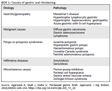
Primary gastric lymphomas are rare, however, the stomach is the most common gastrointestinal (GI) site of involvement in extra-nodal disease.1 Lymphomas may have diverse radiological manifestations, which can mimic a variety of benign diseases and malignant pathologies as enumerated in Box 1.2 Most of the lymphomas are either of the two most common types of B-cell non-Hodgkin lymphoma (NHL), that is, mucosa-associated lymphoid tissue (MALT) lymphoma and diffuse large B-cell lymphoma (DLBCL). The radiological differentiation between the two types is difficult but necessary because the prognosis and management differs accordingly.3 Various imaging modalities such as fluoroscopic upper GI barium imaging, ultrasonography, CT and MRI are used for the pre-treatment assessment and staging of upper GI malignant pathologies. This report describes the classic imaging findings of primary gastric lymphoma and possible differentials with a special emphasis on differentiating between the DLBCL and MALT subtypes.
Case presentation
A 54-year-old male patient presented to our hospital with symptoms of chronic epigastric pain accompanied by postprandial discomfort, early satiety and 2 kg of weight loss in the previous three months. There was no history of haematemesis, melena or vomiting. The liver and kidney function tests were within normal limits.
As a result of the predominant upper GI complaints, the patient underwent an upper GI barium study, which demonstrated circumferential gastric luminal narrowing in the region of the fundus and body without obvious mucosal irregularity (Figure 1a). At a subsequent contrast enhanced CT scan, homogenous minimally enhancing circumferential thickening of the gastric wall (measuring ~2.5 cm) in the region of fundus and body was seen (Figure 1b, c) with associated multiple discrete similarly enhancing supra- and infra-diaphragmatic lymph nodes (Figure 1c). The MRI scan revealed isointense thickening on T1 weighted fat saturated images (Figure 1d), showing homogenous enhancement on post-contrast T1 weighed images (Figure 1e) in the same region with prominent diffusion restriction (Figure 1f, g). In view of the degree of thickening and associated lymphadenopathy, gastric lymphoma was presented as an imaging differential.
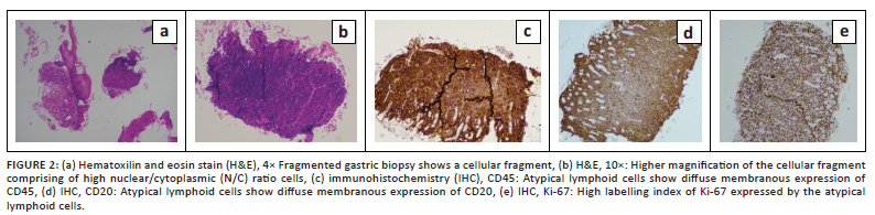
Upper GI endoscopy (Figure 1h) demonstrated a thickened and lobulated appearance of the fundus causing moderate luminal narrowing, however, no ulcerations or mucosal erosions were seen. Histology on the biopsy acquired reported sheets of atypical lymphoid cells in the subepithelium (Figure 2a), intermediate to large in size, exhibiting moderate nuclear pleomorphism, a high nuclear-cytoplasmic ratio, vesicular chromatin, conspicuous nucleoli and a scant amount of cytoplasm (Figure 2b). These cells were found to infiltrate the submucosal glands. On immunohistochemistry, the tumour cells were positive for CD45 (leucocyte common antigen [LCA]) (Figure 2c) and CD20 (Figure 2d) and negative for CK & CD3. Ki-67 labelling index was 90% - 95% (Figure 2e). These findings supported a primary high-grade gastric lymphoma - DLBCL type.
The patient was initiated on R-CHOP regimen (Rituximab, Cyclophosphamide, Doxorubicin, Oncovin [Vincristine], and Prednisone) chemotherapy.
Discussion
Primary gastric lymphomas are diagnosed using the criteria by Dawson et al.4, which includes predominant involvement of the stomach along with nodal involvement confined to its drainage area, no other palpable superficial lymphnodes, a normal chest radiograph, normal total leukocyte counts and no involvement of the other organs such as the liver or spleen. Most GI lymphomas represent the non-Hodgkin's subtype with the previous studies reporting Hodgkin's lymphoma only in few cases.5,6 The incidence of NHL has been increasing over time because of various risk factors, which include HIV infection, Helicobacter pylori infection, coeliac disease, inflammatory bowel disease and immunosuppression after solid organ transplantation.7
Under normal circumstances gastric mucosa is devoid of lymphoid tissue, however, with chronic H. pylori infection there is reactive development of lymphoid tissue within the lamina propria. Mucosa-associated lymphoid tissue lymphoma is a low-grade lymphoma and more than 70% of cases are secondary to chronic H. pylori infection whilst DLBCL, in contrast, is an aggressive and high-grade lymphoma, which has a poor prognosis with high rates of recurrence.3,8 Conventional upper GI barium studies and upper GI endoscopy are limited as they only assess luminal characteristics. This limitation is overcome in upper GI endoscopy by accompanying endoscopic ultrasound, which can assess the extra-luminal characteristics of the lumen and also assist in acquiring targeted biopsies.
Multiplanar reconstruction CT can better assess the gastric wall thickness, mucosal enhancement, lymph node involvement and other organ involvement and thus has become the investigation of choice for assessing lesions of the stomach.9 MRI is used for evaluation of lesions that are difficult to characterise at multidetentor CT. Lymphomas demonstrate significant restriction on diffusion weighted images (DWI) and corresponding apparent diffusion coefficient (ADC) maps because of their hypercellular nature. Positron emission tomography with computed tomography (PET/CT) scan is helpful in disease staging and treatment response assessment due to its high sensitivity.
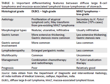
Gastric wall thickness > 1 cm favours the diagnosis of lymphoma.10 In both DLBCL and MALT lymphoma, the antrum and body are the most common areas of involvement, however, heterogeneity of contrast enhancement, extensive thickening, serosal involvement and extra-nodal disease is more common with DLBCL. Multifocal involvement of the stomach, nodular and ulcerative morphology of lesions, which can lead to subsequent gastric stenosis, are more common in DLBCL. Mucosa-associated lymphoid tissue lymphoma on the other hand has an infiltrative nature and thus luminal stenosis is uncommon. Differences between these two types of lymphoma are documented in Table 1.
Differentiation of lymphomatous gastric wall thickening from other malignant conditions, like adenocarcinoma, is essential. Adenocarcinoma is more likely to infiltrate the gastric wall and infiltrate the adjacent structures, whilst preservation of the perigastric fat planes is more probable in lymphoma.7 The lumen of the stomach remains patent even with extensive lymphomatous infiltration of the wall whilst in cases of adenocarcinoma the patient can present with gastric outlet obstruction because of the constricting and scirrhous nature of the pathology.11 The linitus plastica appearance related to infiltration of the submucosa of the gastric wall in scirrhous adenocarcinoma leading to decreased capacity and rigidity of stomach wall can also be seen in non-Hodgkin gastric lymphoma, however, in these cases lymphomatous cells are seen within the submucosa on pathological evaluation.12 Transpyloric spread of disease is also more common in lymphoma than adenocarcinoma however the higher incidence of carcinoma decreases the specificity of this differentiation.13 Lymph nodes in cases of lymphomas are homogenously enhancing and bulky and often seen to extend below the level of the renal hilum whereas they are smaller, necrotic and localised to the local drainage site of the stomach in cases of adenocarcinoma.14,15
In suspected cases of lymphoma and in cases where a diagnostic dilemma persistsupper GI endoscopy investigation is warranted. It can delineate the extent of the pathology and guided biopsy from suspicious areas will allow a histopathological diagnosis. Immunohistochemistry stains can further differentiate the subtypes of lymphoma as was described in the presented case. Once the diagnosis is confirmed, treatment of MALT lymphoma consists of anti-H. Pylori medication, which is initiated as early as possible once the diagnosis is confirmed.9,16 With DLBCL a combination of chemotherapy and radiation therapy can result in complete remission in up to 90% of cases.16
Conclusion
This case demonstrates the usefulness of multimodality imaging and describes the classic imaging findings in the DLBCL type of gastric lymphoma. It is quintessential for radiologists and clinicians to be aware of this entity, differentiating it from other types of lymphomas and other malignant pathologies so that appropriate treatment is not delayed.
Acknowledgements
Competing interests
The authors declare that they have no financial or personal relationships that may have inappropriately influenced them in writing this article.
Authors' contributions
B.S. was responsible for conceptualisation and supervision. S.C. wrote the original draft. S.K., A.A., C.L.B., S.B. helped in proving the necessary investigations and reviewing the original draft to its final form.
Ethical considerations
Informed consent was obtained from the patient for publication.
Funding information
This research received no specific grant from any funding agency in the public, commercial or not-for-profit sectors.
Data availability
Data sharing is not applicable to this article as no data sets were generated or analysed during this study.
Disclaimer
The views expressed in the submitted article are the author's own and not an official position of their institution.
References
1. Kelessis NG, Vassilopoulos PP, Tsamakidis KG, Bai MG, Avital S, Rosenthal RJ. Is gastroscopy still a valid diagnostic tool in detecting gastric MALT lymphomas? A dilemma beyond the eye mucosa-associated lymphoid tissue. Surg Endosc. 2003;17:469-474. https://doi.org/10.1007/s00464-002-8544-0 [ Links ]
2. Agarwala R, Shah J, Dutta U. Thickened gastric folds: Approach. J Dig Endosc. 2018;09(04):149-154. https://doi.org/10.4103/jde.JDE_72_18 [ Links ]
3. De Jong D, Boot H, Van Heerde P, Hart GA, Taal BG. Histological grading in gastric lymphoma: Pretreatment criteria and clinical relevance. Gastroenterology. 1997;112:1466-1474. https://doi.org/10.1016/S0016-5085(97)70026-X [ Links ]
4. Dawson IM, Cornes JS, Morson BC. Primary malignant tumors of the intestinal tract. Br J Surg. 1961;49(213):80-89. https://doi.org/10.1002/bjs.18004921319 [ Links ]
5. Crump M, Gospodarowicz M, Shepherd FA. Lymphoma of the gastrointestinal tract. Semin Oncol. 1999;26:324-337. [ Links ]
6. Isaacson PG. Gastrointestinal lymphomas of T and B-cell types. Mod Pathol. 1999;12:151-158. [ Links ]
7. Ghai S, Pattison J, Ghai S, O'Malley ME, Khalili K, Stephens M. Primary gastrointestinal lymphoma: Spectrum of imaging findings with pathologic correlation. Radiographics. 2007 Sep-Oct;27(5):1371-1388. https://doi.org/10.1148/rg.275065151 [ Links ]
8. Boot H. Diagnosis and staging in gastrointestinal lymphoma. Best Pract Res Clin Gastroenterol. 2010;24(1):3-12. https://doi.org/10.1016/j.bpg.2009.12.003 [ Links ]
9. Choi D, Lim HK, Lee SJ, et al. Gastric mucosa-associated lymphoid tissue lymphoma: Helical CT findings and pathologic correlation. AJR Am J Roentgenol. 2002;178(5):1117-1122. https://doi.org/10.2214/ajr.178.5.1781117 [ Links ]
10. Asai S, Miyachi H, Hara M, Fukagawa S, Shimamura K, Ando Y. Extensive wall thickening in intestinal Burkitt lymphoma. J Ultrasound Med. 2002;21(6):657-661. https://doi.org/10.7863/jum.2002.21.6.657 [ Links ]
11. Ciftci AO, Tanyel FC, Kotiloglu E, Hicsonmez A. Gastric lymphoma causing gastric outlet obstruction. J Pediatr Surg. 1996;31(10):1424-1426. https://doi.org/10.1016/S0022-3468(96)90845-3 [ Links ]
12. Levine MS, Pantongrag-Brown L, Aguilera NS, Buck JL, Buetow PC. Non-Hodgkin lymphoma of the stomach: A cause of linitis plastica. Radiology. 1996;201(2):375-378. https://doi.org/10.1148/radiology.201.2.8888226 [ Links ]
13. Cho KC, Baker SR, Altemann DD, Fuscoo JM, Cho S. Transpyloric spread of gastric tumors: Comparison of adenocarcinoma and lymphomas. AJR Am J Roentgenol. 1996;167(2):467-469. https://doi.org/10.2214/ajr.167.2.8686627 [ Links ]
14. Buy JN, Moss A. Computed tomography of gastric lymphoma. AJR Am J Roentgenol. 1982;138(5):859-865. https://doi.org/10.2214/ajr.138.5.859 [ Links ]
15. Miller FH, Kochman ML, Talamonti MS, Ghahremani GG, Gore RM. Gastric cancer: Radiologic staging. Radiol Clin North Am. 1997;35:331-349. [ Links ]
16. Hellmig S, Bartscht T, Fischbach W, et al. Germline variations of the MALT1 gene as risk factors in the development of primary gastric B-cell lymphoma. Eur J Cancer. 2009;45(10):1865-1870. https://doi.org/10.1016/j.ejca.2009.03.010 [ Links ]
 Correspondence:
Siddhi Chawla
Siddhi.chawla870@gmail.com
Received: 10 Mar. 2022
Accepted: 29 Mar. 2022
Published: 28 June 2022

