


Services on Demand
Article
 English (pdf)
 Article in xml format
 Article references
Indicators

Related links
-
 Cited by Google
 -
 Similars in Google
Share

 Permalink
SA Journal of Radiology
On-line version ISSN 2078-6778
Print version ISSN 1027-202X
S. Afr. J. radiol. (Online) vol.18 n.1 Johannesburg Oct. 2014
http://dx.doi.org/10.4102/sajr.v18i1.654
CASE REPORTS
Imaging of compound palmar ganglion with pathologic correlation
Sourav TalukderI; Anindya BandyopadhayI; Shamick BiswasI; Sumit ChakrabortyI; Suchismita ChakrabartiII
IInstitute of Post-Graduate Medical Education and Research and Seth Sukhlal Karnani Memorial Hospital (IPGME&R and SSKM), India
IIR.G. Kar Medical College and Hospital, India
ABSTRACT
Compound palmar ganglion, or chronic flexor tenosynovitis, most commonly of tuberculous origin, is a rare extrapulmonary manifestation of tuberculosis (TB). The flexor synovial sheath is not a common site for TB but, once involved, causes rapid involvement of all flexor tendons. We discuss the case of a 70-year-old farmer who presented to us with pain and progressive swelling of the palmar aspect of the wrist. On clinical examination, swelling both above and below the proximal wrist crease was found, with positive cross-fluctuation. On ultrasonography and magnetic resonance imaging, features suggestive of compound palmar ganglion were present. The patient underwent surgical resection (extensive tenosynovectomy) and chemotherapy. Post-operative histopatholgical findings correlated with the radiological features.
Introduction
Tuberculous chronic flexor tenosynovitis of the wrist is also termed compound palmar ganglion. Although it is a rare extrapulmonary manifestation of tuberculosis (TB), it is not uncommon in a developing country such as India. The route of infection is either direct inoculation or haematogenous spread from a primary focus such as lungs, lymph nodes, kidney or spine. Clinical diagnosis is often delayed owing to a wide range of differential diagnoses and poor clinical awareness. Sensitive radiological investigations such as ultrasonography (US) and magnetic resonance imaging (MRI) aid in the early diagnosis of, and treatment for, this locally destructive condition.
Ethical considerations
Appropriate consent was taken from the patient for relevant investigations before surgery as per institutional protocol.
Case presentation
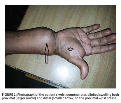
A 70-year-old man, a farmer by profession, presented with swelling in the volar aspect of his right wrist with associated dull aching pain for 6 months. Tingling and numbness was present in the volar aspect of the wrist and the ring and little fingers. There was associated history of evening pyrexia and weight loss. On examination, an hour-glass swelling above and below the proximal palmar crease (Figure 1), with a positive cross-fluctuation test, was noted. Blood parameters were within normal limits except for a high erythrocyte sedimentation rate (ESR) of 70 mm/hr in the first hour.
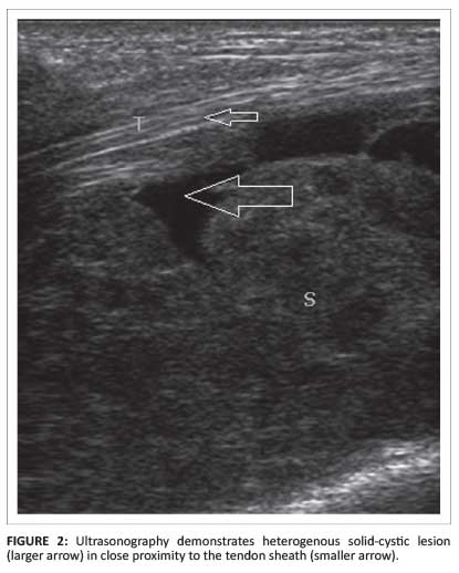
On chest radiography, no obvious abnormality was noted. The Mantoux test was positive (induration of 15 mm). Nerve conduction studies (NCS) revealed features suggestive of median nerve compression; there was increased distal motor latency (5.1 milliseconds) and distal sensory latency (4.2 milliseconds) of the median nerve with decreased median nerve conduction velocity (41 m/s). Radiography of the hand and wrist revealed diffuse soft-tissue swelling without any osseous abnormality. US of the wrist revealed a heterogeneously hypoechoic bilobed mass (Figure 2) containing both solid and cystic components in and around the flexor tendon sheaths, with involvement both above and below the flexor retinaculum.
On power Doppler study, there was no obvious increased vascularity. A 3-Tesla MRI scan confirmed the bilobed mixed solid-cystic nature of the lesion (Figure 3).
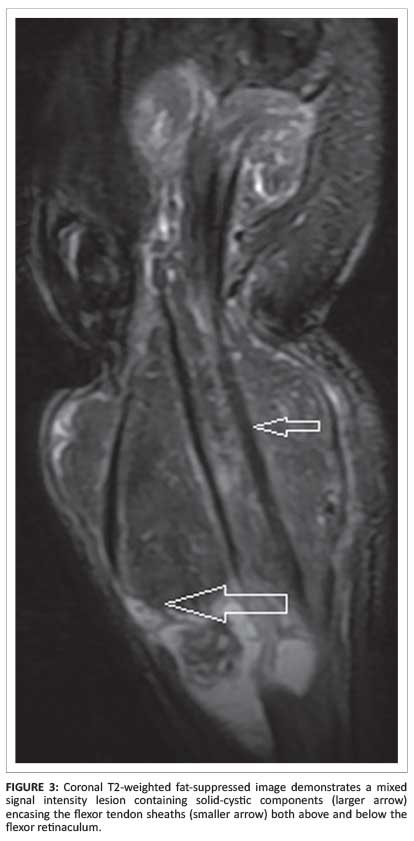
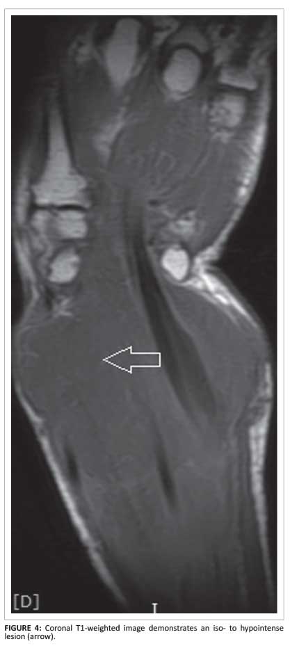
The solid component of the lesion was hypointense on T1-weighted images (Figure 4) and iso- to hypointense on T2-weighted and 3D fast gradient refocussed echo (3DFGRE) imaging.
The synovial fluid was hypointense on T1-weighted, and hyperintense on T2-weighted and 3DFGRE sequences. On postcontrast study, there was irregular rim enhancement of the lesion (Figure 5). A provisional diagnosis of compound palmar ganglion was made, based on the radiological findings.

Management and outcome
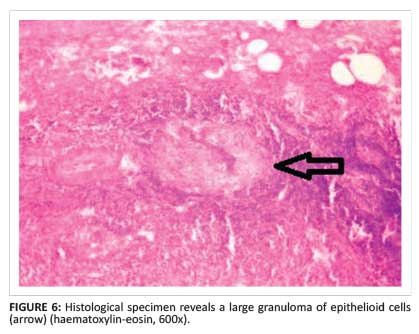
The patient subsequently underwent surgery; the lesion was completely excised along with the involved tendon sheaths. Intra-operative findings revealed characteristic melon seed bodies. A sample was sent for histopathological and polymerase chain reaction (PCR) examination. The histopathology of the resected specimen revealed granulomatous inflammation with central caseous necrosis, surrounded by epithelioid cells and multiple giant cells (Figure 6). PCR was positive for Mycobacterium tuberculosis. The patient received a full course of antituberculous chemotherapy. To date, he remains asymptomatic without any further recurrence.
Discussion
Compound palmar ganglion affects the radial and ulnar bursae. The radial bursa is the synovial sheath that surrounds the tendon of the flexor pollicis longus in its course through the carpal tunnel. It arises 2.5 cm proximal to the flexor retinaculum and continues to form the digital sheath of the thumb. The ulnar bursa includes the little finger tendon sheath which begins at the terminal phalanx and extends proximally halfway up the palm, where it envelops the superficial and deep tendons of the third, fourth and fifth fingers. In 80% of cases, the ulnar bursa communicates with the radial bursa, which accounts for horseshoe-shaped tenosynovitis that simultaneously affects both the bursae.1
TB is widespread in India, affecting various organ systems in people. The most commonly affected extrapulmonary sites are the lymph nodes, genito-urinary tract, bone marrow, central nervous system and musculoskeletal system, which include bones, joints, bursae, tendons and tenosynovium. Infection of the wrist and hand, however, is quite rare, and diagnosis and treatment of this clinical entity is usually delayed; but early diagnosis and treatment is most important, as the lesion is destructive. Pathogenesis of the condition is either direct inoculation or haematogenous spread from a distant focus in the lung, lymph nodes, genito-urinary tract or bones. The lesion has a gradual onset and progresses slowly, so patients usually present at a fairly advanced stage. Severe swelling may compress the median nerve, resulting in neurological symptoms.
Laboratory investigations are mostly normal except for a raised ESR and strongly positive Mantoux test. The chest radiograph is mostly unremarkable. NCS and electromyography (EMG) may reveal features of median nerve compression, if present. NCS reveal delayed distal latencies and slowed conduction velocities as a result of damage to the myelin sheath owing to nerve compression. With sustained or more severe compression, axonal loss may also occur, resulting in a reduction of the median nerve compound motor or sensory action potential amplitude.
EMG may reveal evidence of pathological changes in muscles innervated by the median nerve, by demonstrating either active denervation (e.g. spontaneous activity such as fibrillation potentials, positive sharp waves and fasciculation potentials) or chronic changes that indicate denervation with subsequent reinnervation (e.g. changes in the motor unit action potential amplitudes, durations and recruitment).
Radiological modalities play a pivotal role in the diagnosis of compound palmar ganglion. Plain radiography may demonstrate soft-tissue opacity and evidence of bone destruction. Computed tomography (CT) can show bone destruction and simultaneously demonstrate the solid-cystic nature of the lesion. B-mode US is a very useful tool to visualise, in real time, the proximal and distal extent of the lesion, and its contents and relation to the flexor tendon sheath. Usually, heterogenous solid-cystic components are noted in and around the flexor pollicis longus, flexor digitorum superficialis and flexor digitorum profundus tendons, both proximal and distal to the flexor retinaculum. MRI, by virtue of its excellent soft-tissue delineation, is the best imaging modality in this context. Pre- and postcontrast T1, T2 and 3DFGRE sequences in both the axial and coronal planes are commonly used. As the lesion is predominantly fluid-containing with thickened synovium and tenosynovium, it is hypointense on T1W1; on T2WI, the fluid component is hyperintense and the thickened synovium and tenosynovium appear hypointense. T2*-weighted images also reveal the fluid component to be hyperintense and the solid component to be hypointense.3, 4On postcontrast studies, there is usually rim enhancement of the lesion.
Histopathological testing of the specimen reveals the granulomatous nature of the lesion. There are three histological forms of tuberculous tenosynovitis as a result of the long duration of the disease, depending upon the resistance of the individual and varying virulence of the micro-organism. In the earliest stage, the tendon is replaced by vascular granulation tissue. Later on, the sheath is obliterated by fibrous tissue. Fluid is contained within the sheath, and rice bodies may appear as a result of caseation. If healing by fibrosis fails to curtail the pathologic process, extensive caseation and granulation occur.5
The treatment of compound palmar ganglion consists of antituberculous drugs along with excision of the lesion.6 Complete excision and evacuation of the lesion along with excision of the inflamed tendon sheaths is done via the volar aspect.6
Conclusion
Radiological imaging is important in assisting with the early diagnosis of tuberculous compound palmar ganglion; it is otherwise delayed owing to the rarity of the condition and its nonspecific clinical presentation. The diagnosis is, however, confirmed by histopathological testing. Early diagnosis and proper treatment can cure this otherwise destructive condition.
Acknowledgements
Competing interests
The authors declare that they have no financial or personal relationship(s) that may have inappropriately influenced them in writing this article.
Authors' contributions
S.T. (IPGME&R and SSKM Hospital) was responsible for the conception and design, drafting the article, revising it critically for intellectual content, US and MR image acquisition, analysis and interpretation and final approval of the version to be published. S.B. (IPGME&R and SSKM Hospital) was responsible for the conception and design, revising the article critically for intellectual content, US and MR image acquisition, analysis and interpretation and final approval of the version to be published. A.B. (IPGME&R and SSKM Hospital) was responsible for the conception and design, revising the article critically for intellectual content, US and MR image acquisition, analysis and interpretation and final approval of the version to be published. S.C. (IPGME&R and SSKM Hospital) was responsible for the conception and design, revising the article critically for intellectual content, US and MR image acquisition, analysis and interpretation and final approval of the version to be published. S.C. (R.G. Kar Medical College and Hospital) was responsible for the conception and design, revising the article critically for intellectual content, pathological specimen collection, slide preparation and interpretation and final approval of the version to be published.
References
1. Pimm LH, Waugh W. Tuberculous tenosynovitis. J Bone Joint Surg Br. 1957;39-B(1):91-101. [ Links ]
2. Kumar KA, Kanthimathi B, Krishnamurthy CS, Sujai S. Compound palmar ganglion: A tubercular manifestation of flexor tenosynovitis of the wrist. International Journal of Case Reports and Images. 2012;3(2):28-31. http://dx.doi.org/10.5348/ijcri-2012-02-93-CR-7 [ Links ]
3. Hsu CY, Lu HC, Shih TT. Tuberculous infection of the wrist: MRI features. Am J Roentgenol. 2004;183(3): 623-628. http://dx.doi.org/10.2214/ajr.183.3.1830623 [ Links ]
4. Parmar R, Mehta K, Gandhi R, Mohite N. compound palmar ganglion. International Journal of Health Sciences and Research. 2013;3(12):172-176. [ Links ]
5. Lall H, Nag SK, Jain VK, Khare R, Mittal D. Tuberculous extensor tenosynovitis of the wrist with extensor pollicis longus rupture: A case report. Journal of Medical Case Reports. 2009;3:142. http://dx.doi.org/10.1186/1752-1947-3-142 [ Links ]
6. Krishna D, Bisht RS, Sikarwar V, Chand. Compound palmar ganglion: A case report. IOSR Journal of Pharmacy. 2012;2(6):20-22. http://dx.doi.org/10.9790/3013-26502022 [ Links ]
 Correspondence:
Sourav Talukder
Bagbazar, Nilkantha Sarkar
Road Bye Lane (west)
Chandannagar, Hooghly
West Bengal, India
Email: talukdersourav@gmail.com
Received: 02 June 2014
Accepted: 09 Oct. 2014
Published: 11 Dec. 2014

