


Services on Demand
Article
 English (pdf)
 Article in xml format
 Article references
Indicators

Related links
-
 Cited by Google
 -
 Similars in Google
Share

 Permalink
SA Journal of Radiology
On-line version ISSN 2078-6778
Print version ISSN 1027-202X
S. Afr. J. radiol. (Online) vol.18 n.1 Johannesburg Oct. 2014
http://dx.doi.org/10.4102/sajr.v18i1.601
ORIGINAL ARTICLES
A retrospective study of computed tomography angiography versus digital subtraction angiography in penetrating neck trauma at Groote Schuur Hospital, South Africa
Paul Scholtz; Steve Beningfield; Sally Candy
Division of Radiology, Groote Schuur Hospital, Cape Town, South Africa
ABSTRACT
BACKGROUND: Penetrating neck trauma is commonly encountered in South African trauma units, and is associated with high mortality and morbidity rates. The imaging protocol for stable patients with penetrating neck trauma remains controversial. There is only sparse data validating the use of computed tomography angiography (CTA) in the evaluation of penetrating neck trauma in South Africa.
OBJECTIVES: To assess the sensitivity and specificity of CTA versus digital subtraction angiography (DSA) in detecting arterial injury and secondarily evaluate the ability of CT to assess non-arterial injury.
METHOD: Using hospital and radiology databases, 23 patients were identified who had undergone both CTA and DSA for penetrating neck trauma. The data was retrospectively anonymised and randomised. A radiologist experienced in the interpretation of both trauma CTA and DSA re-reported all the imaging and the findings were compared and analysed.
RESULTS: Twenty-four arterial injuries were detected. The sensitivity of CTA for detecting arterial injury was 78% and the specificity 83%. The ability of CTA to delineate wound track and detect non-arterial visceral injury was also confirmed.
CONCLUSION: CTA is an attractive initial diagnostic investigation that, along with clinical evaluation, effectively guides further investigation and intervention. It is important for the radiologist to understand the limitations of CTA and have a low threshold for DSA in equivocal cases.
Introduction
South African violent crime and trauma rates are high,1 and penetrating neck trauma is common in South African trauma centres. These injuries have high mortality and morbidity rates of between 3% and 6%, with most fatalities due to major neck vessel injuries.2
The role and type of imaging of stable patients with penetrating neck trauma is controversial. Given the probability of vascular, aerodigestive tract and/or neurological injury, important clinical decisions have to be made regarding further investigation or intervention. Although digital subtraction angiography (DSA) is the gold standard for detecting arterial vascular injury, it is an invasive procedure with potential complications, such as puncture-site haematoma, iatrogenic vascular damage and embolic complications, including stroke.3 DSA is also resource intensive, although it does permit therapeutic endovascular options during the same procedure in some cases.
Because computed tomography (CT) has become an integral part of emergency units worldwide, CT angiography (CTA) provides an attractive alternative to DSA. CT also has the ability to determine the direction and course of any wound tracks that can suggest particular damage, along with direct evidence of visceral, bony or aerodigestive tract injury.
Although frequently used, the role of CTA remains unclear. Sparse data exist in South Africa regarding the use of CTA in evaluating penetrating neck trauma.
This study aims to retrospectively compare and analyse the findings of CTA and DSA in patients with penetrating neck trauma at Groote Schuur Hospital, a tertiary referral university hospital in Cape Town, South Africa, serving a drainage population of approximately 2 million. The hospital has a busy trauma unit, with a high incidence of penetrating trauma due to gunshot and stab wounds. From July 2004 to June 2005 (12 months), 203 patients with penetrating neck injuries were admitted into the trauma unit, of whom 21% sustained gunshot wounds and 78% stab wounds.4
Method
Formal ethics approval was obtained from the Research Ethics Committee, Health Sciences Faculty, University of Cape Town.
Using the hospital and radiological databases, all patients with penetrating neck trauma who underwent both DSA and CTA (in either order) during the period from March 2009 to March 2012 were identified for the study. In these patients, CTA and DSA were performed for various reasons, including clinical suspicion, imaging features of injury on the other modality, surgical planning and/or endovascular treatment.
CTA was performed using a 16-slice Siemens Emotion multidetector CT scanner in the radiology department. Intravenous access was obtained using a cubital or forearm cannula of at least 18 gauge placed in an upper limb vein, if possible contralateral to the side of injury. A volume of 100 mL of non-ionic contrast medium (Iohexol 350) was administered using an injector pump at a rate of 6 mL/s. Caudocranial helical acquisition was planned using the lateral scout view of the neck from at least the aortic arch to the Circle of Willis. The scan was initiated using a bolus tracker system placed over the aortic arch triggering at 100 Hounsfield units. A saline chaser was not utilised. Typical reconstruction parameters were 1 mm slice thickness, a pitch of 1.5, 110 kVp and 100 mA.
Digital subtraction angiography was performed using a single C-arm ceiling-suspended DSA system (Toshiba Infinix VC-i). For initial diagnostic purposes, aortic arch and relevant selective brachiocephalic and branch vessel arteriograms where performed. Approximately 100 mL of non-ionic contrast (Iopromide 300) was used on average.
The CTA and DSA images were retrospectively anonymised and randomised. A radiologist experienced in the interpretation of both trauma CTA and DSA and who was blinded to the clinical data and prior opinions, reported on all the imaging. Various window settings, multiplanar reconstructions (MPR) and maximum intensity projections (MIP) were permitted. Bone subtraction was used when considered warranted in CTA. Both CTA and DSA were evaluated for both vascular and non-vascular pathology. Unsubtracted DSA images were not always available due to image archiving technicalities.
All vessels within the field of view were assessed. Any identified vascular injuries on CTA and DSA were localised and described. Injury types were categorised as occlusion, contrast extravasation, pseudo-aneurysm, arteriovenous fistula (AVF), intimal flap or a combination of these. Non-vascular evaluation in both modalities included the soft tissues, aerodigestive tract, bones, lungs and mediastinum. Contrast swallows were also included. Where possible, the wound tract was identified on CTA.
Clinical data that was retrospectively analysed included patient demographics, penetrating agent, zone of injury, indications for investigation, associated injuries and management.
CTA and DSA findings were compared and analysed. DSA findings were considered the standard of reference for arterial pathology, whilst CTA was considered the reference for soft-tissue damage. True positives required the correct definition and appropriate clinical correlation of injury type and location. Sensitivity, specificity and positive and negative predictive values were specifically calculated for arterial vascular injuries.
Results
Over the 3-year period, 23 patients (18 male and 5 female) were included in the study. All imaging was considered to be of diagnostic quality, and complete clinical data was available on folder and radiological review.
The delays between CTA and DSA ranged from an immediate follow-on procedure to up to 72 hours. All but one patient underwent CTA prior to DSA; the single follow-on CTA was performed due to concern of an injury at the origin of the left common carotid artery on DSA.
The mean age was 30 years, with a range of 19-51 years. Stab wounds accounted for 20 (87%) and gunshots for 3 (13%) of the injuries.
The location of the entry sites of penetrating neck injury was allocated into the three standard anterior triangle anatomical zones (Figure 1). There were no injuries involving the posterior triangle.

Injury locations amounted to 8 (35%) in zone 1, 14 (61%) in zone 2 and 1 (4%) in zone 3. Sixteen (70%) of the patients suffered injuries to the left neck and six (26%) to the right, whilst one (4%) had bilateral injuries.
Various clinical signs for vascular injury were recorded in the clinical notes (Figure 2). Three patients had no detectable significant clinical signs of injury at the time of scanning.

Foley-catheter tamponade using the inflated balloon to temporarily arrest bleeding within the wound tract was employed in six (26%) patients.
Contrast swallows were performed on 14 (61%) of the patients on the basis of clinical symptoms and signs, with a single oesophageal injury detected. A further oesophageal injury was detected intra-operatively (this patient had not undergone a contrast swallow as the patient was intubated and had other existing indications for surgical exploration).
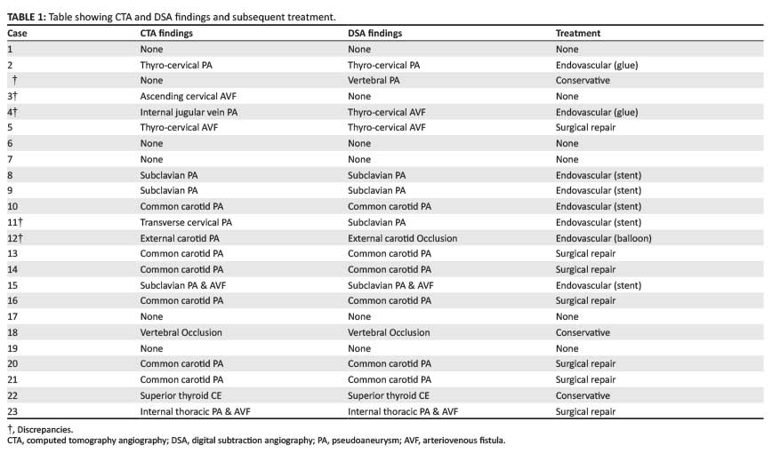
Digital subtraction angiography demonstrated 18 arterial injuries in 17 patients, with one patient having both a vertebral and an inferior thyroid artery injury (Table 1).
In comparison to DSA, the sensitivity of CTA for detecting arterial injury was 78%, the specificity 83%, the positive predictive value (PPV) 93% and the negative predictive value (NPV) 56%.
Conservative management was undertaken in three (17%) patients. Endovascular management was used to manage eight (44%) injuries, of whom five were treated with covered stents, two were embolised with cyanoacrylate glue and one was embolised using a detachable balloon. Surgical vascular repair was performed in seven (39%) cases.
During the in-hospital course, one patient suffered a middle cerebral artery infarction seven days after primary surgical repair of a common carotid injury, and another died of hypoxic brain injury two days after surgical repair of a common carotid injury.
Discussion
Classically, stable patients with injuries penetrating the platysma muscle mandated exploratory surgery.5 Since the 1990s, however, selective non-operative management (SNOM) has gained favour and has become the rule.4,5 Yet the optimal management and imaging strategy for stable patients with penetrating neck trauma remains controversial.
Vascular injury is suspected clinically when certain 'hard' signs are present. The most reliable pointers to vascular injury include a bruit or thrill, expanding or pulsating haematoma, pulsating haemorrhage (including a history thereof), pulse deficit and/or major neurologic deficit.
'Soft' signs are less reliable and include systemic hypotension, identifiable injury in proximity to vessels and a moderate to large stable haematoma. Injuries crossing the midline or those that require use of Foley-catheter balloon tamponade also may influence the decision to perform angiography. Other clinical signs included in our study included rapid drainage of blood from an intercostal drain and a widened or abnormal mediastinal contour on chest radiography.
The most reliable clinical sign to predict arterial injury was pulsatile haemorrhage. Three patients had no vascular signs on presentation. One of these patients had a significant injury (subclavian artery pseudo-aneurysm with an AVF) requiring endovascular stenting, but the other two were normal on imaging. All patients with Foley-catheter balloon tamponade suffered arterial injuries.
Several groups have compared CTA with DSA for detection of arterial injury in penetrating neck trauma; reported sensitivities ranged between 80% and 100%, with specificities above 90%.6,7,8,9 Our study demonstrated a CTA sensitivity of 78% and specificity of 83%. Four false-negative findings and one false-positive finding occurred with CTA.
Of the false-negative findings, a small (3 mm) pseudo-aneurysm in case 2 seen on DSA of the vertebral artery was not detected on CTA (Figure 3). The lesion could also not be detected on retrospective review of the CTA using MPR and MIP imaging. Subtle lesions may not be well demonstrated on CT as compared to DSA due to the inferior spatial resolution of CT, lack of bone subtraction and pulsation artefact. Although the clinical significance of these 'minor' lesions may be controversial, studies show that many of these lesions either resolve or remain asymptomatic.10The small vertebral artery aneurysm was managed conservatively and therefore no significant management difference occurred.

Another false-negative CTA result in case 4 was due to an AVF arising from the inferior thyroid branch of the left thyrocervical trunk (Figure 4). This injury had been misdiagnosed as an internal jugular vein injury on presentation. Clinical concern of an injury persisted due to the presence of a bruit. The patient was therefore referred for DSA, confirming the AVF, for which endovascular embolisation was performed. As CTA is not a dynamic study, the single acquisition during the contrast bolus may include physiological venous 'contamination' masking early venous filling due to an AVF. DSA is arguably required when an AVF is clinically suspected but not detected on CTA.

In case 12, a transection of the external carotid artery was misinterpreted as a pseudo-aneurysm on CTA as the ascending pharyngeal artery was interpreted as spasm of the continuation of the external carotid artery (Figure 5). When this was referred for DSA and endovascular embolisation, it was revealed as an ECA transection. After debate, the vessel was occluded with a detachable balloon.

Incorrect localisation of a pseudo-aneurysm accounted for the last false-negative result in case 11. CTA reported the lesion to involve a cervical branch of the subclavian artery, whereas on DSA the lesion arose from the subclavian artery itself, near the cervical vessel origin. The pseudo-aneurysm was managed with a covered endovascular stent of the subclavian artery. It is questionable whether this led to a significant management difference, as either lesion was amenable to endovascular management.
The only false-positive result was the over-interpretation of what was thought to be a small AVF from the ascending cervical artery to the internal jugular vein on CTA, with DSA revealing no injury. Although an AVF may have occluded spontaneously, this was felt to be unlikely.
CTA was performed after DSA in one patient in whom suspicion of an injury at the origin of the left common carotid artery was suspected on DSA. CTA confirmed the injury as a pseudo-aneurysm at the common carotid artery origin.
The ability of CT to evaluate structures other than the arterial vasculature adds further benefits. Assessment of the aerodigestive tract, viscera (lungs, thyroid), veins and bony structures (especially the spine) assist in clinical management. Determining the trajectory of the wound track further aids in the evaluation of patients with penetrating neck trauma, because the organs lying along the path can be considered to have a higher likelihood of injury (Figure 6).

The assessment of bullet and bone fragments not only aid in identifying the wound track but may also be helpful in assessing the direction of travel of a projectile such as a bullet. Careful evaluation of the bone should show bevelling toward the direction of travel, which is important in forensic analysis.11
In penetrating neck trauma, it is also important to deliberately assess the airway on CT. Close attention has to be paid to airway narrowing due to surrounding haematoma or direct injury. Patient 5, with an AVF detected on CTA, acutely deteriorated on the DSA table, requiring intubation and emergency surgery due to airway compromise as a result of a large mediastinal haematoma (Figure 7). Signs of direct tracheal injury include tracheal wall defects or deformity, excessive surgical emphysema, pneumomediastinum and pneumothorax (Figure 8). A low threshold for endoscopy is required to accurately assess suspected airway injury.


Multiple types of artefacts have to be taken into account during CTA and DSA, including those induced by patient size, motion, incoming dense venous contrast and post-traumatic fragments (bullet shrapnel, knife or bone fragments). Contrast inflow artefact is usually due to use of upper limb venous injections resulting in streak artefact at the thoracic inlet. This may interfere with the visualisation of the proximal arteries exiting the thorax, especially the ipsilateral subclavian artery. Injection of contrast into the contralateral side of injury or using femoral venous access is helpful in reducing this artefact. The use of a saline chaser also reduces the amount of streak artefact.
Multiple limitations were present in the study. Most other studies used two experienced radiologists during the retrospective review, with the consensus reached as the definitive impression. Little short-term to long-term patient follow-up was present. Unsubtracted images on DSA were not always available due to archiving technicalities; this reduced the ability to evaluate for foreign bodies, bony detail and soft tissues, which are important for thorough assessment.
Although sensitivity and specificity in this study is lower than reported, CTA remains attractive as the initial diagnostic investigation. It is readily available, non-invasive and less resource-intensive, although it may take longer to analyse than DSA.
To our knowledge, this is the first study documenting the use of 16-slice multidetector CT in penetrating neck trauma. Larger, prospective trials are required for further evaluation.
Conclusion
Selective non-operative management is the rule in management of stable patients with penetrating neck trauma. CTA is an attractive initial diagnostic investigation that, along with clinical evaluation, effectively guides further investigation and intervention. High sensitivities and specificities for detecting arterial vascular injury have been documented. The added advantages of CTA include the ability to detect wound trajectory and determine visceral, bony and aerodigestive tract injury. However, it is important for the radiologist to understand the limitations of CTA and have a low threshold for conventional angiography in equivocal cases, or where there is a strong suspicion of an AVF.
Acknowledgements
Competing interests
The authors declare that they have no financial or personal relationship(s) that may have inappropriately influenced them in writing this article.
Authors' contributions
P.S. (Groote Schuur Hospital) was the lead researcher and was responsible for the concepts, design, literature review and preparation of the manuscript. P.S. and S.B. (Groote Schuur Hospital) conducted the study. S.B. and S.C. made conceptual contributions and were involved with the manuscript editing.
References
1. Centre for the Study of Violence and Reconciliation. Why does South Africa have such high rates of violent crime? Supplement to the final report of the study on the violent nature of crime in South Africa. Johannesburg: CSVR; 2009 [cited 2012 April 04]. [ Links ] Available from: http://www.csvr.org.za/docs/study/7.unique_about_SA.pdf
2. Brywczynski JJ, Barrett TW, Lyon JA, Cotton BA. Management of penetrating neck injury in the emergency department: A structured literature review. Emerg Med J. 2008;25:711-715. http://dx.doi.org/10.1136/emj.2008.058792 [ Links ]
3. Schroeder JW, Baskaran V, Aygun N. Imaging of traumatic arterial injuries to the neck with an emphasis on CTA. Emerg Radiol. 2005;13(3):158-163. [ Links ]
4. Thoma M, Navsaria PH, Edu S, Nicol AJ. Analysis of 203 patients with penetrating neck injuries. World J Surg. 2008;32:2716-2723. http://dx.doi.org/10.1007/s00268-008-9766-7 [ Links ]
5. MacFarlane C, Benn CA. Penetrating neck trauma: A review. Trauma. 2002;4:79-90. http://dx.doi.org/10.1191/1460408602ta226oa [ Links ]
6. LeBlang SD, Núñez DB, Rivas LA, Falcone S, Pogson SE. Helical computed tomographic angiography in penetrating neck trauma. Emerg Radiol. 1997;4:200-206. http://dx.doi.org/10.1007/BF01508171 [ Links ]
7. Múnera F, Soto JA, Palacio DM, Velez SM, Medina E. Diagnosis of arterial injuries caused by penetrating trauma to the neck: Comparison of helical CT angiography and conventional angiography. Radiology. 2000;216(2):356-362. http://dx.doi.org/10.1148/radiology.216.2.r00jl25356 [ Links ]
8. Múnera F, Soto JA, Palacio DM, et al. Penetrating neck injuries: Helical CT angiography for initial evaluation. Radiology. 2002;224:366-372. http://dx.doi.org/10.1148/radiol.2242010973 [ Links ]
9. Inaba K, Múnera F, McKenney M, et al. Prospective evaluation of screening multislice helical computed tomographic angiography in the initial evaluation of penetrating neck injuries. J Trauma. 2006;61:144-149. http://dx.doi.org/10.1097/01.ta.0000222711.01410.bc [ Links ]
10. Frykberg ER, Vines FS, Alexander R. The natural history of clinically occult arterial injuries: A prospective evaluation. J Trauma. 1989;29:577-583. http://dx.doi.org/10.1097/00005373-198905000-00006 [ Links ]
11. Wilson AJ. Gunshot injuries: What does a radiologist need to know? Radiographics. 1999;19:1358-1368. http://dx.doi.org/10.1148/radiographics.19.5.g99se171358 [ Links ]
 Correspondence:
Paul Scholtz
Division of Radiology
Room 16, C16, New Groote
Schuur Hospital, Private Bag
7935, Observatory 7935
Cape Town
Received: 07 Jan. 2014
Accepted: 22 May 2014
Published: 20 Oct. 2014

