


Services on Demand
Article
 English (pdf)
 Article in xml format
 Article references
Indicators

Related links
-
 Cited by Google
 -
 Similars in Google
Share

 Permalink
African Journal of Health Professions Education
On-line version ISSN 2078-5127
Afr. J. Health Prof. Educ. (Online) vol.12 n.2 Pretoria Jun. 2020
http://dx.doi.org/10.7196/AJHPE.2020.v12i2.1261
SHORT COMMUNICATION
Exposure technique factors in digital X-ray imaging systems: Demonstrating the effect of mAs
S Lewis
MTech: Radiography. Department of Medical Imaging and Radiation Sciences, Faculty of Health Sciences, University of Johannesburg, South Africa
Why was the idea necessary?
Digital X-ray imaging systems have a wide, dynamic exposure latitude that allows almost 500 times the exposure necessary to produce optimal diagnostic images.[1, 2] Consequentially, patients may receive more exposure to ionising radiation than necessary to produce an image of optimal diagnostic quality. A recent study showed that ~54% of radiographers understood and used indicators of exposure in digital X-ray imaging systems.[3] With just more than half of radiographers understanding and using indicators of exposure in digital X-ray imaging systems, enhancing radiographers' understanding of exposure technique factors (kilovoltage peak (kVp), milliampere and the exposure time (mAs)) in digital X-ray imaging systems is needed to curtail unnecessary exposure to ionising radiation.
Ethical approval for the study was obtained from the Faculty of Health Sciences Research Ethics Committee, University of Johannesburg (ref. no. REC-234-2019).
What was tried?
A structured tutorial demonstrating the effect of mAs on image quality in digital X-ray imaging systems was tried. Four radiographs were taken of a hand phantom, using a constant kVp, focal film distance (FFD), focal spot size, 4-sided collimation and the same computed radiography (CR) (a type of digital X-ray imaging system) cassette. Only the mAs changed for each exposure (exposure refers to each time the radiograph is taken and the phantom hand is 'exposed' to ionising radiation) (Fig. 1A-D). Before the tutorial, students needed to predict the image quality for each exposure. After the tutorial, students compared their predicted outcomes with the actual outcomes. Dose area products (DAPs), exposure indicators (Els) and image quality for each exposure without any image post-processing were tabulated. Students then evaluated and reported the effect of mAs. The resultant images and exposure technique factors are indicated in Fig. 1.
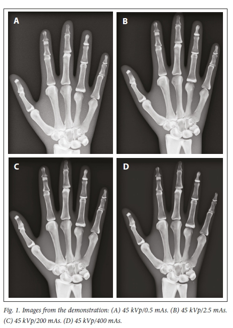
The effect of mAs on image quality in digital X-ray imaging systems was observed. It was noticeable that at higher than standard optimum mAs, the image quality was preserved (acceptable mAs for a posteroanterior (PA) projection of a hand for the X-ray unit used in the tutorial was 2.5 mAs (Fig. 1B)). However, with higher mAs, there was a congruent increase in the dose to the phantom (DAP). Students predicted that for the exposure technique factors used in Fig. 1C and Fig. 1D, they would not be able to observe an image, but acceptable images were obtained. Students predicted a contrary outcome, despite learning the theory of the wide dynamic exposure latitude of digital X-ray imaging systems and seeing images similar to those in Fig. 1 in the literature. Therefore, using a blended teaching approach provided an opportunity for students to experiment with varied mAs to enhance their understanding of the effect of mAs on image quality in digital X-ray imaging systems.
Acknowledgements. Thank you to the students who voluntarily participated in the tutorials.
Author contributions. Sole author.
Funding. None.
Conflicts of interest. The author lectures the students who participated in the tutorial.
References
1. Moore QT, Don S, Goske MJ, et al. Image gently: Using exposure indicators to improve pediatric digital radiography. Radiol Technol 2012;84(1):93-99. [ Links ]
2. Don S, Whiting BR, Rutz LJ, Apgar BK. New exposure indicators for digital radiography simplified for radiologists and technologists. Am J Radiol 2012;199(6):1337-1341. https://doi.org/10.2214/AJR.12.8678 [ Links ]
3. Lewis S, Pieterse T, Lawrence H. Evaluating the use of detector dose indicators in digital X-ray imaging systems. Radiography (Lond) 2019;25(3):e58-e62. https://doi.org/10.1016/j.radi.2019.01.003 [ Links ]
 Correspondence:
S Lewis
shantell@uj.ac.za

