


Services on Demand
Article
 English (pdf)
 Article in xml format
 Article references
Indicators

Related links
-
 Cited by Google
 -
 Similars in Google
Share

 Permalink
South African Journal of Child Health
On-line version ISSN 1999-7671
Print version ISSN 1994-3032
S. Afr. j. child health vol.11 n.4 Pretoria Dec. 2017
http://dx.doi.org/10.7196/sajch.2017.v11i4.1420
CASE REPORT
doi:10.7196/sajch.2017.v11i4.1420
Diandric triploidy in a liveborn infant with 3-4 syndactyly and a neural tube defect
C E SpencerI; B MofokengII; A TurnerIII; F NakwaIV; A KrauseV
IMB ChB, DCH, FCMG, MMed (Med Genet); Division of Human Genetics, School of Pathology, Faculty of Health Sciences, University of the Witwatersrand and National Health Laboratory Service, Johannesburg, South Africa
IIMB ChB; Division of Human Genetics, School of Pathology, Faculty of Health Sciences, University of the Witwatersrand and National Health Laboratory Service, Johannesburg, South Africa
IIIMSc (Med) (Hum Genet); Division of Human Genetics, School of Pathology, Faculty of Health Sciences, University of the Witwatersrand and National Health Laboratory Service, Johannesburg, South Africa
IVMB BCh, FCPaeds, MMed (Paeds), Cert (Neonatol), Cert (Paed Neurol); Department of Paediatrics, Division of Neonatology, Chris Hani Baragwanath Academic Hospital, Faculty of Health Sciences, University of the Witwatersrand, Johannesburg, South Africa
VMB BCh, PhD; Division of Human Genetics, School of Pathology, Faculty of Health Sciences, University of the Witwatersrand and National Health Laboratory Service, Johannesburg, South Africa
ABSTRACT
Triploidy is a chromosomal abnormality caused by an additional set of haploid chromosomes. It is a common cause of early first-trimester miscarriages. Only very rarely are babies with triploidy born alive and even more rarely do they survive beyond the first few days of life. We present here a case of a term baby with confirmed paternal (diandric) triploidy and some unusual features, who survived for 50 days, and review the literature on those who survived.
Triploidy is a chromosomal abnormality caused by an additional haploid set of chromosomes. It has been reported to occur in 1 to 3% of all conceptuses with most of these resulting in spontaneous miscarriage at an early gestation.[1] Only 1 in 10 000 fetuses with triploidy will survive to term, with only 1 in 1 200 of these surviving after birth.[2] The literature reports on only a small number of long-term surviving infants,[1] which is defined as those surviving >45 days.[2] Triploidy can occur due to maternal (digynic) or paternal (diandric) additional haploid chromosomes and, depending on the parent of origin, the phenotype can be predicted to some degree. Potential karyotype complements are 69XXX, 69XXY or 69XYY. This report presents a case of paternal (diandric) triploidy with structural abnormalities, minimal dysmorphic features and long survival of 50 days.
Case report
A 4-day-old male was referred to the Division of Human Genetics, National Health Laboratory Service and the University of the Witwatersrand for a clinical genetic assessment, as he had multiple congenital abnormalities and the diagnosis was uncertain. Ethics approval for publication was obtained from the University of the Witwatersrand Human Research ethics committee (ref. no. M1604107).
The patient was the only child to his non-consanguineous parents. He had three maternal half-siblings, the eldest of whom has an intellectual disability and a clubfoot of unknown cause. There is no further family history of physical disability, neural tube defects or miscarriages.
The baby was born after an uneventful pregnancy, during which his 37-year-old G4P4 HIV-positive mother took antiretroviral medication, smoked six cigarettes per day and received adequate dosing of penicillin for a positive rapid plasma reagin (RPR) result. She attended regular antenatal clinics but no ultrasound investigations were conducted during the pregnancy. The baby was born at the local clinic at 39 weeks' gestation by normal vertex delivery. His birth parameters were as follows: weight 1 670 g (Z-score <-3); length 35 cm (Z-score <-3); and head circumference 30 cm (Z-score <-3). He had severe and asymmetric intrauterine growth restriction. His Apgar scores were good but he was transferred to the academic referral hospital after birth as congenital abnormalities were noted and respiratory distress was present. Upon admission, his mild respiratory distress was presumed to be due to a congenital pneumonia and he was initiated on antibiotics.
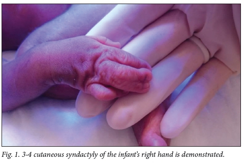
On examination, he was noted to have a square head with a prominent forehead, prominent eyes and down-slanted palpebral fissures. He had mild facial asymmetry. His lips were thin with a smooth philtrum and his mouth was small with down-turned corners. He had bilateral 3-4 complete cutaneous syndactyly of his hands (Fig. 1). A lumbosacral myelomeningocele was present and there was no spontaneous movement of his lower limbs.
Investigations revealed normal kidneys on ultrasound and also a normal cranial ultrasound. His cardiac echogram showed a ventriculoseptal defect (VSD), atrioseptal defect (ASD) and a patent ductus arteriosus (PDA), with severe tricuspid regurgitation (TR) and a dilated right heart.
A quantitative fluorescent polymerase chain reaction (QF-PCR) (Elucigene QST*Rplusv2, United Kingdom) for aneuploidy demonstrated likely triploidy, which was confirmed by a karyotype demonstrating 69XXY in 11 out of 11 counted cells. The same QF-PCR investigation was used to determine the parent of origin of the additional haploid chromosome complement. Only maternal DNA was available and thus some markers were not informative. Of the 19 markers tested 11 were informative and confirmed that the additional haploid complement was not maternal in origin, leading us to conclude that this baby in fact had paternal (diandric) triploidy. Three of the informative markers showed a single paternal allele of increased peak area ratio and 6 had two distinct paternal alleles.
The parents were counselled regarding the diagnosis and the poor prognosis. Management options were discussed with them and a decision was made not to offer extraordinary lifesaving measures in the event of a cardiac or respiratory arrest. The baby received orogastric feeds, antibiotics and dressings for his large myelomeningocele. He was also later treated for cardiac failure. At about 3 weeks of age his condition was stable and a decision was made to close his myelomeningocele in order to facilitate the mother's wish to care for him at home. This surgery was delayed owing to a confirmed Acinetobacter baumanii meningitis. His condition deteriorated and he was transferred to NICU for nasal continuous positive airway pressure and treatment of his infection. He passed away in the NICU at 50 days of age owing to sepsis.
Discussion
Triploidy is a major cause of miscarriages.[3] Three different mechanisms can lead to triploidy: (i) a non-disjunction event in meiosis 1 or 2 of spermatogenesis; (ii) non-disjunction in meiosis 1 or 2 of oogenesis; and (iii) dispermy. Dispermy is considered to be the most common cause of triploidy.[4]
Two different phenotypes are recognised based on whether the additional set of chromosomes is maternal (digynic) or paternal (diandric) in nature. Type 1 is diandric and recognised by a large, cystic placenta with a well-formed fetus and a normal or microcephalic head. Type 2 is digynic with a small non-cystic placenta and a growth-retarded fetus with relative macrocephaly.'4 In more recent larger studies this phenotypic distinction between maternal and paternal triploidy has been challenged.[5] Aside from the features defining type 1 and type 2 triploidy, the literature describes a multitude of other malformations. These include facial dysmorphism, central nervous system abnormalities in 50% of cases, cardiac defects in 31% of cases on second-trimester ultrasound, limb abnormalities and genito-urinary and gastrointestinal defects.[6] Type 1 fetuses are reported to be more likely to miscarry early in pregnancy, whereas type 2 cases often survive until later in the pregnancy and are more likely to live beyond birth.[7] In our case, the placenta was not available for examination and therefore we cannot comment on its features.
An investigation of the markers in the baby and the mother in this case showed that the infant had diandric triploidy and, for 6 of the 9 informative markers, two different alleles of paternal origin were identified. Thus, the mechanism causing triploidy was more likely to have been either dispermy or a non-disjunction event in meiosis 1 and very unlikely to have been due to an event in meiosis 2. Triploidy owing to a non-disjunction event in meiosis 2 will have a preponderance of similar-sized alleles, as two similar sister chromatids separate during this process. The determination that this baby had diandric triploidy was unexpected in view of the fact that the traditional clinical phenotype of a severely growth-restricted baby was compatible with a type 2 triploidy and also that the baby survived beyond birth, which is more common in digynic triploidy cases.[4] Subsequent literature suggests that this needs further investigation with larger sample sizes, and that the parent of origin cannot be used to provide a prognosis for the pregnancy or postnatal survival.[3]
In our case, the infant presented with severe and asymmetric growth restriction, cardiac lesions and minimal dysmorphic features. It was, however, the combination of the 3-4 syndactyly in a growth-restricted infant with a neural tube defect that prompted the consideration of triploidy as the diagnosis. Syndactyly of the third and fourth digits is an uncommon abnormality and only associated with a limited number of genetic syndromes. This case report highlights the need to consider triploidy in live-born infants with 3-4 cutaneous finger syndactyly.
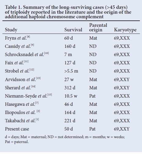
Live birth is exceedingly rare in cases of triploidy. This male infant survived 50 days, which is considered a 'long survival' in the literature. Since 1977 there have only been 11 published reports of babies with triploidy living beyond 45 days.[1] Since this baby survived for 50 days, he represents the 12th case reported in the literature who survived beyond 45 days (Table 1). Infants affected with triploidy usually succumb to respiratory problems or pneumonia. It has been proposed that survival has improved owing to successful treatment of infections. The association between digynic triploidy and an increased chance of survival is perhaps now even less clear than previously thought, as this case represents a second example of diandric triploidy with long survival compared with 6 cases with digynic triploidy.
Despite the fact that some infants with triploidy live longer, their outcome is still extremely poor. The longest surviving child was only 10.5 months old and had severe neurological and developmental impairment.'141 Because of the poor outcomes, extreme measures to prolong life are not usually considered. The management should however be discussed in a multidisciplinary setting and in conjunction with the parents in order to respect their views and decisions.
Conclusion
This case report describes a term, severely growth-retarded infant with minimal facial dysmorphism, cutaneous 3-4 syndactyly, cardiac defects and a neural tube defect. He was confirmed to have triploidy that was likely diandric in origin. This case history illustrates that although most cases of triploidy end in miscarriage, live birth is possible. The diagnosis should be considered in growth-restricted babies with cutaneous 3-4 syndactyly and other suggestive features. There are only a handful of reported cases of triploidy in which infants survived beyond 45 days - our case was the 12th such description. With this report we add to the literature describing the clinical features of those babies with triploidy surviving more than 45 days and also question whether parental origin of the triploidy can reliably be used to predict likelihood of survival.
Acknowledgement. The authors wish to thank the family described in this case report for their permission to use this information.
Author contribution. All authors were involved in the management of this patient. CS drafted the initial manuscript and BM, FN, AT and AK reviewed and edited it.
Funding. None.
Conflict of interest. None.
References
1. Takabachi N, Nishimaki S, Omae M, et al. Long-term survival in a 69,XXX triploid premature infant. Am J of Med Genet A 2008;146A(12):1618-1621. https://doi.org/10/1002/amjg.a.32352 [ Links ]
2. Iliopoulos D, Vassiliou G, Sekerli E, et al. Long survival in a 69,XXX triploid infant in Greece. Genet Mol Res 2005;4(4):755-759. [ Links ]
3. Baumer A, Balmer D, Binkert F, Schinzel A. Parental origin and mechanisms of formation of triploidy: A study of 25 cases. Eur J Hum Genet 2000;8(12):911-917. https://doi.org/10.1038/sj.ejhg.5200572 [ Links ]
4. McFadden DE, Kalousek DK. Two different phenotypes of fetuses with chromosomal triploidy: correlation with parental origin of the extra haploid set. Am J Med Genet 1991;38(4):535-538. https://doi.org/10.1002/ajmg.1320380407 [ Links ]
5. Joergensen MW, Niemann I, Rasmussen AA, et al. Triploid pregnancies: genetic and clinical features of 158 cases. Am J Obstet Gynecol 2014;211(4):370 e1-19. https://doi.org/10.1016/j.ajog.2014.03.039 [ Links ]
6. Chen CP, Chern SR, Tsai FJ, Hsu CY, Ko K, Wang W. Prenatal diagnosis and molecular analysis of triploidy in a fetus with intrauterine growth restriction, relative macrocephaly and holoprosencephaly. Taiwan J Obstet Gynecol 2009;48(3):323-326. http://doi.org/10.1016/S1028-4559(09)60318-1 [ Links ]
7. Hasegawa T, Harada N, Ikeda K, et al. Digynic triploid infant surviving for 46 days. Am J Med Genet 1999;87(4):306-310. https://doi.org/10.1002/(sici)1096-8628(19991203)87:4%3C306::aid-ajmg5%3E3.0.co;2-6 [ Links ]
8. Fryns JP, van de Kerckhove A, Goddeeris P, van den Berghe H. Unusually long survival in a case of full triploidy of maternal origin. Hum Genet 1977;38(2):147-155. https://doi.org/10.1007/bf00527396 [ Links ]
9. Cassidy SB, Whitworth T, Sanders D, Lorber CA, Engel E. Five month extrauterine survival in a female triploid (69,XXX) child. Ann Genet 1977;20(4):277-279. [ Links ]
10. Schrocksnadel H, Guggenbichler P, Rhomberg K, Berger H. Complete triploidy (69,XXX) surviving until the age of 7 months. Wien Klin Wochenschr 1982;94(12):309-315. [ Links ]
11. Faix RG, Barr M, Jr., Waterson JR. Triploidy: Case report of a live-born male and an ethical dilemma. Pediatrics 1984;74(2):296-299. [ Links ]
12. Strobel SL, Brandt JT. Abnormal hematologic features in a live-born female infant with triploidy. Arch Pathol Lab Med 1985;109(8):775-777. [ Links ]
13. Arvidsson CG, Hamberg H, Johnsson H, Myrdal U, Anneren G, Brun A. A boy with complete triploidy and unusually long survival. Acta Paediatr Scand 1986;75(3):507-510. [ Links ]
14. Sherard J, Bean C, Bove B, et al. Long survival in a 69,XXY triploid male. Am J Med Genet 1986;25(2):307-312. http://doi.org/10.1002/ajmg.1320250216 [ Links ]
15. Niemann-Seyde SC, Rehder H, Zoll B. A case of full triploidy (69,XXX) of paternal origin with unusually long survival time. Clin Genet 1993;43(2):79-82. https://doi.org/10.1111/j.1399-0004.1993.tb04432.x [ Links ]
 Correspondence:
C E Spencer
careni.spencer@nhls.ac.za
Accepted 31 July 2017

