


Servicios Personalizados
Articulo
 Inglés (pdf)
 Articulo en XML
 Referencias del artículo
Indicadores

Links relacionados
-
 Citado por Google
 -
 Similares en Google
Compartir

 Permalink
South African Journal of Child Health
versión On-line ISSN 1999-7671
versión impresa ISSN 1994-3032
S. Afr. j. child health vol.7 no.3 Pretoria ene. 2013
RESEARCH
Internipple measurements in Indian neonates
M M A FaridiI; P DhingraII
IMD, DCH, MNAMS, FIAP, FNNFDivision of Neonatology, Department of Paediatrics, University College of Medical Sciences, University of Delhi, and Guru Tegh Bahadur Hospital, Delhi, India
IIMD, Division of Neonatology, Department of Paediatrics, University College of Medical Sciences, University of Delhi, and Guru Tegh Bahadur Hospital, Delhi, India
ABSTRACT
BACKGROUND: Anthropometric parameters such as the distance between the nipples and the internipple index are important signs of some genetic disorders. Indian data on these measurements are scarce.
OBJECTIVES: To determine internipple distance and the internipple index and their correlation with gender, birth weight, length, chest circumference and gestational age in term Indian newborns.
METHODS: Internipple distance was measured and the internipple index was calculated in 1 077 full-term newborn infants of both genders within 72 hours of birth at a tertiary care hospital in North India. The chest circumference was measured and the internipple index was calculated. Values for male and female infants were compared and correlated with weight, length, chest circumference and gestational age.
RESULTS: The mean internipple distance (± standard deviation) was 8.5±1.4 cm and the mean internipple index was 27.04±3.5% in male and female infants taken together. The 3rd and 97th percentile values for internipple distances were 7.3 cm and 9.5 cm, respectively. Male infants had a larger mean internipple distance than females (p=0.03), but the two groups had a similar mean internipple index. Weight, length, chest circumference and gestation had a positive correlation with internipple distance and internipple index in both genders (p<0.05).
CONCLUSION: The mean internipple distance at birth in term Indian infants was 8.5 cm. Nipples can be considered widely spaced if >9.5 cm apart (>97th percentile) and narrowly spaced if <7.3 cm apart (<3rd percentile). Internipple distance tended to be significantly greater in male neonates than in females.
Various genetic syndromes, birth defects and acquired diseases are characterised by abnormal shape and size of the chest and site of the nipples. These abnormalities evident in longitudinal measurement as a short sternum, or in horizontal measurement as deviations in chest circumference and widely spaced nipples. Kyphosis and pectus excavatum are two such acquired or congenital deformities, and what appeared to be widely spaced nipples have been noted in several birth defects, including Turner syndrome, Noonan syndrome and Edward syndrome;[1] however, this finding was not always confirmed when actual measurements were made.[2] Closely spaced nipples have often been noted in Jeune syndrome (asphyxiating thoracic dystrophy) and cerebrocostomandibular syndrome.[3] Internipple distance, head circumference, inner and outer canthal distance and anteroposterior and transverse ocular distance are some important measurements when diagnosing dysmorphology.[4]
The internipple index (internipple distance (cm) x 100 ÷ circumference of chest (cm)) has been proposed as a means of assessing the distance between the nipples.[5] The internipple index stays constant during various gestational ages, and an index >28% (>2 standard deviations (SD)) at any gestational age in the immediate postnatal period is considered consistent with widely spaced nipples.[6] Feingold and Bossert][7 were the first to publish certain selected anthropometric parameters, including internipple distance, based on measurements of 2 403 American children aged 0 - 14 years. These values are generally considered standards. Hassan et al.[8] studied 133 newborns of various gestational ages, finding a progressive increase in internipple distance with gestational age. Ejiwunmi et al.[9] showed that Nigerian neonates had shorter ears, more closely spaced nipples and a lower ratio of internipple distance to chest circumference compared with neonates in the USA. Racial and geographical differences may affect these less commonly used anthropometric parameters, and insufficient data on the subject are available. We therefore undertook the present study to establish normal standards for internipple measurements in term Indian newborn infants.
Objectives
To measure internipple distances and indices in term newborns within first 3 days of life, and to correlate these measurements with gender, birth weight, body length, chest circumference and gestational age.
Methods
After obtaining institutional ethical committee approval, a cross-sectional study of 1 077 newborn male and female infants was done at a tertiary hospital in North India. All clinically stable babies born at term (37 - 41 completed weeks' gestation) and weighing ≥2 500 g were recruited after the purpose and procedure of the study had been explained to the parents and their written informed consent had been obtained. The babies were drawn from the postnatal wards and the neonatal intensive care unit.
Gestational age was calculated from the first day of the mother's last menstrual period, and was also assessed clinically using the Ballard Score.[10] If there was disparity of 2 weeks or more, the gestational age according to the Ballard Score was used. Neonates with any gross congenital anomalies or dysmorphic features were excluded from the study.
All measurements were taken between 6 and 72 hours of age, 45 - 60 minutes after feeding while the baby was quiet, by a single observer (PD) to avoid inter-observer bias.
Anthropometric measurements
The infants were weighed without clothes on a digital scale (Goldtech digital) with a sensitivity of 10 g. Total body length was measured to the nearest 1 mm on a flat surface, using an infantometer by standard method. Internipple distance was measured between the centres of the nipples using a divider, with blunted legs to prevent injury, and measuring the distance with a non-stretchable measuring tape with 1 mm graduations. Chest circumference was measured at the level of the nipples as close as possible to the end of expiration, using a non-stretchable measuring tape. The internipple index was calculated using the formula given above.
Statistical analysis
Data were analysed using SPSS software version 17. Means and standard deviations (SDs) were calculated for all the measurements and for both genders. We used a curve-fitting method to find the relationship between internipple distance and weight, length, chest circumference and gestation. In all cases the linear relationship was better than the nonlinear relationship such as quadratic, exponential, cubic and logarithmic.
To assess the independent effect of the above measurements, we used multiple linear regression analysis with a stepwise method, taking p<0.05 to include independent variables into the model and p>0.1 to remove them from it. We also assessed the correlation between birth weight, length, chest circumference and gestational age to rule out co-linearity. All the bivariate correlations between measurements were <0.8, suggesting there was no co-linearity, and all of them were included in the model.
Internipple measurements corresponding to the <3rd and >97th percentiles were taken as significant deviations from the normal and regarded as abnormally narrow or wide.
Results
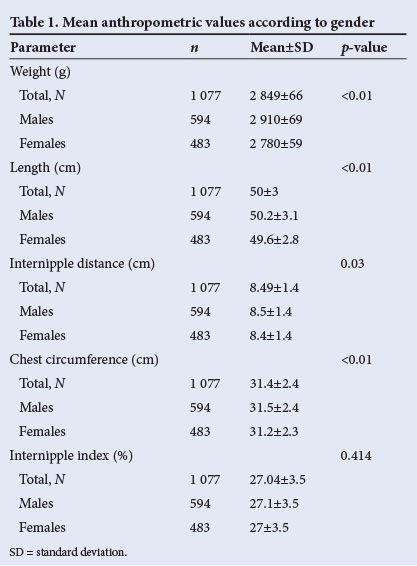
Mean weights, lengths, internipple distances, chest circumferences and internipple indices for the study group as a whole and for the males and females separately are set out in Table 1. Male infants had greater distances between the nipples and larger chest circumferences than females, and these differences were statistically significant (p=0.03 and p<0.01, respectively). Internipple indices were similar in males and females (p=0.414).
There was a significant positive correlation of weight (r=0.478), length (r=0.542), chest circumference (r=0.659) and gestational age (r=0.128) with internipple distance (p<0.05). When stepwise regression analysis was applied, all four parameters were found to be significant. The standardised coefficient showed that chest circumference was highly correlated with internipple distance after adjusting for weight, length and gestation (β=0.530), whereas weight was negatively associated with internipple distance after adjusting for length, chest circumference and gestational age. The overall adjusted R2 was 0.487, which explained the approximately 50% variance in internipple distance values.
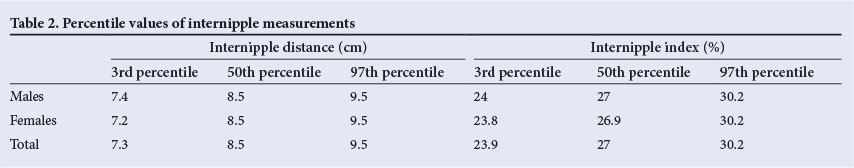
Percentile values for internipple distance and internipple index are given in Table 2. Internipple distances of 7.3 cm (7.4 cm for males and 7.2 cm for females), 8.5 cm and 9.5 cm corresponded to the 3rd, 50th and 97th percentiles, respectively. The internipple index was 24.0% for males and 23.8% for females at the 3rd percentile, 27.0% for males and 26.9% for females at the 50th percentile, and 30.2% in both genders at the 97th percentile.
Discussion
Abnormal position of the nipples is one of the many parameters that define dysmorphology. Widely spaced nipples are seen in several genetic disorders and other conditions, such as Turner syndrome,[1] and closely spaced nipples are associated with Jeune syndrome and cerebrocostomandibular syndrome.[3] According to Collins,[2] the subjective observation of wide nipple spacing is not sufficient, and should be confirmed by objective measurement. There are few studies on internipple distance and the internipple index in the early neonatal period. Available literature shows that these measurements vary.[3,6,9,11]
Internipple distance varies with gestational age and chronological age, but the internipple index, which is highest in the neonatal period, decreases until 2 - 4 years of age and then rises to remain almost constant after 18 years in males.[7,8,12] Few authors have suggested that an internipple index of >28% in children should be regarded as indicating widely spaced nipples.
Apart from race and ethnicity,[12] methods of measurement and instruments used can also lead to variations in internipple distance. To minimise error, the internipple distance and chest circumference should be measured as close as possible to the end of expiration; failure to do so may result in a falsely high chest circumference and a correspondingly low internipple index.
The mean internipple distance (8.5 cm) in our study was consistent with figures reported in the literature[9,12-15] (Table 3), and with a study on both term and preterm neonates that evaluated anthropometric measurements as a tool to assess gestation (Dewan et al. - unpublished data, 2012). However, Kulkarni and Rajendran[16] reported less distance between the nipples than that in our study. The mean internipple index in our study was higher than in the earlier studies, the 97th percentile for internipple index being 30.2%. This may be because our babies were relatively lean (although they all weighed >2 500 g at birth), as a smaller chest circumference increases the internipple index. According to our study, an internipple distance of 9.5 cm or an internipple index of >30% in the neonatal period indicates widely spaced nipples.
Internipple distance is directly proportional to gestational age and increases as the pregnancy continues. Kulkarni and Rajendran[16] studied internipple distance in newborn babies from 26 to 42 weeks of gestation and reported a mean internipple distance of 5.17 cm at 28 weeks compared with 7.51 cm at 41 weeks. Similar observations were made by Merlob,[3] Sivan et al.[11] and Fok et al.[14] Our study also found a positive correlation between gestational age and internipple distance, although we included only term newborns from 37 to 41 weeks' gestation, and the majority of our subjects were born at 38 weeks.
Internipple distance and the internipple index were significantly greater in male than female infants in our study, but other authors have not found these measurements to differ according to gender.[3,5,9,10,12] Chinese male infants had more distance between the nipples than females, but the difference was not statistically significant.[14]
Conclusions
The 50th percentiles for internipple distance and internipple index in the first week after birth were 8.5 cm and 27.0%, respectively, in term Indian newborn infants of appropriate weight for gestational age. There was a positive correlation between internipple distance and index and weight, length, chest circumference and gestational age.
Nipples situated >9.5 cm apart or with an internipple index >30% (>97th percentile) in the first week of life in term Indian infants may be considered widely spaced. Similarly, an internipple distance <7.3 cm and an internipple index <23.9% (<3rd percentile) may be considered to indicate narrowly spaced nipples.
Author contributions. MMAF conceived and designed the study, analysed the data and drafted the manuscript. PD collected data, helped in data analysis and contributed to writing the manuscript. The final manuscript was approved by both authors.
References
1. Jones KL. Smith's Recognizable Patterns of Human Malformation. 6th ed. Philadelphia: Elsevier Saunders, 2006:13, 76, 124, 652. [ Links ]
2. Collins E. The illusion of widely spaced nipples in the Noonan and the Turner syndromes. J Pediatr 1973;83(4):557-561. [http://dx.doi.org/10.1016/S0022-3476(73)80214-8] [ Links ]
3. Merlob P. Congenital malformations and developmental changes of breast: A neonatological view. J Pediatr Endocrinol Metab 2003;16(4):471-485. [http://dx.doi.org/10.1515/JPEM.2003.16.4.471] [ Links ]
4. Guihard-Costa AM, Menez F, Delezoide AL. Standards for dysmorphological diagnosis in human fetuses. Pediatr Dev Pathol 2003;6(5):427-434. [http://dx.doi.org/10.1007/s10024-003-1004-6] [ Links ]
5. Pelz L. The intermamillary index in children. Kinderarztl Prax 1972;40(6):257-258. [ Links ]
6. Mehes K, Kitzveger E. Inner canthal and intermamillary indices in the newborn infant. J Pediatr 1974;85(1):90-92. [http://dx.doi.org/10.1016/S0022-3476(74)80295-7] [ Links ]
7. Feingold M, Bossert WH. Normal values for selected physical parameters: An aid to syndrome delineation. Birth Defects Orig Artic Ser1974;10(13):1-16. [ Links ]
8. Hassan Z, Karna P, Dolanski EA. Intermamillary indices in premature infants. Am J Perinatol 1988;5(1):54-56. [http://dx.doi.org/10.1055/s-2007-999654] [ Links ]
9. Ejiwunmi AB, Okanlawon OA, Ojo OO. Interpupillary and internipple distances and ear lengths in Nigerian newborns. Ann Trop Paediatr 1984;4(2):103-106. [ Links ]
10. Ballard JL, Khoury JC, Wedig K, Wang L, Eilers-Walsman BL, Lipp R. New Ballard Score, expanded to include extremely premature infants. J Pediatr 1991;119(3):417-423. [http://dx.doi.org/10.1016/S0022-3476(05)82056-6] [ Links ]
11. Sivan Y, Merlob P, Reisner SH. Sternum length, torso length and internipple distance in newborn infants. Pediatrics 1983;72(4):523-525. [ Links ]
12. Leung AK, Kao CP, Sauve RS, Fang JH, Leong AG, Liu EK. Internipple distance and internipple index. J Natl Med Assoc 2004;96(8):1092-1096. [ Links ]
13. Chen H, Espiritu C, Casquejo C, Boriboon K, Woolley P Jr. Internipple distance in normal children from birth to 14 years and in children with Turner's, Noonan's, Down's and other aneuploidies. Growth 1974;38(4):421-426. [ Links ]
14. Fok T F, Hon K L, Wong E, et al.; Hong Kong Neonatal Measurement Working Group. Trunk anthropometry of Hong Kong Chinese infants. Early Hum Dev 2005;81(9):781-790. [http://dx.doi.org/10.1016/j.earlhumdev.2005.06.002] [ Links ]
15. Kapoor S, Bhushan S, Ghosh VB, Pandey RM, Kalaivani M. Normative data for anthropometric parameters used in delineation of dysmorphic features in North Indian children. Indian J Pediatr 2012;79(5):619-631. [http://dx.doi.org/10.1007/s12098-011-0572-0] [ Links ]
16. Kulkarni ML, Rajendran NK. Internipple distance in the newborns. Indian Pediatr 1992;29(5):619-620. [ Links ]
 Corresponding author: M M A Faridi (drmmafaridi@gmail.com; catchme852@yahoo.co.in)

