


Services on Demand
Article
 English (pdf)
 Article in xml format
 Article references
Indicators

Related links
-
 Cited by Google
 -
 Similars in Google
Share

 Permalink
SA Orthopaedic Journal
On-line version ISSN 2309-8309
Print version ISSN 1681-150X
SA orthop. j. vol.22 n.4 Centurion 2023
http://dx.doi.org/10.17159/2309-8309/2023/v22n4a3
HAND
Diagnostic accuracy of preoperative clinical examination in zone V flexor injuries
Emmanuel D OseiI, *; Mokgopo C SathekgaII; Philani NtombelaII; Abdirashid A AdenIII
IDepartment of Orthopaedic Surgery, University of the Witwatersrand, Helen Joseph Hospital, Johannesburg, South Africa; Orthopaedic Surgery, St. Martin's Catholic Hospital, Agroyesum, Ghana
IIDepartment of Orthopaedic Surgery, University of the Witwatersrand, Johannesburg, South Africa
IIIDepartment of Orthopaedic Surgery, University of the Witwatersrand, Helen Joseph & Rahima Moosa Hospital Complex; Netcare Garden City Hospital, Johannesburg, South Africa
ABSTRACT
BACKGROUND: Zone V flexor tendon injuries are devastating and may result in significant morbidity. There may be remarkable differences between preoperative clinical and the intraoperative findings of zone V flexor injuries. In this study, we assessed the demographics of patients presenting with zone V flexor injuries and the diagnostic accuracy of preoperative clinical examination.
METHODS: This was a prospective study of patients who presented with zone V flexor injuries at an emergency department of a tertiary academic hospital for a period of one year. The demographic data were analysed and the preoperative clinical examination findings were compared to the definitive intraoperative findings to assess the accuracy of the former. The one-sample test for proportion was used to assess if the difference between the two findings was statistically significant.
RESULTS: Zone V flexor injuries occurred predominantly in males under 40 years of age. Assault was the leading cause of these injuries. Alcohol intake was a significant factor for the causes of these injuries. Almost one-third (33%) of lacerated anatomical structures were missed and 91% of partially lacerated anatomical structures were inaccurately diagnosed. The mean number of errors of the clinical examination was 3.55; only 9% of the clinical examination had no error and 42% had four or more errors.
CONCLUSION: There was a significant difference between preoperative clinical examination and intraoperative findings and we recommend that all zone V flexor injuries extending beneath the subcutaneous tissue should be explored in theatre.
Level of evidence: Level 4.
Keywords: diagnostic accuracy, zone V flexor injuries, preoperative clinical examination
Introduction
The zone V flexor region is anatomically defined as the region from the proximal end of the carpal tunnel to the musculotendinous junction in the forearm. It is densely packed with 12 tendons (flexor carpi ulnaris [FCU], flexor carpi radialis [FCR], flexor pollicis longus [FPL], palmaris longus [PL], flexor digitorum superficialis [FDS] and flexor digitorum profundus [FDP] to the index, middle, ring and little fingers), three nerves (median nerve, ulnar nerve, superficial radial nerve) and two major arteries (ulnar and radial arteries) and their satellite veins.1 The tendons, nerves and arteries are vital to the meaningful function of the human wrist, fingers and the hand. The superficial location of the tendons and neurovascular structures in zone V flexor region with its widely exposed surface area makes it increasingly vulnerable for penetrating, accidental, homicidal and suicidal injuries.2,3
Clinical examination of hand injuries gives the operating surgeon an idea of the injured structures. Often surgeons discover a completely different set of intraoperative findings than the documented clinical examination findings. This poses a great challenge with regard to theatre time planning and utilisation, resulting in an increased number of cases being cancelled. In some instances, some injuries are missed in the emergency department and the patient is sent home only to come back with hand dysfunction.4
There is limited information on the diagnostic accuracy of the clinical examination of zone V flexor injuries. In our literature search, there was only one study that assessed the diagnostic accuracy of clinical examination in zone V flexor injuries carried out by Gibson et al.5 Most of the findings in their study were based on a retrospective review with its inherent design limitations.
Our prospective study sought to assess the accuracy of the preoperative clinical examination of zone V flexor injuries by the orthopaedic registrars in the emergency department and to analyse the demographic data of patients who presented with these injuries. It provides rich empirical data for surgeons on accuracy and reliability of preoperative clinical examination of zone V flexor injuries performed by orthopaedic registrars.
Methods
Ethics clearance was obtained. All patients aged 18 years and above with zone V open flexor injuries that presented to the emergency department of a tertiary academic hospital from 1 March 2018 to 30 April 2019 were enrolled into the study after an informed consent. The patients were interviewed and their demographic data documented on a questionnaire. In the emergency department, the patients were examined for each of the eleven tendons (excluding PL), three nerves and two arteries and assessed as being lacerated, partially torn or intact and documented on the clinical examination finding sheet of the questionnaire. After consent was obtained for exploration in theatre, the operating surgeon documented the definitive intraoperative findings of all the structures as being lacerated, partially lacerated or intact on the intraoperative findings sheet of the questionnaire.
The inclusion criteria for the study were patients with injuries in the zone V flexor region with laceration extending beneath the subcutaneous tissue and presenting within 24 hours after sustaining the injury. The exclusion criteria were patients with previous wound exploration, mental disability, Glasgow coma scale (GCS) of less than 15, crush injury, traumatic amputation, associated fractures, bites and inability to give an informed consent.
The raw data were entered on an Excel sheet and analysed using Stata version 15.0. Frequency, proportions and percentages were used to report and describe the demographics and clinical characteristics of the study. The mean and standard deviation for numerical variables were calculated.
Accuracy and inaccuracy of diagnosing a lacerated, partial tear, and an intact anatomical structure were assessed. This was done by comparing the preoperative clinical examination findings to definitive intraoperative findings. It was done for each of the 16 anatomical structures in all the injuries that were examined. Accurate diagnosis of an anatomical structure was computed when the findings of the preoperative clinical examination was exactly the same as the definitive intraoperative findings. Inaccurate diagnosis of an anatomical structure was computed when the preoperative clinical examination finding of an anatomical structure was different from the definitive intraoperative findings. The overall accuracy of preoperative clinical examination was assessed by computing the preoperative clinical examination findings of the 16 anatomical structures which were exactly the same as the definitive intraoperative findings in the 58 injuries examined and expressed as a percentage.
The one-sample test for proportion was used to assess if there was a statistically significant difference between intraoperative and preoperative clinical examination findings for lacerated, partial tear, intact and the total anatomical structures in the 58 injuries examined.
Results
During the study period, 61 patients with zone V flexor injuries were admitted at the casualty unit. Five patients were excluded from the study, of which two patients refused surgery after extensive counselling, two patients had a GCS of 14 and one patient was rushed to theatre before preoperative clinical examination was carried out. Fifty-six patients were enrolled in the study with 58 injuries as two patients had bilateral zone V flexor injuries. Forty-five (80%) out of the 56 patients were males with a 4.1:1 male to female ratio. The mean age was 32.2 years (18-67 years), with 84% (47/56) below the age of 40 years. Of the injuries sustained, 66% (38/58) and 60% (35/58) occurred on the right upper limb and the dominant hand of the patient respectively.
Figure 1 illustrates the employment status of the patients. Forty-four (79%) out of the 56 patients were actively working, engaged in contract-based or informal jobs or studying. Figure 2 illustrates the day of the week in which the injury occurred. Thirty-seven out of the 58 injuries (64%) occurred from Friday night to Sunday night.


Figure 3 demonstrates the mechanism of injury. Most of the injuries (52 out of 58) were assault, accidental and industrial injuries. Figure 4 describes alcohol intake by the patients when the injury occurred. The mechanism of injury among patients who had taken alcohol at the time of injury is described in Figure 5. Inferring from Figures 3 and 5, 13 out of the 23 patients (57%) who were assaulted and seven out of the 19 patients (37%) who had accidental injuries had taken alcohol.


The preoperative clinical examination findings and definitive intraoperative findings are summarised in Table I. FCU, ulnar nerve and ulnar artery (ulna triad) were the most frequently injured structures among the tendon, nerve and artery groups intraoperatively, respectively. Collectively, 268 anatomical structures were found to be lacerated intraoperatively while 238 anatomical structures were assessed clinically to be lacerated. Similarly, 55 anatomical structures were found to be partially lacerated intraoperatively while 49 anatomical structures were assessed clinically to be partially lacerated. Intraoperatively, there were more lacerated FDS tendons than lacerated FDP tendons; however, the number of FDP partial injuries was more than FDS partial injuries.

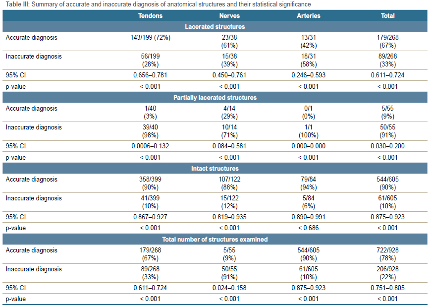
Twenty-eight out of the 58 zone V flexor injuries (48%) in this study could be classified as spaghetti wrist using the minimum definition as described by Kumar-Kempelingaiah et al.6 The number of structures injured in the spaghetti wrist ranged from three (one nerve, one artery and one tendon) to 14 (11 tendons, two nerves and one artery). Injuries to the ulnar triad structures were found in 21 out of the 58 injuries (36%) examined. There were ten cases of combined ulnar and median nerve injuries, and 31 out of the 58 injuries had complete laceration of either radial or ulnar arteries. Table II summarises the accurate and inaccurate diagnosis of each of the 16 anatomical structures in the 58 injuries examined. Table III summarises the accurate and inaccurate diagnosis of the anatomical structures in terms of tendons, nerves and arteries and their statistical significance.
Accurate and inaccurate diagnosis of lacerated structures
FDS to the middle finger had the highest rate of inaccurate (missed) diagnosis of lacerated structures, while FDP to the index finger had the lowest rate of missed diagnosis. There was a higher rate of missed diagnosis of total lacerated FDS tendons than total lacerated FDP tendons. A hundred per cent (100%), 50% and 20% of lacerated superficial radial nerve, median nerve and ulnar nerve injuries respectively were missed on preoperative clinical examination. The arteries had the highest rate of missed diagnosis of lacerated structures (58%). The difference between the intraoperative and preoperative examination findings of lacerated tendons, nerves, arteries and total lacerated anatomical structures was statistically significant.
Accurate and inaccurate diagnosis of partially injured structures
The overall rate of inaccuracy of diagnosing partial lacerations was very high (91%). Sixty-four per cent (35/55) of the partially lacerated structures were assessed preoperatively as intact. The difference between the intraoperative and preoperative examination findings of partially lacerated structures was statistically significant.
Accurate and inaccurate diagnosis of intact structures
The differences between the intraoperative and preoperative findings of intact tendons, nerves and total intact anatomical structures were statistically significant; however, that of intact arteries was not statistically significant. The rate of inaccurate diagnosis of intact FDS tendons was higher than the inaccurate diagnosis of intact FDP tendons.
Accurate and inaccurate diagnosis of anatomical structures examined
FPL had the highest and FCU the lowest rate of accuracy of clinical examination respectively. The accuracy of the clinical examination of FDP was higher than FDS. Superficial radial nerve and ulnar nerve had the highest and lowest rates of accuracy of clinical examination respectively among the three nerves. Radial artery had a higher rate of accuracy of clinical examination than ulnar artery. The accuracy of clinical examination of tendons, nerve and arteries were similar. The differences between the intraoperative and preoperative examination findings of all the tendons, nerves and arteries examined were statistically significant. The difference between the intraoperative and preoperative findings of the anatomical structures examined was statistically significant.
Number of errors per clinical examination
Table IV describes the total number of errors in each injury examined. The number of errors per each examination in the 58 injuries ranged from zero to eight. The mean number of errors was 3.55. Only five out of 58 (9%) preoperative clinical examinations had no error. Forty-one per cent of the number of injuries examined had four or more errors.

Discussion
In our study, zone V flexor injuries occurred predominantly in males and patients below 40 years of age. Assault was the leading cause of these injuries in our study. This finding could be explained by the high crime rate in South Africa.7,8 Accidental injury was the second commonest mechanism of injury. There were three cases of self-inflicted injuries following an anger burst from frustration. There were also three cases of parasuicide, all of which occurred in females.
Thirty-nine per cent (39%) of the patients admitted that they had taken alcohol when the injury occurred. This finding is comparable to the finding of Kabak et al. where 42.9% of the patients in their study were intoxicated when they sustained the injuries.3 Assault and accidental injuries were the main mechanisms of injury among patients who had taken alcohol and sustained zone V flexor injuries. Excessive alcohol intake was a significant association among patients whose mechanisms of injury were assault and accidental injuries.
From our study, 64% of the zone V flexor injuries occurred from Friday night to Sunday night. This is in keeping with a study by Schuurman et al. where 66.7% and 60% of intentional and unintentional injuries in Cape Town, South Africa, occurred during weekends.9
From our study, tendons were injured more commonly than nerves and arteries; FCU tendon was injured more frequently than ulnar nerve and ulnar artery among the ulnar triad structures; FDS tendons were also more frequently injured than FDP tendons. This is due to the superficial location of the former as opposed to the latter.
The most common combined injury pattern observed in this study was ulnar triad injuries in keeping with various studies.10-13 This is due to the proximity of these anatomical structures, their superficial and ulna location in the wrist. In cases of assault, the ulnar side of the forearm is used for protection, hence more easily lacerated.
Lacerated FDS tendon injuries were frequently missed more than FDP tendon injuries. Accurate diagnosis of FDS tendon injuries requires adequate knowledge about the functional anatomy of the FDS and FDP tendons and clinical examination skills. FDS flexes the proximal interphalangeal joint (PIPJ). The PIPJ is also flexed by FDP. In order to accurately examine FDS clinically, one needs to eliminate the effect of FDP. This is done by fully extending the other unexamined fingers and then asking the patient to flex the examined finger. To be sure that the effect of FDP has been eliminated, the distal interphalangeal joint (DIPJ) of the examined finger should not be seen flexing during its examination. However, this may be difficult to carry out by a freshly injured patient who is in pain.
The missed diagnosis of FCR and FCU was very high. This could be attributed to lack of knowledge of the proper clinical skills of these anatomical structures by the examining doctors. Both FCU and FCR are flexors of the wrist. In the presence of injury to either of these tendons, the intact FCU or FCR can flex the wrist though not with full strength. FCR and FCU needs to be examined by flexing the wrist against resistance to radial and ulna deviation respectively. If either of these tendons is not thoroughly examined in this fashion, their isolated injuries will be missed on clinical examination.
The high rate of missed diagnosis of lacerated nerves can be explained by the hypothesis put up by Lynch et al. where a nerve impulse can jump a transection gap until 72 hours when Wallerian degeneration occurs.14
The high rate of missed diagnosis of lacerated arteries could be due to assessment of the arteries by palpation only and not performing Allen's test. When Allen's test is not carried out to assess arterial injury, the rate of missed diagnosis could be very high due to good and rich backflow through the palmar arches.5,15
The rate of accurate diagnosis of partially injured anatomical structures in this study was very low (9%). This low rate was expected because of residual function of the partially injured structures.16,17 In this study, partial injuries of two median nerves, two ulnar nerves and an FDP tendon to the middle finger were diagnosed by clinical examination as compared to none in Patel et al. study.18
The overall rate of inaccurate diagnosis of intact anatomical structures was low (10%). Among the intact anatomical structures, nerves had the highest rate of inaccurate diagnosis. This could be attributed to the fact that nerves are very sensitive. When a nerve is recently contused, it has impaired sensation and motor function during the early hours and days and can be easily diagnosed clinically as a laceration or partial tear.
FDS to the little finger had the highest rate of overdiagnosis among the tendons. FDS to the little finger has the least strength among all the FDS and FDP. So even in the presence of an intact FDS to the little finger, a clinician can easily assess it as a partial tear.
Gibson et al. explains that some clinicians are biased during their clinical examination of anatomical structures.5 This is due to their knowledge of the proximity of some anatomical structures and not objectively assessing each structure individually and on merit. For example, when examining the ulnar triad structures, an examiner will mark FCU as lacerated without examining it when he or she finds the ulnar nerve injured. This introduces observer bias and increases the rate of overdiagnosis. The high overdiagnosis rate of FCU can be attributed to this.
Patient factors such as pain, anxiety, drunken state and lack of cooperation are also contributing factors to inaccurate diagnosis of intact structures. The patient does not carry out the movement of the fingers and other instructions well. This will be interpreted by the examining doctor as a malfunctioning (lacerated or partial tear) anatomical structure.
The accuracy of examining tendons, nerves and arteries was much the same, ranging from 78% to 79%. The difference in accuracy occurred when the anatomical structures were grouped into lacerated, partial injuries and intact structures. Generally, the rate of accuracy of clinical examination of FDP tendons was higher than FDS tendons due to the relatively high level of skills required in the examination of FDS tendons. The high rate of inaccurate diagnosis of FCU was due to the high rate of inaccurate diagnosis of its lacerated and intact tendons.
The number of errors per each clinical examination ranged from zero to eight. This is dependent on the nature of the injury, the clinical skills of the examining doctor and the patient being examined. About 41% of the 58 clinical examinations had four or more errors per examination. Approximately 9% of the examinations had no error on clinical examination.
In order to improve the accuracy of clinical examination of zone V flexor injuries, the examining doctor needs to familiarise him or herself with the anatomy and proper clinical examination of the tendons, nerves and arteries in zone V flexor region. Allen's test should always be used in assessing arterial injuries. Each anatomical structure should be assessed individually and objectively regardless of the pattern of injury, and assumptions should not be made. The examining doctor needs to be thorough and meticulous in his or her examination. The patient should be pain-free, calm and sober with a high level of cooperation. It must, however, be stated that errors in clinical examination of all zone V flexor injuries are very difficult to reduce to zero. However, the error margin can be reduced in some injuries.
Conclusion
There was a statistically significant difference in the clinical examination of zone V flexor injuries in this study, especially examination of lacerated and partially injured structures. There should, therefore, be a high index of suspicion and exploration of zone V flexor injuries to avoid missed injuries and the consequent morbidities and litigations associated therewith.
Acknowledgements
We acknowledge and thank Charles Adjei Manful and Kwabena Asare for their extensive input into the data analysis.
Ethics statement
The authors declare that this submission is in accordance with the principles laid down by the Responsible Research Publication Position Statements as developed at the 2nd World Conference on Research Integrity in Singapore, 2010. Prior to the commencement of the study, hospital permission was obtained from the CEO of Chris Hani Baragwanath Academic Hospital and ethics clearance from Human Research Ethics Committee (Medical) of the University of the Witwatersrand, ethics clearance certificate number M1711120. All procedures were in accordance with the ethical standards of the responsible committee on human experimentation (institutional and national) and with the Helsinki Declaration of 1975, as revised in 2008. Informed written consent was obtained from all patients for being included in the study. Consent was obtained from patients for the use of clinical photographs and these images were adequately anonymised.
Declaration
The authors declare authorship of this article and that they have followed sound scientific research practice. This research is original and does not transgress plagiarism policies.
Author contributions
EDO: study conceptualisation, study design, data capture, data analysis, first draft, subsequent draft preparation, manuscript preparation and revision
MCS: study conceptualisation, study design, data capture, data analysis, manuscript draft, preparation and revision
PN: study conceptualisation, study design, data capture, data analysis, manuscript, preparation and revision
AAA: study conceptualisation, study design, data analysis, manuscript draft, preparation and revision
ORCID
Osei ED https://orcid.org/0000-0003-4544-7749
Sathekga MC https://orcid.org/0000-0002-8532-5001
Ntombela P https://orcid.org/0000-0002-4301-1899
Aden AA https://orcid.org/0000-0001-5589-2972
References
1. Ozinko M, Otei O, Ekpo R, Isiwele E. Spaghetti wrist injury: epidemiology and management in Calabar, Southern Nigeria. IOSR J Dent Med Sci. 2017;16:87-91. [ Links ]
2. Yüksel F, Peker F, Açikel C, ÇelIkoz B. Secondhand management of "spaghetti wrist": do not hesitate to explore. Ann Plast Surg. 2002;49:500-505. [ Links ]
3. Kabak S, Halici M, Baktir A, et al. Results of treatment of the extensive volar wrist lacerations: 'the spaghetti wrist.' Eur J Emerg Med. 2002;9:71-76. [ Links ]
4. Morrison CM, Thompson NW, Herbert KJ, Brennen MD. Missed injuries in the acutely traumatised hand. Ulster Med J. 2003;72:22-25. [ Links ]
5. Gibson T, Schnall S, Ashley E, Stevanovic M. Accuracy of the preoperative examination in Zone 5 wrist lacerations. Clin Orthop Relat Res. 1999 Aug;(365):104-110. [ Links ]
6. Kumar-Kempelingaiah A, Naraynsingh V, Ali TF. (2003). Epidemiology and classification of extensive volar wrist lacerations: the 'spaghetti wrist.' The Internet Journal of Third World Medicine. 2003; \1(2):1-9. [ Links ]
7. Matzopoulos R, Prinsloo M, Wyk VP, et al. Injury-related mortality in South Africa: a retrospective descriptive study of postmortem investigations. Bull World Health Organ. 2015;93:303-313. [ Links ]
8. Jabar A, Matzopoulos R. Violence and injury observatories: Reducing the burden of injury in high-risk communities. South Afr. Crime Q. 2017;59:47-57. [ Links ]
9. Schuurman N, Cinnamon J, Walker BB, et al. (2015). Intentional injury and violence in Cape Town, South Africa: an epidemiological analysis of trauma admissions data. Glob Health Action. 2015 Jun 12;8:27016. [ Links ]
10. Noaman HH. Management and functional outcomes of combined injuries of flexor tendons, nerves, and vessels at the wrist. Microsurgery 2007;27:536-43. [ Links ]
11. Vaughn CJ, Raghavan SS, Hansen SL, Terry MJ. Obstacles to the care of patients with multicomponent volar wrist lacerations at a county hospital. Ann Plast Surg. 2016;76:S238-S240. [ Links ]
12. Chin G, Weinzweig N, Mead M, Gonzalez M. 'Spaghetti wrist': management and results. Plast Reconstr Surg. 1998;102:96-102. [ Links ]
13. Yazdanshenas H, Naeeni A, Ashouri A, et al. Treatment and postsurgery functional outcome of spaghetti wrist. J Hand Microsurg. 2016;8(3):127-33. [ Links ]
14. Lynch G, Quinlan D. Jump function following nerve division. Br J Plast Surg. 1986;39:364-66. [ Links ]
15. Nassab R, Kok K, Constantinides J, Rajaratnam V. The diagnostic accuracy of clinical examination in hand lacerations. Int J Surg. 2007;5:105-108. [ Links ]
16. Ashfaq F, Beg MSA, Sadiq M, Ghani S. Accuracy of physical examination in glass lacerations to hand and wrist. Med Channel. 2013;19:17-20. [ Links ]
17. Gupta A, Kleinert HE. Evaluating the injured hand. Hand Clin. 1993;9: 195-212. [ Links ]
18. Patel J, Coull R, Harris PA, Percival NJ. Hand lacerations: An audit of clinical examination. J Hand Surg. 1998;23:482-84. [ Links ]
Received: March 2023
Accepted: July 2023
Published: November 2023
* Corresponding author: drdwomoh@yahoo.com
Editor: Dr Duncan McGuire, University of Cape Town, Cape Town, South Africa
Funding: No funding was received for this study.
Conflict of interest: The authors declare they have no conflicts of interest that are directly or indirectly related to the research.

