


Servicios Personalizados
Articulo
 Inglés (pdf)
 Articulo en XML
 Referencias del artículo
Indicadores

Links relacionados
-
 Citado por Google
 -
 Similares en Google
Compartir

 Permalink
SA Orthopaedic Journal
versión On-line ISSN 2309-8309
versión impresa ISSN 1681-150X
SA orthop. j. vol.21 no.2 Centurion 2022
http://dx.doi.org/10.17159/2309-8309/2022/v21n2a5
ORTHOPAEDIC ONCOLOGY AND INFECTIONS
Modular prosthetic reconstruction for primary bone tumours of the distal tibia in ten patients
Walid MuglaI; Henrik CF BauerII; Jonathan VogelIII; Keith V HoskingIV; Neil CampbellV; Thomas L HiltonVI, *
ISarcoma Unit, Groote Schuur Hospital; Faculty of Medicine and Health Sciences, Department of Surgery, Division of Orthopaedics, University of Cape Town, Cape Town, South Africa
IIDepartment of Molecular Medicine and Surgery, Karolinska Institute, Stockholm, Sweden; visiting professor in the Faculty of Medicine and Health Sciences, Department of Surgery, Division of Orthopaedics, University of Cape Town, Cape Town, South Africa
IIIFaculty of Medicine and Health Sciences, Department of Surgery, Division of Orthopaedics, University of Cape Town, Cape Town, South Africa
IVVincent Pallotti Life Orthopaedic Hospital, Cape Town; Faculty of Medicine and Health Sciences, Department of Surgery, Division of Orthopaedics, University of Cape Town, Cape Town, South Africa
VLRS Implants, Cape Town, South Africa
VIGroote Schuur Hospital and Life Vincent Pallotti Orthopaedic Hospital, Cape Town; Faculty of Medicine and Health Sciences, Department of Surgery, Division of Orthopaedics, University of Cape Town, Cape Town, South Africa
ABSTRACT
BACKGROUND: Below-knee amputation (BKA) is the safest treatment for benign aggressive and malignant bone tumours of the distal tibia, yielding good oncological and functional results. However, in selected patients where limb salvage is feasible and amputation unacceptable to the patient, limb salvage using a distal tibial replacement (DTR) can be considered. This study aims to present the oncological and functional results of the use of the latter treatment method in our unit.
METHODS: A retrospective folder review was performed for all ten patients who received a modular DTR between 1 January 2005 and 31 January 2019 for a primary bone tumour, either benign aggressive or malignant. Six were female and the mean age was 31 (12-75) years. There were five patients with giant cell tumour of bone, four with osteosarcoma and one with a low-grade chondrosarcoma. The patients with osteosarcoma had neoadjuvant chemotherapy before surgery. Function was assessed by the Musculoskeletal Tumor Society (MSTS) score.
RESULTS: Two patients had local recurrence treated with a BKA and one other patient died of metastases three years postoperatively. At a mean follow-up of three years, the remaining eight patients had a mean MSTS score of 83% (67-93%). There were no radiological signs of loosening, and no revision surgeries.
CONCLUSION: Endoprosthetic replacement of the distal tibia for primary bone tumours can be a safe treatment option in very selected cases.
Level of evidence: Level 4
Keywords: distal tibia, endoprosthetic replacement, osteosarcoma, giant cell tumour, limb salvage, amputation
Introduction
Limb-sparing surgery for primary bone tumours of the distal tibia is fraught with difficulties due to the paucity of soft tissue coverage and difficulties in creating a durable fixation of the prosthetic components.1 Wide surgical margins and acceptable function of the ankle joint can seldom be achieved.1,2 Therefore, below-knee amputation (BKA) is the surgical method of choice. While oncologically safe, it also provides excellent function with the ever-improving external prosthetics.3 In selected cases where a wide surgical margin is possible and amputation unacceptable to the patient, limb salvage may be attempted.3 With the advent of additive manufacturing and improvements in polyethylene components and manufacturing, distal tibial replacement (DTR) design has provided solutions to previous problems and reduced implant cost by creating an 'off-the-shelf' prosthesis rather than an expensive and time-consuming custom prosthesis.3,4 The aims of this study are to present the oncological and functional assessment of ten patients treated with resection of the distal tibia and reconstruction with a DTR. Our objectives are to do this through a retrospective folder review of all patients treated in this manner in our unit.
Patients and methods
A medical record and image review was performed of ten patients who underwent a DTR between 1 January 2005 and 31 January 2019 for Enneking benign aggressive or malignant primary bone tumour.5 No patient was excluded due to missing data or lost to follow-up.
Data capture included patient demographics, procedural complications, revision procedures, local recurrence, tumour metastases and death. The histological diagnosis was established by core needle biopsy using a Jamshidi™ 12G needle.6 Functional outcome was assessed using the Musculoskeletal Tumor Society (MSTS) score. The MSTS scoring system is a specific scoring system to determine the physical and mental health of patients with extremity sarcoma. The system assigns numerical values (0-5) for six categories. A numerical score and per cent rating is calculated to allow for comparison of results.7 Recommendations regarding amputation and limb salvage were made at multidisciplinary team conferences.
There were six females and four males, with a mean age of 31 (12-75) years. Five patients had a giant cell tumour (GCT) of bone (Figure 1), four an osteosarcoma, and one a low-grade chondrosarcoma. The four osteosarcoma patients had neoadjuvant chemotherapy, and none of the GCT patients had preoperative demosumab.

Description of the prosthesis
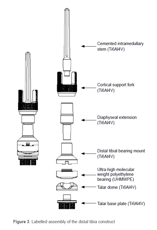
The distal tibia replacement used in this study is an LRS Distal Tibia Replacement (www.lrs.com). It is a modular reconstruction system that allows for different resection lengths of the distal tibia in 10 mm increments. The implant is not side specific.
The talar side of the prosthesis creates a metal (titanium Ti6Al4V) talar dome. It is made up of two parts: the talar base plate, and the talar dome. The base plate is 3D printed in titanium, incorporating a trabecular mesh structure for bone ingrowth. It is based on cementless fixation. There are three 8 mm pegs which are impacted into the talus. All surfaces in contact with the talus contain the trabecular mesh structure to encourage bone ingrowth. The talar dome is attached to the base plate by a morse taper. The dome is titanium with a titanium oxide ceramic surface. It has a 'saddle' shape similar to that of a native talus, to provide tibial tracking and a degree of varus-valgus support. The orientation of the dome can be adjusted prior to impaction onto the talar base plate.
The talar dome articulates with an ultra-high molecular weight polyethylene (UHMWPE) bearing to replicate the natural range of motion of the ankle. The prosthesis is not constrained, except for the congruent 'saddle' fit of the talar dome and the polyethylene bearing surface. The bearing sizing is available in 3 mm increments to allow for balancing of the implant and soft tissues. The bearing is impacted onto a titanium mount which then attaches to diaphyseal extensions whose number and length are matched to fill the defect left by the resection.
The implant is secured into the tibia by a cemented titanium intramedullary stem, with additional fixation provided by a trabecular 3D-printed extra-cortical fork to limit rotation of the implant in the bone and encourage bone ingrowth.
Surgical technique
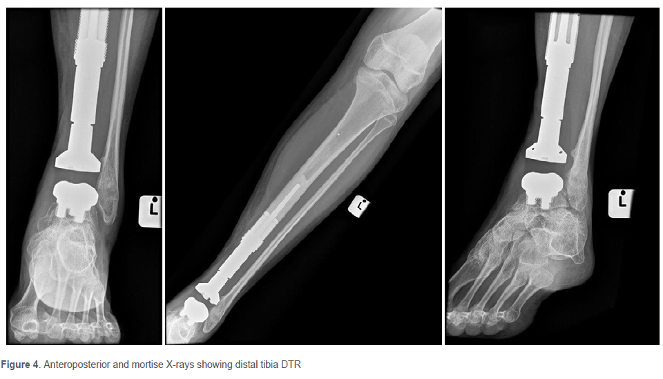
The patient is positioned supine, and an above-knee tourniquet is applied. An anteromedial approach is performed to access the distal tibia and ankle joint (Figure 2). The biopsy site is included in the resected specimen. The tendons of tibialis anterior and extensor digitorum communis along with the neurovascular bundle are dissected away from the tumour, and the deltoid ligament, ankle syndesmotic ligament and capsule are cut. This allows for the distal tibia to be delivered from the leg. The remaining soft tissue is dissected off the tibia. The tibial diaphysis may be transected proximally before the ankle ligaments are cut to allow for easier manipulation of the distal tibia. Once the specimen is removed, it is placed nearby to assist with measurement of the length of prosthesis to be inserted. The talus is then cut transversely with an oscillating saw. A high-speed burr and a guide are used to create three peg holes which will accept the uncemented talar baseplate and titanium pegs (Figure 3). The articulation of the prosthetic ankle joint consists of the titanium tibial dome and polyethylene bearing. The distal tibia body and appropriately sized extra-cortical fork and diaphyseal extensions are attached to an intramedullary stem which is then cemented into the proximal tibia after sequential reaming of the proximal tibia shaft and trialling for length. Care must be taken during reduction not to fracture the fibula which is left intact and provides lateral support to the construct. The ankle is immobilised in a below-knee backslab for two weeks. The patient is then placed into a moon boot or below-knee plaster for a further four weeks (see Figure 4 for postoperative X-ray). Thereafter, the patient begins physiotherapy consisting of graduated weight bearing and active and passive ankle dorsiflexion and plantar flexion.
Results
One patient died three years after treatment due to metastatic disease. Two patients had local recurrence, one of whom also had a deep infection, and both were treated with a BKA. After amputation, both patients remain disease free (Table I).
Functional outcome and complications
After a mean follow-up of 43 months (6-116), of the eight patients who did not undergo a BKA, the mean MSTS score was 83% (70-93). Two patients complained of mild ongoing pain around their lateral malleolus and had an antalgic gait on examination. There were no radiological signs of loosening, and no revision surgeries. They scored modestly in their MSTS assessment which has grades, none, modest and severe. The patients' pain was controlled with oral analgesia only.
Discussion
This retrospective study of ten patients with primary bone tumours of the distal tibia shows that acceptable oncological and functional results can be achieved in the short to medium period of follow-up. Nevertheless, BKA will remain the treatment of choice, providing safe oncological margin and excellent function.3
In South Africa, the management of primary bone tumours of the appendicular skeleton with limb ablation is often met with strong opposition due to cultural and traditional beliefs. These usually preclude amputation, often with increased morbidity and mortality of the patient.8 Brown et al. described the challenges associated with cross-cultural communication in this regard, and highlighted the family-centred decision-making unit, which often refuses a limb ablation.8 In these circumstances, an alternative treatment, potentially with higher oncological risks, needs to be considered to prevent morbidity and possible mortality that may result from rejection of medical treatment. We, therefore, propose that in South Africa, and many other countries across the African continent, an attempt at limb-sparing surgery and distal tibial replacement may be considered.
In resource-limited countries like South Africa, BKA is often recommended as it is supposedly cheaper than megaprosthetic replacement, and also minimises complications and repeat surgery. However, in these countries adequate external prosthetics cannot be assured during the patient's whole life span. Grimer et al. have also showed that in the long run, limb-sparing surgery, in general, is cost effective when compared to amputations due to the accrued cost of repair and replacement of artificial limbs.9 Furthermore, with modular systems of megaprosthetics, as reported here, unit costs should come down compared to custom-made implants.
There are only a few reports of DTR in primary bone tumours. Interestingly, none of the reports have more than six patients and all are at least ten years old.1,2,10,11 Similar to our study, they report a good functional outcome, reasonable complication rates and prosthesis longevity (Table II). Infection and recurrence were the most common causes of secondary amputation. Mechanical failure was reported, whereas we did not have any cases of mechanical failure in our series.
In our series of ten patients, two were amputated because of tumour recurrence and infection. For comparison, the final amputation rate after limb-sparing surgery for tumours of the proximal tibia is around 10%.12 In the proximal tibia, there are similar problems to the distal tibia of soft tissue coverage and restoring active joint function. The reason why amputation is seldom the procedure of choice for the proximal tibia is probably that a knee disarticulation or through-thigh amputation is considered more debilitating than a below-knee amputation.
The most common mechanical complication of ankle joint replacement is aseptic loosening of the talar tray.11 We had no cases of mechanical loosening at final follow-up. Abudu et al. and Shekkeris et al. both described loosening of the tibial baseplate in one patient each, and Lee et al. reported talar collapse in one.1,3,13 The uncemented, grown titanium design of the implant may prove to reduce the risk of talar prosthetic complications but the follow-up and number of patients is too small to be conclusive.10,11
Future research is needed to determine how this procedure can be of benefit in those instances where patients refuse amputation at any cost for cultural reasons but will accept limb-sparing surgery. This is difficult due to the small number of patients that may have this procedure and a national and international sarcoma registry would assist in providing more data on the subject. Engagement with cultural leaders would also help with earlier presentation of these patients to sarcoma centres and allow limb-sparing surgery.
Conclusion
Reconstruction of the distal tibia after resection for primary bone tumours with a distal tibial megaprosthesis yields good functional results with a high MSTS score and acceptable oncological outcomes with only a 20% local recurrence rate in the short to medium term. Therefore, this procedure can be considered as an alternative to limb ablation in selected cases.
Ethics statement
The authors declare that this submission is in accordance with the principles laid down by the Responsible Research Publication Position Statements as developed at the 2nd World Conference on Research Integrity in Singapore, 2010. Ethical approval number HREC 734/2019. For this retrospective study, formal consent was not required.
Declaration
The authors declare authorship of this article and that they have followed sound scientific research practice. This research is original and does not transgress plagiarism policies.
Author contributions
WM: study conceptualisation, study design, data capture, state patients' follow-up and scoring, first draft preparation, manuscript preparation HCFB: data analysis, manuscript preparation
JV: data capture, first draft preparation
KVH: private patients' data capture and score
NC: description of the prosthesis and pictures
TLH: involved in all aspects of this article
ORCID
Mugla W https://orcid.org/0000-0002-2961-7296
Bauer HCF https://orcid.org/0000-0002-3557-0252
Vogel J https://orcid.org/0000-0003-1156-6168
Hosklng KV https://orcid.org/0000-0002-3557-0252
Hilton TL https://orcid.org/0000-0002-6178-5062
References
1. Abudu A, Grimer RJ, Tillman RM, Carter SR. Endoprosthetlc replacement of the distal tibia and ankle joint for aggressive bone tumours. Int Orthop. 1999;23(5):291-94. https://doi.org/10.1007/s002640050374. [ Links ]
2. Natarajan MV, Annamalai K, Williams S, et al. Limb salvage in distal tibial osteosarcoma using a custom mega prosthesis. Int Orthop. 2000;24(5):282-84. https://doi.org/10.1007/s002640000172. [ Links ]
3. Shekkeris AS, Hanna SA, Sewell MD, et al. Endoprosthetic reconstruction of the distal tibia and ankle joint after resection of primary bone tumours. J Bone Joint Surg Br. 2009;91(10):1378-82. https://doi.org/10.1302/0301-620x.91b10.22643. [ Links ]
4. Hilton T, Campbell N, Hosking K. Additive manufacturing in orthopaedics: clinical implications. SA Orthop J. 2017;16(2):63-67. [ Links ]
5. Enneking WF, Spanier SS, Goodman MA. A system for the surgical staging of musculoskeletal sarcoma. Clin Orthop Relat Res. 2003;(415):4-18. https://doi.org/10.1097/01.blo.0000093891.12372.0f. [ Links ]
6. Kundu ZS. Classification, imaging, biopsy and staging of osteosarcoma. Indian J Orthop. 2014;48(3):238-46. https://doi.org/10.4103/0019-5413.132491. [ Links ]
7. Enneking WF, Dunham W, Gebhardt MC, et al. A system for the functional evaluation of reconstructive procedures after surgical treatment of tumors of the musculoskeletal system. Clin Orthop Relat Res. 1993;(286):241-46. [ Links ]
8. Brown O, Goliath V, Van Rooyen R, et al. Communicating about prognosis with regard to osteosarcoma in a South African cross-cultural clinical setting: strategies and challenges. SA Orthop J. 2019;18(4):46-51. [ Links ]
9. Grimer RJ, Carter SR, Pynsent PB. The cost-effectiveness of limb salvage for bone tumours. J Bone Joint Surg Br. 1997;79(4):558-61. https://doi.org/10.1302/0301-620x.79b4.7687. [ Links ]
10. Harris N. Total ankle arthroplasty. Pract Proced Elect Orthop Surg Pelvis Low Extrem. 2013;6(8):269-72. [ Links ]
11. Gross CE, Palanca AA, DeOrio JK. Design rationale for total ankle arthroplasty systems: an update. J Am Acad Orthop Surg. 2018;26(10):353-59. https://doi.org/10.5435/jaaos-d-16-00715. [ Links ]
12. Summers SH, Zachwieja EC, Butler AJ, et al. Proximal tibial reconstruction after tumor resection: A systematic review of the literature. JBJS Rev. 2019;7(7):e1. https://doi.org/10.2106/jbjs.rvw.18.00146. [ Links ]
13. Lee SH, Kim HS, Park YB, et al. Prosthetic reconstruction for tumours of the distal tibia and fibula. J Bone Joint Surg Br. 1999;81(5):803-807. https://doi.org/10.1302/0301-620x.81b5.9588. [ Links ]
Received: May 2021
Accepted: November 2021
Published: May 2022
* Corresponding author: thomas@drthilton.com
Editor: Prof. Theo le Roux, University of Pretoria, Pretoria, South Africa
Funding: No funding was received for this study.
Conflict of interest: Authors KVH and TLH are consultants for LRS. NC is the biomechanical engineer and managing director at LRS Implants. NC provided information based solely on the manufacturing of the implant. The remaining authors declare that they have no conflicts of interest that are directly or indirectly related to this research.

