


Servicios Personalizados
Articulo
 Inglés (pdf)
 Articulo en XML
 Referencias del artículo
Indicadores

Links relacionados
-
 Citado por Google
 -
 Similares en Google
Compartir

 Permalink
SA Orthopaedic Journal
versión On-line ISSN 2309-8309
versión impresa ISSN 1681-150X
SA orthop. j. vol.16 no.3 Centurion ago./sep. 2017
http://dx.doi.org/10.17159/2309-8309/2017/v16n3a6
TRAUMA
Intramedullary nailing of subtrochanteric femur fractures caused by low velocity gunshots
S SwanepoelI; D ChiversII; W LeongIII; M LaubscherIV; G McCollumV; S MaqungoVI
IMBChB(Pret), Registrar, Orthopaedic Surgery, Groote Schuur Hospital, University of Cape Town
IIMBBCh(Wits), FC Orth(SA), MMed Ortho(UCT), Consultant Orthopaedic Surgeon, Port Shepstone Regional Hospital
IIIMBChB(UKZN), Medical Officer, Hong Kong
IVMBChB(UFS), Dip PEC, FC Orth(SA), MMed Ortho(UCT), Consultant Orthopaedic Surgeon, Groote Schuur Hospital
VMBCI-ib(uCT), Dip Pec(SA), MMed Ortho(UCT), FC Orth(SA), Consultant Orthopaedic Surgeon, Groote Schuur Hospital
VIMBChB(Natal), MMed Ortho(UCT), FC Orth(SA), Consultant Orthopaedic Surgeon, Professor and Head of the Orthopaedic Trauma Service, Division of Orthopaedic Surgery, University of Cape Town, Groote Schuur Hospital
ABSTRACT
BACKGROUND: Subtrochanteric femur fractures remain challenging injuries to treat. There is paucity of literature evaluating their outcomes and complications following low-velocity civilian gunshots. The purpose of this study is to evaluate the results of intramedullary nailing of subtrochanteric femur fractures secondary to low-velocity gunshots.
METHODS: A retrospective review of clinical and radiological data was performed on all patients who sustained subtrochanteric femur fractures (AO type 32C3) caused by low-velocity civilian gunshots treated at a single institution between March 2008 and December 2014. Data was analysed to determine the time to union, post-operative complication rates and patient outcomes. Radiographic evidence of healing was defined as bridging callus on three of four cortices on two orthogonal views.
RESULTS: Fifty-one patients (48 men and two women) were identified. Mean age was 28 years (range 16-50 years). The predominant method of fixation was a cephalomedullary nail in 43 patients (84%), and eight patients had locking into the lesser trochanter. Thirty patients with a mean follow-up period of 3.3 weeks (0-6 weeks) were lost to follow-up. Twenty-one patients had adequate radiographic and clinical follow-up data suitable for analysis. The mean follow-up period was 24.8 weeks (range 2-40 weeks) for this group. The average time to union was 17.2 weeks (7-40 weeks). Seven fractures (37%) had delayed union. None of the patients required additional surgery. Union was achieved in all cases and no implant failure occurred.
CONCLUSION: This study demonstrates that intramedullary nail fixation of subtrochanteric femur fractures caused by low-velocity civilian gunshots is an acceptable option for the treatment of these injuries with good union rates and low complication rates.
Key words: femur fracture, subtrochanteric, civilian gunshot wound
Introduction
Subtrochanteric femur fractures caused by low-velocity gunshot wounds present a considerable challenge to the orthopaedic surgeon.
Gunshot injuries represent an increasing proportion of admissions to trauma centres with up to 127 000 non-fatal firearm injuries treated each year at South African state hospitals. A study conducted at Groote Schuur Hospital found that almost 42% of patients presenting with firearm-related injuries required surgery.1
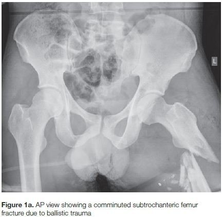
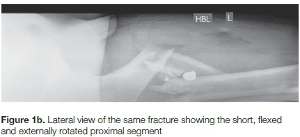
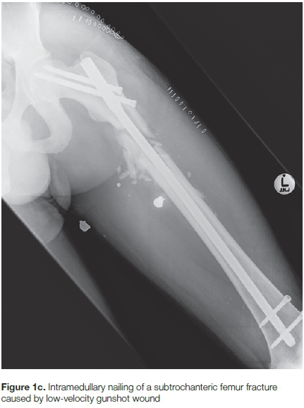
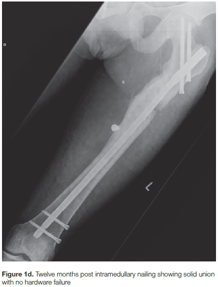
Femur fractures are the most common orthopaedic injury due to gunshots2 and locked intramedullary nailing is the mainstay of treatment for diaphyseal femur fractures.3 Femur fractures from ballistic injuries, in particular, are often comminuted which may complicate pre-operative planning and affect outcomes (Figures 1a and 1b). The goals of treatment in these injuries are to restore and maintain normal length and alignment and to prevent rotation (Figures 1c and 1d) while at the same time avoiding excessive soft tissue disruption.4 However, there remains a paucity of literature regarding the management of subtrochanteric femur fractures caused by low-velocity gunshot wounds.
Subtrochanteric fractures involve a zone between the inferior border of the lesser trochanter and the junction of the proximal and middle one-third of the femur (approximately a 5 cm segment).
The difficulties encountered in the treatment of subtrochanteric fractures are related to the unique anatomy and high bending forces transmitted across the subtrochanteric region of the proximal femur5 with significant rates of complications reported with their surgical treatment. Despite numerous studies to improve technology and implant designs and outcomes of these injuries, malunion, nonunion and implant failure can occur with some frequency.6
In this retrospective review, we evaluated the results of intramedullary nailing of subtrochanteric femur fractures caused by low-velocity gunshot injuries. To our knowledge, there are no clinical series published on intramedullary nailing for subtrochanteric femur fractures secondary to civilian gunshot wounds.
Methods
We performed a retrospective review of clinical and radiographs records of patients who sustained a subtrochanteric femur fracture caused by low-velocity gunshot wounds (AO/OTA type 32C3)7 and who were stabilised using a locked intramedullary nail between March 2008 and December 2014. The study was performed at an academic Level 1 trauma centre. Institutional review board approval was obtained prior to review of patient records. Patients were Identified through a review of the institution's trauma database.
All civilian patients with subtrochanteric femur fractures due to gunshot wounds treated with intramedullary nailing were included. We included all patients who were followed up to clinical and radiographic union. Demographic data included patient age and sex, medical comorbidities and smoking status. Injury data included AO fracture classification, time to surgery, orthopaedic implant used, neurovascular status and other associated injuries.
Radiographs obtained at the time of injury, immediately postoperatively and with subsequent follow-up visits were reviewed to determine fracture type and the progression of fracture union.
The main outcome measure was fracture union. Clinical union was defined as full weight bearing without pain. Radiographic healing was defined as three cortices with bridging callus on two orthogonal views. Fractures healing after four months were defined as delayed unions while fractures not demonstrating any progression of callus formation by six months or those with hardware failure were defined as non-unions.
We used a univariate method for data analysis which consisted of the Fisher exact test due to our small study population. The primary outcome variable was fracture union and the secondary outcome variables were delayed union, infection and implant failure. Fractures with delayed union were compared to those without. Statistical significance was set at p<0.05.
Results
A total study population of 51 patients was identified. There were 48 men and two women with an average age of 28 years (range 16-50 years). Forty-two patients (82%) sustained isolated subtrochanteric femur fractures and the remaining nine patients (18%) sustained additional injuries due to multiple gunshots including abdominal, pelvic, soft tissue and other extremity injuries. Six patients (12%) had an associated sciatic nerve palsy. The sciatic nerve palsies were all treated with non-surgical management. Only one patient had a full recovery of function.
After the assessment of the patients' injuries was completed, a single injection of 1 200 000 units penicillin G benzathine (Bicillin) was administered according to our protocol. All injuries were low-velocity gunshot wounds with cefazolin administered as perioperative antibiotic prophylaxis for 24 hours as a routine in our unit. No formal debridement was done to the entrance and exit wounds. These wounds were not closed. One patient sustained a vascular injury to the superficial femoral artery. A surgical repair was done for the transected vessel and the fracture was nailed after the vascular repair.
One patient had a fasciotomy at the time of the intramedullary nailing for compartment syndrome not associated with a vascular injury.
Patients were treated by several orthopaedic registrars with varying levels of experience. The predominant method of fixation was a reamed cephalomedullary nail in 43 patients (84%). Eight fractures (16%) were treated with a standard proximal interlocking nail with locking into the lesser trochanter.
The majority of patients were treated within a mean of 24.2 hours from the time of admission (range 5-48 hours). Twenty patients (39%) had regular tobacco use and two patients had additional medical comorbidities.
Of the six patients who had a sciatic nerve palsy only one patient had complete recovery of function at six weeks post-operatively. One patient presented with a superficial incisional infection according to the Centres for Disease Control and Prevention criteria for surgical site infection. No wound dehiscence occurred. This settled with local wound cleansing and oral antibiotics. There were no cases of deep infection.
Thirty patients with a mean follow-up period of 3.3 weeks (0-6 weeks) were lost to follow-up.
Twenty-one patients (41%) had adequate radiographic and clinical follow-up data till union. The mean clinical follow-up period for this group was 24.8 weeks. From this group, 14 of 21 fractures (67%) healed within four months of surgery and seven fractures (37%) had delayed union. None of the patients required additional surgery. Union was achieved in all cases (Figure 1d). The average time to union was 17.2 weeks (range 7-40 weeks).
No association of statistical significance was found between delayed union and smoking (p=0.620), presence of multiple injuries (p=1.0), time to surgery (p=0.151) or the presence of medical comorbidities (p=1.0). No implant failure occurred.
None of the fractures that had adequate clinical follow-up developed a non-union.
Discussion
Orthopaedic surgeons are being exposed to an increasing incidence of ballistic trauma as gunshot wounds and their associated fractures are becoming more common in today's urban hospitals.
Injuries inflicted by gunshot wounds are an immense financial burden on the South African healthcare system. In 2011 South Africa had a gun-related mortality rate of 14.0 per 100 000. By comparison, the United States reported a firearm mortality rate of 10.4 per 100 000. While the mortality rate attributable to firearms in South Africa is high, the burden of non-fatal firearm-related injuries is far worse.8 In 2005, Allard et al. postulated that South African state hospitals treat approximately 127 000 firearm victims each year.1
In general, civilian gunshot wounds are considered low energy. However, an increasing frequency of high-energy injuries in civilian populations is being reported due to greater availability of military-type firearms, especially among the criminal element.9 As physicians we need to understand that the differentiation between high- and low-velocity injuries is crucial, because treatments are distinct.
According to the formula KE=1/2 mv2 (where KE=kinetic energy, m=mass and v=velocity) , the kinetic energy of a bullet is proportional to the square of its velocity.10 Therefore increases in velocity have exponential effects on the energy of the injury.
Muzzle velocities of 1 000 to 2 000 feet per second are characterised as low velocity and these firearms include pistols and other handguns. These firearms mainly cause direct injury to tissue as there is little to no blast or cavitation wave effect on the target. Weapons such as the AK-47 and M-16 military assault rifles fire with a muzzle velocity of greater than 2 000 feet per second and are classified as high velocity. These military-type firearms carry so much energy that they cause both direct and indirect injuries due to the shock-wave effect.11
The bullet energy is not the sole determinant of the extent of injury as the path, and size and composition of the projectile, and distance between firearm and target will affect the ballistics of the projectile. Most bullets are composed of a lead core, which may be combined with other metals to allow for less deformation with firing and to improve accuracy on long-range targets. Missile fragmentation occurs readily on impact with hollow-point bullets and can further increase the zone of destruction.
The most common area of fracture due to gunshot wound is the femur.8 Initially, femur fractures due to gunshot were treated by delayed intramedullary nailing. The rationale was that a delay of 10 to 14 days would allow the missile track to seal.4
Nowotarski and Brumback12 reviewed 39 femoral shaft fractures in 37 patients due to low-velocity gunshot wounds. The patients were treated with antegrade interlocked nailing within 18 hours of injury. The results were favourable and the early fixation of these injuries had no effects on outcome, so immediate intramedullary nailing of gunshot injuries of the femur has become an accepted standard of care with significant economic advantages and clinical benefits as shown by Molinari et al.13
The patients in our study were treated with early antegrade intramedullary nailing. This is similar to previously reported studies of immediate interlocked nailing of femoral fractures due to gunshot wounds.
Literature reports up to a 43% incidence of associated injuries.2 We found that 18% of the patients in our group had associated injuries. These include a whole array of additional soft tissue and extremity injuries secondary to the gunshot wounds to significant life-threatening injuries, including abdominal and pelvic injuries.
The main objectives of treatment are to provide a stable fixation with a satisfactory fracture reduction and to decrease the surgical insult to the patient. The anatomy of the subtrochanteric region of the femur creates several issues that affect fracture healing and successful surgical management.
There are three major muscular deforming forces applied to this area: the hip abductors; short external rotators; and iliopsoas. All pull on the proximal fragment resulting in the very familiar flexed, abducted and externally rotated position. This results in an overall varus and apex anterior deformity at the fracture site. This deformity complicates attempts at closed reduction. This causes a high concentration of stresses and deforming forces in this portion of the bone. The subtrochanteric area is made of mainly cortical bone with diminished blood flow to this region when compared to the meta-physeal bone of the intertrochanteric region.14 These factors contribute to the complex nature of managing these subtrochanteric fractures as has been made evident throughout the orthopaedic literature.
Intramedullary nailing has the biomechanical advantage over plates in this situation. Their intramedullary position reduces the moment arm on the nail by reducing the distance over which the bending forces act compared with a plate on the lateral cortex. This also allows for decreased surgical dissection and the possibility of earlier weight-bearing and has therefore gained popularity among orthopaedic surgeons. However, obtaining and maintaining an accurate reduction and starting point for nail insertion are key factors for successful management of these complicated fractures as the short proximal segment with its wide canal can make fixation with intramedullary devices challenging. The angle formed by the axis of the femoral neck and femoral shaft is 127° to 130°. If this angle is decreased, as would occur with a varus reduction of the fracture, the distance between the head and shaft is increased, which increases the moment arm and the bending forces across the fracture site and may produce varus collapse and result in a higher possibility of implant failure and non-union.15
These challenges are further complicated by the often-comminuted nature of the fracture as a result of civilian gunshot or ballistic injury.
Historically operatively treated subtrochanteric fractures are known to have a high complication rate. They are prone to deformity and fixation failure with up to a 21% complication rate.
Intramedullary nailing has significantly reduced their complications; however, they still occur frequently with recent literature showing a re-operation rate of 4.7% from all complications with the use of contemporary intramedullary nailing.16 However no data exists on the complication of intramedullary nailing of subtrochanteric femur fractures due to gunshots. There were minimal post-operative complications in our patients, no cases of clinical malrotation, and our overall results showed an acceptable union rate. Little data exists regarding the complication rates and outcomes of intramedullary nailing in these comminuted and possibly contaminated sub-trochanteric femur fractures caused by low velocity ballistic trauma. The incidence of delayed union in the cohort with adequate follow-up until union was 37%. However, all fractures went on to clinical and radiographic union. No association of statistical significance was found between delayed union and smoking, presence of multiple injuries, time to surgery or the presence of medical comorbidities. No implant failure occurred.
In the AO classification system of long bone fractures, the standard classification used by trauma surgeons and physicians dealing with skeletal trauma worldwide, subtrochanteric femur fractures are classified as diaphyseal fractures and appear under the classification for the diaphyseal segment. A horizontal transverse line at the inferior border of the lesser trochanter divides the proximal femur in a trochanteric and subtrochanteric zone. The value of this classification system when dealing with ballistic trauma is reduced as it takes no account of the soft tissue injury. Studies have already suggested poor reproducibility of the Seinsheimer, AO/OTA and Russell-Taylor classification systems used for subtrochanteric femur fractures and they seem to be of little value in predicting the outcome of intramedullary nailing.17 The majority of fractures (80%) in our study were classified according to the AO classification as 32C3 with various fragment configurations ranging from three intermediate fragments to fractures with extensive shattering.
The study limitations include the retrospective design with no control group, the use of multiple surgeons using variable locking configurations for each intramedullary nail and the limited follow-up. Eighty per cent of patients returned for clinical follow-up at two weeks. However only 41% had adequate radiographic and clinical follow-up data till union. This is historically a difficult patient group to deal with as they are often poorly compliant. Average follow-up in our study was 24.8 weeks, with the majority of patients following up in the first 6- to 8-week period.
Conclusion
Our study addresses the paucity of literature currently available concerning the management of subtrochanteric femoral fractures caused by low-velocity ballistic trauma. This study demonstrates that intramedullary nail fixation of subtrochanteric femur fractures caused by low-velocity civilian gunshots is an acceptable option for the treatment of these injuries. Our results showed a high rate of union with rare complications; however, the results should be interpreted with caution due to the high attrition rate.
Compliance with ethics guidelines
Institutional review board approval was obtained prior to the review of patient records.
The content of this article is the original work of the authors.
References
1. Allard D, Burch VC. The cost of treating serious abdominal firearm-related injuries in South Africa. South African Med J. 2005;95(8):591-94. [ Links ]
2. Cannada LK, Jones TR, Guerrero-Bejarano M, et al. Retrograde intramedullary nailing of femoral diaphyseal fractures caused by low-velocity gunshots. Orthopedics. 2009;32(3):162. http://www.ncbi.nlm.nih.gov/pubmed/19309067. Accessed February 6, 2016. [ Links ]
3. Tejan J, Lindsey RW. Management of civilian gunshot injuries of the femur. A review of the literature. Injury. 1998;29 Suppl 1(1): SA18-SA22. doi:10.1016/S0020-1383(98)90034-1. [ Links ]
4. Wiss DA, Brien W, Becker V. Interlocking nailing for the treatment of femoral fractures due to gunshot wounds. J Bone Joint Surg [Am] 1991;73-A:598-606. [ Links ]
5. Shukla S, Johnston P, Ahmad MA, Wynn-Jones H, Patel AD, Walton NP. Outcome of traumatic subtrochanteric femoral fractures fixed using cephalo-medullary nails. Injury. 2007;38(11):1286-93. doi:10.1016/j.injury.2007.05.013. [ Links ]
6. De Vries JS, Kloen P, Borens O, Marti RK, Helfet DL. Treatment of subtrochanteric nonunions. Injury. 2006;37(2):203-11. doi:10.1016/j. injury.2005.09.017. [ Links ]
7. Marsh JL, Slongo TF, Agel J, et al. Fracture and Dislocation Classification Compendium - 2007: Orthopaedic Trauma Association Classification, Database and Outcomes Committee. J Orthop Trauma. 2007;21 Supplement 10 pp: S1-S133. Accessed March 2, 2016. [ Links ]
8. Maqungo S, Martin C, Thiart G, Mccollum G, Roche S. The burden of orthopaedic gunshot injuries on healthcare resources in South Africa. Bone Joint J 2014;6(Supp 19):38. [ Links ]
9. Zawitz MW. Guns used in crime. Bureau of Justice Statistics1995;(July). [ Links ]
10. Bono CM, Heary RF. Gunshot wounds to the spine. Spine J. 2004;4(2):230-240. doi:10.1016/S1529-9430(03)00178-75. [ Links ]
11. Jakoi A, Iorio J, Howell R, Zampini JM. Gunshot injuries of the spine. Spine J. 2015;15(9):2077-85. doi:10.1016/j.spinee.2015.06.007. [ Links ]
12. Nowotarski P, Brumback RJ. Immediate interlocking nailing of fractures of the femur caused by low- to mid-velocity gunshots. J Orthop Trauma. 1994;8(2):134-41. http://www.ncbi.nlm.nih.gov/pubmed/8207570. Accessed February 6, 2016. [ Links ]
13. Molinari RW, Yang EC, Strauss E, Einhorn TA. Timing of internal fixation in low-velocity extremity gunshot fractures. Contemp Orthop. 1994;29(5):335-339. http://europepmc.org/abstract/med/10150253. Accessed February 6, 2016. [ Links ]
14. Bourgeon R, Pantin JP, Frailong J. Intramedullary nailing of subtrochanteric fractures. Afr Fr Chir. 2014;4(2):144-46. doi:10.1097/BTO.0b013e31817bf2a1. [ Links ]
15. Sims SH. Femoral fractures. Orthop Clin North Am. 2002;33(1):113-26. [ Links ]
16. Joglekar SB, Lindvall EM, Martirosian A. Contemporary management of subtrochanteric fractures. Orthop Clin North Am. 2015;46(1):21-35. doi:10.1016/j.ocl.2014.09.001. [ Links ]
17. Guyver PM, McCarthy MJH, Jain NPM, Poulter RJ, McAllen CJP, Keenan J. Is there any purpose in classifying subtrochanteric fractures? The reproducibility of four classification systems. Eur J Orthop Surg Traumatol. 2014;24(4):513-18. doi:10.1007/s00590-011-0780-3. [ Links ]
 Correspondence:
Prof Sithombo Maqungo
Division of Orthopaedic Surgery, H49 Old Main Building
Groote Schuur Hospital, Tel: +27832341723
Email: sithombo@msn.com
Received: July 2016
Accepted: October 2016
Published: August 2017
Funding: No benefits of any form have been or are to be received from a commercial party related directly or indirectly to the subject of this article.
Conflict of interest: The authors have no conflicts of interest to declare.

