


Servicios Personalizados
Articulo
 Inglés (pdf)
 Articulo en XML
 Referencias del artículo
Indicadores

Links relacionados
-
 Citado por Google
 -
 Similares en Google
Compartir

 Permalink
SA Orthopaedic Journal
versión On-line ISSN 2309-8309
versión impresa ISSN 1681-150X
SA orthop. j. vol.10 no.2 Centurion ene. 2011
ARTICLE
Multifocal tuberculous spondylitis with rib involvement - a case report and review of the literature
JWM KigeraI; N OrwothoII
IMBChB. Department of Orthopaedics, Makerere University, Kampala, Uganda
IIMBChB, MMed(Ortho). Orthopaedic Surgeon, Spine Unit, Department of Orthopaedics, Mulago Hospital, Kampala
ABSTRACT
Tuberculosis is a common diagnosis in the developing world. Its incidence is increasing in the developed world due to immigrants from endemic regions, increased cases of immune-compromised patients, multi-drug resistance and low socio-economic status. Tuberculous spondylitis is the commonest extra-pulmonary manifestation of tuberculosis. The rib is only very rarely involved. We present a case of multifocal tuberculous spondylitis in the T6-9 and L3 regions with involvement of the ninth rib.
Key words: tuberculosis, spine, ribs, surgery, multifocal
Introduction
Tuberculosis is common in the developing world and its incidence is increasing. Three to five per cent of patients have skeletal tuberculosis, of whom about half have tuberculous spondylitis.1 Spinal tuberculosis is associated with great potential morbidity and mortality hence the importance of proper and quick diagnosis.2
Case report
A 32-year-old man was admitted with a three-month history of back pain and a week's history of inability to use the lower limbs. He had no history of trauma and denied history of fever, cough, night sweats or weight loss. He is a long distance taxi driver and had initially attributed his lower back symptoms to fatigue due to long hours of driving. Examination revealed tenderness in the mid and lower thoracic spine region with reduced lower limb power judged to be grade 3 and sensation was reduced to grade 1 in the ASIA classification system.3
Examination of the respiratory system was normal. Laboratory investigations revealed an ESR of 70 mm in the first hour and negative serology to HIV. The haemoglobin level and white blood cell counts were within the reference ranges.
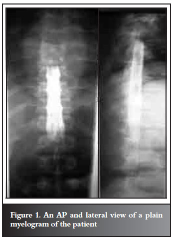
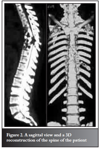
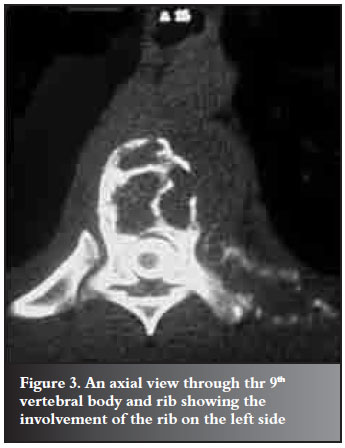
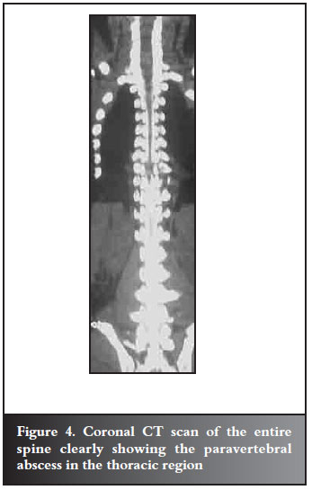
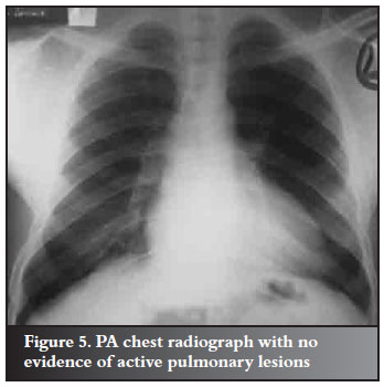
Plain myelography showed multiple hypodense lytic lesions in the thoracic spine region from T6-9 associated with a paravertebral shadow. There was anterior collapse with a kyphotic deformity. It also revealed blockage of contrast media at the T8/9 level (Figure 1). Computerised tomography demonstrated the involvement of the thoracic vertebrae as seen on plain radiographs and revealed further lesion in the posterior aspect of the left ninth rib and a lesion in the third lumbar vertebral body (Figures 2, 3 and 4). The chest radiograph did not reveal any active pulmonary lesions (Figure 5).
A diagnosis of tuberculous spondylitis with osteitis of the rib was made and the differential diagnosis of metastatic malignancy, lymphoma, multiple myeloma, chordoma, sarcoidosis, brucellosis, fungal disease and bacterial spondylitis were entertained.
An open biopsy was planned to obtain tissue for histology and microbiology. This was also intended to drain any paravertebral abscess that was to be encountered as part of decompression of the neural structures.
The spine was approached posteriorly with the incision centred over T9. On exposure of the transverse process a pocket of pus was encountered and we drained about 30 ml. Macroscopic appearance was that of a caseous nature. Samples were obtained for bacteriology. Biopsy of the transverse process of T9 was also taken for histology. A costotransversectomy was done and aggressive surgery was avoided due to the extensive nature of the lesion.
The bacteriology report did not report any organisms on staining or on culture. Histology showed caseating epitheloid tubercles with Langerhans' giant cells pathognomonic of tuberculosis.
A diagnosis of tuberculosis was made and the patient started on chemotherapy according to the Uganda National Tuberculosis and Leprosy programme treatment guidelines.
The patient has not shown any neurological deterioration and the quality of sensation has improved. Good response has been noted on chemotherapy and he is currently ambulating with the aid of crutches.
Discussion
Osteoarticular tuberculosis is the commonest manifestation of extra-pulmonary tuberculosis occurring in about 1-5% of all TB cases. Multifocal skeletal tuberculosis has osteoarticular lesions that occur simultaneously at two or more locations and are usually associated with disseminated disease.4 The initial route of entry of M. tuberculosis is usually the respiratory tract, followed by haematogenous dissemination. Spread to the spine can occur via secondary haematogenous seeding from a silent focus in the body (e.g. gut, kidney, and tonsil) or spread from involved contiguous para-aortic lymph nodes. Haematogenous seeding can occur via intercostal and lumbar arteries and the Batson's plexus.
The tubercle bacillus begins its destruction in cancellous bone and eventually extends to the cortex. The infection gradually spreads to adjacent vertebrae via the disc space. In advanced stages of the disease, progressive vertebral collapse occurs, resulting in kyphosis and gibbus formation.
TB spine usually involves the thoracic and lumbar regions and infrequently the cervical spine. Though up to 100% of patients with spinal TB will present with back pain, only about 19-29% will have musculoskeletal and neurological signs.5 Multifocal involvement of the spine is seen in less than 5%.6 Rib tuberculosis is seen in only 0.1% of all tuberculosis infections.7 Multifocal spine involvement has been noted by several authors and raises diagnostic difficulty.8-10 Our case demonstrates that in endemic countries it is possible to encounter the more rare presentations of the disease and TB should remain at the top of the list of differential diagnosis.
Multifocal tuberculous spondylitis is a rare presentation and should always be in the list of differential diagnosis of a multifocal spine lesion. The combination with a lesion of the ribs is even rarer. All clinicians should acquaint themselves with these manifestations and the difficulties of making a diagnosis. Further research is needed to ascertain the true prevalence of these disease presentations.
Acknowledgements
Surgeons and residents of the Spine Unit, Mulago Hospital.
References
1. Council MR. Medical Research Council National Survey of Tuberculosis Notifications in England and Wales in 1983: characteristics of disease. Tubercle 1988; 68:19-33. [ Links ]
2. Ormerod L, Charlett A, Gilham C. Geographical distribution of tuberculosis in national surveys of England and Wales in 1988 and 1993: report of the Public Health Laboratory Service/British Thoracic Society/Department of Health Collaborative Group. Thorax 1998; 53:176-81. [ Links ]
3. Association ASI. Standard neurological classification of spinal cord injury. 2006 [cited; Available from: http://www.asia-spinalinjury.org/publications/2006_Classif_worksheet.pdf [ Links ]
4. Ozol D, Koktener A, Uyar ME. Active pulmonary tuberculosis with vertebral and rib involvement- a case report. South Med J 2006; 99(2):171-3. [ Links ]
5. Cormican L, Harmal R, Messenger J, Milburn HJ. Current difficulties in the diagnosis and management of spinal tuberculosis. Postgrad Med J 2006; 82 : 46-51. [ Links ]
6. Moujtahid M, Essadki B, Lamine A. Multifocal bone tuberculosis: apropos of a case. Rev Chir Orthop Reparatrice Appar Mot. 1995(81):553. [ Links ]
7. Tatelman M, Drouillard EJP. Tuberculosis of the ribs. AJR 1953; 70:923-35. [ Links ]
8. Albert Neher, Werner Kopp, Gabriele Berna, Johannes Frank, Martin Kohlha. Advanced multifocal tuberculous spondylitis without disk involvement and with multi-drugresistant bacilli. Clinical Infectious Diseases. 2007; 45:109-12. [ Links ]
9. Yilmaz MH, Kantarci F, Mihmanli I, Kanberoglu K. Multifocal skeletal tuberculosis. Southern Medical Journal. 2004; 97(8):785-7. [ Links ]
10. Turgut M. Multifocal extensive spinal tuberculosis (Pott's disease) involving cervical, thoracic and lumbar vertebrae. British Journal of Neurosurgery. 2001; 15(2):142-6. [ Links ]
 Reprint requests:
Dr JWM Kigera
P O Box 7062
Kampala Uganda
Tel +256 414 542332
Email: jameskigera@yahoo.co.uk
All the authors participated in the design and preparation of the manuscript. The patients were informed that the material would be used for publication.

