


Servicios Personalizados
Articulo
 Inglés (pdf)
 Articulo en XML
 Referencias del artículo
Indicadores

Links relacionados
-
 Citado por Google
 -
 Similares en Google
Compartir

 Permalink
Journal of the South African Veterinary Association
versión On-line ISSN 2224-9435
versión impresa ISSN 1019-9128
J. S. Afr. Vet. Assoc. vol.94 no.1 Pretoria 2023
http://dx.doi.org/10.36303/jsava.548
ORIGINAL RESEARCH
Seroprevalence and associated risk factors for Toxoplasma gondii infection of goats and sheep in the Khomas region of Namibia
A SamkangeI, II; S ChitangaII, III, IV; GN Tjipura-ZaireV; VG MutjavikuaII; JW SmithII; L NevesI, VI; T MatjilaI
IDepartment of Veterinary Tropical Diseases, Faculty ofVeterinary Science, University of Pretoria, South Africa
IISchool ofVeterinary Medicine, Faculty of Health Sciences and Veterinary Medicine, University of Namibia, Namibia
IIIDepartment of Biomedical Sciences, School of Health Sciences, University of Zambia, Zambia
IVSchool of Life Sciences, College of Agriculture, Engineering and Sciences, University of KwaZulu-Natal, South Africa
VDirectorate ofVeterinary Services, Namibia
VICentro de Biotecnologia, Universidade Eduardo Mondlane, Mozambique
ABSTRACT
This study aimed to determine the seroprevalence levels of Toxoplasma gondii in small ruminants (goats and sheep) and the associated risk factors in the Khomas region of Namibia. A total of 299 and 345 sheep and goat sera from 22 farming establishments were tested, respectively. An IDEXX Toxotest Ab®, a commercial ELISA kit, was used to screen for IgG antibodies to T.gondii. Overall, 3.68% (11/299) of the sheep sera were positive, and 61.54% (8/13) of the sheep flocks tested had at least one positive animal. Only one of the 345 goat sera from 19 flocks was positive, giving animal-level and herd-level prevalences of 0.29% and 5.26%, respectively. Sheep flocks had significantly greater animal-level and flock-level prevalences than goats (p < 0.05) and were 13.14 times more likely to be seropositive (OR = 13.14; CI 95%: 1.686-102.382) than goat flocks. A questionnaire was also administered to identify any putative risk factors associated with seropositivity. Eight risk factors were evaluated, including the total number of goats, total number of sheep, farm size, average rainfall, presence of wild Felidae (African lions, caracals, cheetahs and leopards), presence of domesticated and stray cats and history of abortions in the flocks. Seropositivity to T. gondii in sheep was positively associated with the total number at the farming establishment, history of abortions and farm size (p < 0.05), but not with goats. The study determined that sheep in the Khomas region were probably more exposed to T. gondii infection than goats. It also found T. gondii seroprevalences that were much lower than those in similar studies from other countries in the sub-region and elsewhere.
Keywords: seroprevalence, risk factors, T. gondii, goats, sheep, Khomas, Namibia
Introduction
Toxoplasma gondii is an intracellular protozoan pathogen presumed to occur worldwide (Al-Malki 2021). It is associated with livestock reproduction losses through abortions, stillbirths, mummifications, infertility, early embryonic death and foetal resorption (Al-Malki 2021; Omonijo et al. 2022; Tagwireyi et al. 2019), especially in sheep and goats (Abbas et al. 2020). Additionally, sheep are the most susceptible to T. gondii infection among livestock (Cenci-Goga et al. 2013). However, buffalo, cattle and horses are relatively resistant to toxoplasmosis (Abbas et al. 2020; Belluco et al. 2016). In addition, toxoplasmosis is considered an important zoonosis, and it is estimated that one-third of the world's human population is infected with T. gondii (Abbas et al. 2020; Innes 2010; Kolören & Dubey 2020; Tegegne et al. 2016; Zhang et al. 2013).
T. gondii has a complex life cycle which involves the felids (domestic and wild cats) as definitive hosts, in which sexual reproduction takes place and it culminates in the shedding of millions of unsporulated oocysts into the environment through faeces (Belluco et al. 2016; Dubey 1998). Depending on environmental conditions of temperature and humidity, the oocysts may take two to three days to sporulate and become infective (Dubey et al. 2011). The intermediate hosts, virtually all warm-blooded animals, including humans, get infected when they ingest food or water contaminated with sporulated oocysts (Pinto-Ferreira et al. 2019). Tissue cysts in raw or undercooked meat, tachyzoites in unpasteurised milk and milk products like cheese are also important sources of human infection (Al-Malki 2021; Pinto-Ferreira et al. 2019). Asexual reproduction occurs in the intermediate hosts and culminates in the formation of tissue cysts in neural and muscular tissues (Tenter et al. 2000). The cycle is completed when the definitive host consumes the tissues of the intermediate hosts (Tenter et al. 2000). Although T. gondii infection is usually subclinical in humans (Kalogeropoulos et al. 2022), the parasite can occasionally cause acute toxoplasmosis outbreaks (Dubey 2021; Kolören & Dubey 2020). It can also lead to severe complications in immunocompromised individuals and congenital disabilities in foetuses if pregnant women are infected (Kalogeropoulos et al. 2022).
T. gondii seroprevalence rates in small ruminants (sheep and goats) have been reported in different parts of the world, such as 12-36% in sheep in the Nordic-Baltic region (Olsen et al. 2019), 42.47% in goats in India (Bachan et al. 2018), 48.6% in sheep and 30.7% in goats in Greece (Tzanidakis et al. 2012), 41.1% in sheep in Costa Rica (Villagra-Blanco et al. 2019), and 26.1% in sheep in Israel (Mazuz et al. 2023). In southern Africa, the seroprevalence rates reported in sheep and goats in Zimbabwe were 8.8% (n = 216) and 7.1% (n =156), respectively, and in South Africa, the figures ranged from 4.3% (n = 600) to 64.5% (n = 121) in sheep and 53.9% (n = 128) in goats (Omonijo et al. 2022; Tagwireyi et al. 2019). However, Namibia's only documented studies regarding T. gondii infection involved wildlife and humans (Joubert & Evans 1997; Seltmann et al. 2020; Van der Colf et al. 2020) but not in livestock.
The risk factors linked to the infection in other countries include the presence of definitive hosts (cats), flock size, age, geographical factors, farm management factors and biosecurity (Caballero-Ortega et al. 2008; Stelzer et al. 2019). Therefore, this study aimed to determine the seroprevalence levels of T. gondii in small ruminants (goats and sheep) and the associated risk factors in the Khomas region of Namibia.
Materials and methods
Study area
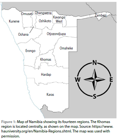
The study area was Namibia's Khomas region, located in the central part of the country (Figure 1). Namibia's sub-tropical climate varies from arid to semi-arid, and it is the driest country in sub-Saharan Africa (Mwazi & Shamathe 2007). The country's central highlands receive an annual rainfall range of 300400 mm and have an altitude of 1 900 m (Kandiwa et al. 2017). The vegetation is predominantly shrub-veld, and ambient temperatures range from 7 oC in winter and up to 33 oC in summer (Kandiwa et al. 2019). The Khomas region has approximately 560 livestock farming units, mainly commercial, with some resettlement farms and communal settlements (Directorate of Veterinary Services 2018).
Study animals
The study animals were adult goats and sheep. This region has approximately 91 000 sheep and 20 000 goats distributed among some of the estimated 560 farming establishments (Directorate of Veterinary Services 2018) that primarily farm beef cattle. All the animals were extensively reared.
Sampling and data collection
Sample size determination was done according to Pfeiffer (2002), using estimated herd-level and animal-level prevalences of 20% and 10%, respectively (Omonijo et al. 2022; Tagwireyi et al. 2019), at 95% confidence level and 5% precision. Convenience sampling was used because most of the farming establishments in the Khomas region keep beef cattle, with only a few rearing small ruminants. At the farm level, multistage sampling was employed, where adult sheep and goats were segregated into distinct groups within each flock. These separate groups were then assigned individual numbers and a random number generator was utilised to select the specific numbers for sampling according to the pre-determined sample size for each flock.
A questionnaire was administered during sample collection to determine the possible risk factors for T. gondii infection. The questionnaire solicited information on the following risk factors: total number of animals on the farm (sheep and goats), farm size, average rainfall, presence of sylvatic Felidae, history of abortions, presence of domesticated cats, and the presence of stray cats. The selection of these risk factors was based on available literature sources in light of the prevailing conditions in the Khomas region of Namibia. Furthermore, to validate it, the questionnaire was administered to a small group of six participants as a pilot test before its use in the main study.
Blood was collected from the jugular veins into plain Vacutainer® blood tubes using 20-gauge needles. In the laboratory, the clotted blood samples were centrifuged at 1,957 x g for 10 min to separate the sera. Next, sera were pipetted into 2 ml cryovials (Labocare™, EREZ Medical UK, London), which were then stored at -20 °C until they were tested.
Serological analysis
The IDEXX Toxotest® (IDEXX Laboratories, Inc, Maine 04092, USA) test kit, an indirect enzyme-linked immunosorbent assay (ELISA), was used to test for specific anti-T. gondii IgG antibodies in sheep and goat sera. The ELISA test has a sensitivity and specificity of 98.8% and 92.8%, respectively (Patel et al. 2017).
The test was performed according to the manufacturer's instructions. All reagents were brought to 18-26 oC and mixed. First, diluted positive and negative controls and diluted test sera were added to antigen-coated plates, mixed and then incubated at 37 oC for 1 hr. After incubation, the excess solution was discarded, and the wells were washed thrice with Wash Solution. Conjugate was then added to all the wells, after which the plates were tightly sealed and subsequently incubated for an additional one hour at 37 oC. Again, the excess conjugate solution was discarded, and the wells were washed thrice with Wash Solution. Next, a TMB Substrate was added to each well, and the plates were incubated at 18-26 oC for 15 min. A Stop Solution was then added to each well. Finally, the results were read at a wavelength of 450 nm using an ELISA reader within 2 hr after adding the Stop Solution.
The positive and negative controls were then validated, and ratios of a sample to positive control were interpreted as follows: a ratio of less than 20% was interpreted as negative; a ratio of at least 20% but less than 30% was suspect; a ratio of at least 30% but less than 100% was interpreted as weak positive. Finally, a ratio of at least 100% was interpreted as positive.
Data analysis
The serology and questionnaire data were captured in Microsoft Excel 2013 spreadsheet, in which all the statistical calculations were done. Descriptive statistics were then used to calculate herd-level and animal-level prevalence (P) rates. The z-test for comparing two proportions and odds ratios (OR) were used to compare the prevalence rates between sheep and goats. Finally, linear regression analysis and the Chi-square test were used to evaluate the risk factors. Linear regression analysis was used to evaluate continuous risk factor variables, and the dependent variable was seropositivity, whilst the independent variables were farm size, average annual rainfall, and the number of sheep and goats. The Chi-square test was used to evaluate the following categorical risk factor variables: the presence of sylvatic Felidae, history of abortions, the presence of domesticated cats, and the presence of stray cats.
Results
Eleven of the 299 sheep sera from 13 farming establishments were positive, giving an animal-level prevalence of 3.68% (Confidence Interval [CI] 95%: 2.07-6.47) (Table I).
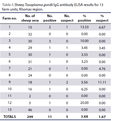
Another five sheep (P 1.67%; CI 95%: 0.72-3.85) were suspicious, giving a combined (positive & suspicious) animal-level prevalence of 5.35% (CI 95%: 3.32-8.51). Eight of the thirteen establishments tested had at least one positive sheep, giving a herd-level prevalence of 61.54% (CI 95%: 35.52-82.29) (Figure 2).
Of the 345 goat sera tested from 19 farming establishments, only one was positive, giving an animal-level prevalence of 0.29 % (CI 95%: 0.05-1.62) and a herd-level prevalence of 5.26% (CI 95%: 0.90-24.60) (Table II). Seven goats (2.03%) were suspicious, giving a combined (positive & suspicious) animal-level prevalence range of 2.32% (CI 95%: 1.20-4.50) and a herd-level prevalence of 31.58% (CI 95%: 8.90-51.10).
Sheep had a significantly greater individual-level prevalence than goats (p = 0.0015) and were 13.14 times more likely to be positive (OR = 13.14; CI 95%: 1.686-102.382) than goats. In addition, the sheep's herd-level prevalence was significantly greater than for goats (p = 0.0005), and sheep farming establishments were 28.8 times more likely to have at least one positive animal than goat flocks (OR = 28.8; CI 95%: 2.879-288.103).
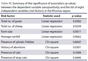
Table III shows the list of eight potential risk factors investigated using questionnaires. Seropositivity to T. gondii was positively associated with the total number of sheep at the farming establishment, a history of abortions and farm size (p < 0.05). However, there was no significant association between seropositivity and the number of goats at the establishment, the presence of Felidae (African lions, caracals, cheetahs, leopards), the presence of domesticated cats, the presence of stray cats and the average rainfall (p > 0.05).
Discussion
To the authors' knowledge, this is Namibia's first study in sheep and goats on T. gondii infection. The study determined animal-level prevalence rates of 3.68% and 0.29% in sheep and goats, respectively, which are lower compared to South Africa, India, Iran and Italy, where seroprevalence rates ranged from 27% to 64.46% (Bachan et al. 2018; Gazzinos et al. 2015; Sharif et al. 2015; Tagwireyi et al. 2019). These low prevalence rates in the Khomas region of Namibia might be due to its harsh climate conditions. The country experiences a predominantly hot and dry climate, characterised by infrequent and unpredictable rainfall. As a result, almost 92% of its land area falls under very-arid, arid, or semi-arid categories, ranking the country as the second driest region after the Sahara Desert (The World Bank Group 2021).
Exposure to temperature extremes and UV radiation have been shown to inactivate T. gondii oocysts. For instance, T. gondii oocysts can survive 32 days at 35 oC but only nine days at 40 oC (Robert-Gangneux & Darde 2012). In addition, continuous exposure to UV irradiation has been shown to inactivate > 99.9% of the T. gondii oocysts exposed (Dumetre et al. 2008). These conditions are not uncommon in the Namibian climate (Samkange et al. 2020; The World Bank Group 2021). Such conditions create an unfavourable environment for the T. gondii oocysts to sporulate (Yan et al. 2016). Therefore, T. gondii infections tend to be high in warm and humid environments but low in hot and dry climates (Meerburg & Kijlstra 2009).
Some studies have shown that animals reared under extensive conditions have lower seroprevalence rates than those reared under intensive or semi-intensive conditions (Tagwireyi et al. 2019; Tzanidakis et al. 2012). In contrast, a study conducted by van der Puije and colleagues showed opposite findings, indicating that sheep raised under extensive conditions had a significantly higher seroprevalence than those raised semi-intensively (Van der Puije et al. 2000). The conditions in which animals are raised, fed, and watered in different production systems can affect the risk of contamination with T. gondii oocysts and, therefore, exposure (Stelzer et al. 2019). For example, intensive farming systems, which often involve high levels of confinement, may have lower exposure to oocysts than extensive or other systems that allow outdoor access for animals (Stelzer et al. 2019). However, if feed supplements are stored in places where they may attract domestic or feral cats, which then go into these feed stores in search of rodent prey, the feed could end up being contaminated with cat faeces and, therefore, T. gondii oocysts. This potentially explains why intensive farming is not always associated with lower toxoplasmosis prevalence (Stelzer et al. 2019). However, all small ruminants in the current study were grazed extensively, which meant less exposure to viable T. gondii oocysts, most likely due to adverse weather conditions.
Interestingly, a recent study among pregnant women in Windhoek, which is in the Khomas region of Namibia, also found a similarly low seroprevalence rate of 2.61% (n = 344), a figure that was lower compared to other developing countries, and the authors attributed this to geographical and climatic factors (Van der Colf et al. 2020). Furthermore, in an earlier study, only 0.961% (n = 312) of blood donors in central Namibia were seropositive to T. gondii (Van der Colf et al. 2014). Humans can acquire T. gondii infection by consuming undercooked or raw meat (Tonouhewa et al. 2017), particularly mutton and goat meat that contains tissue cysts (Al-Kappany et al. 2018; Al Hamada et al. 2019). Additionally, unpasteurised goat milk can also be a source of infection for humans (Jones et al. 2009). However, beef and cow milk are not significant sources of infection because cattle can effectively eliminate T. gondii (Esteban-Redondo & Innes 1997). However, other authors contend that the exact impact of beef on the epidemiology of T. gondii infections in humans remains uncertain (Dubey et al. 2020a). Another way humans can acquire T. gondii infection is by consuming fresh produce contaminated with oocyst-infected cat faeces. Fresh fruits, raw vegetables used in salads, and similar products can be contaminated with these oocysts, which can survive for months in the environment (Lass et al. 2012; Marques et al. 2020). The low seroprevalence rates found in sheep and goats in this study imply that the risk of human exposure to T. gondii tissue cysts from small ruminant animal products and oocysts from the environment could be low. Some authors have described sheep and goats as biological indicators for T. gondii oocysts in the environment (Al-Kappany et al. 2018). This possibly explains why human infections are also low. Hence, the findings of the current study are in agreement with previous human studies conducted in Namibia, indicating low seroprevalence rates and demonstrate that T. gondii infections in sheep, goats and humans in the Khomas region are very low compared to the rest of the world, in which one-third of the globe's human population is estimated to be infected (Tegegne et al. 2016).
Recent reviews have compared worldwide seroprevalences in sheep and goats using different serological tests. The seroprevalence rates in sheep ranged from 0 to 98.9%, whereas in goats, they ranged from 1 to 95.2% (Dubey et al. 2020b; Dubey et al. 2020c), and the serological results were not affected by the type of test used. The current study found that sheep had significantly higher animal-level and herd-level prevalences than goats. Similar trends have been reported in many parts of the world, including Northern Italy (Gazzinos et al. 2015), Africa, the Caribbean Islands, Greece, Portugal, Spain, Poland (Moskwa et al. 2018; Stelzer et al. 2019), Lebanon (Khalife et al. 2022) and Colombia (Martinez-Rodriguez et al. 2020). In addition, studies in Iran and South Africa also found higher seroprevalences in sheep than in goats, although they were not statistically significant (Bahreh et al. 2021; Tagwireyi et al. 2019). However, studies in the USA and Egypt recorded a higher seroprevalence in goats than sheep (Al-Kappany et al. 2018; Guo et al. 2016). Therefore, with few exceptions, the current study agrees with reports from most studies from other parts of the world which indicate that sheep are more infected than goats. The explanation could be that sheep are typical grazers, whereas goats are browsers (Yisehak et al. 2016), which makes the sheep more exposed to T. gondii oocysts than goats.
Toxoplasmosis is a significant cause of abortion for sheep and goats (Abbas et al. 2020; Dubey & Jones 2008). The significant association between seropositivity to T. gondii and a history of abortions found in the current study is in agreement with several studies done elsewhere: Northern Iraq (Al Hamada et al. 2019); Central Ethiopia (Gebremedhin et al. 2013) and a review by Stelzer et al. (2019). However, Abdelbaset et al. (2020) did not find such an association in Egypt.
The total number of sheep at the farming establishment is another significant risk factor determined in the current study. Since sheep appear more at risk of T. gondii infection than goats, as established in this study, it is logical that the higher the number of sheep at an establishment, the greater the risk of toxoplasmosis. An alternative explanation could be that when the level of environmental contamination is minimal, having more animals increases the probability of contracting the infection compared to having fewer animals. However, this disagrees with an Italian study where seropositivity decreased with increasing flock size (Cenci-Goga et al. 2013). It is worth noting, however, that this Italian research studied large flock sizes numbering 300-400 sheep, unlike the current study in which flock sizes were much smaller. Additionally, climatic conditions in Tuscany, Italy, are very different from the semi-arid Namibian conditions, which could affect the epidemiology of the disease.
Farm size was also positively associated with T. gondii seropositivity, implying that larger farming establishments had a significantly higher seropositivity rate in the Khomas region. A similar relationship was also observed in southern Italy (Vesco et al. 2007). Additionally, larger flock sizes have been associated with higher seroprevalences (Caballero-Ortega et al. 2008; Stelzer et al. 2019), which might be indirectly related to farm size. Some larger farms in Namibia at the livestock/wildlife interface would be expected to have more wildlife Felidae in certain places, which have been reported to be seropositive to T. gondii (Seltmann et al. 2020). Therefore, the chances of such felids contaminating the pastures would be higher on larger farms.
Conclusion
The small ruminants in the Khomas region have been exposed to T. gondii infection, although the seroprevalence is lower than that of other countries in the region and elsewhere. Sheep, which are grazers, appear more exposed to T. gondii infections than goats which tend to feed on browse. The authors recommend T. gondii screening for all ruminant abortion samples, including cattle, submitted to the Central Veterinary Laboratory. The data would either confirm or disprove the widely held belief that toxoplasmosis is not a significant disease in cattle. In addition, the country's toxoplasmosis status should be continuously monitored.
Ethical approval
This study was approved by the University of Namibia Ethics Committee (Reference number: NEC0007) and the University of Pretoria's Research Ethics Committees (reference numbers REC087-21 & HUM00/0322).
ORCID
A Samkange https://orcid.org/0000-0003-0646-6250
S Chitanga https://orcid.org/0000-0002-5384-2493
GN Tiipura-Zaire https://orcid.org/0000-0003-2069-6386
VG Mutjavikua https://orcid.org/0000-0001-6911-0476
JW Smith https://orcid.org/0000-0001-6183-6328
L Neves https://orcid.org/0000-0002-5435-5996
T Matiila https://orcid.org/0000-0002-1101-2897
References
Abbas, I.E., Villena, I., Dubey, J.P., 2020, A review on toxoplasmosis in humans and animals from Egypt, Parasitology 147(2), 135-159. https://doi.org/10.1017/S0031182019001367. [ Links ]
Abdelbaset, A.E., Hamed, M.I., Abushahba, M.F.N., et al., 2020, Toxoplasma gondii seropositivity and the associated risk factors in sheep and pregnant women in El-Minya Governorate, Egypt, Veterinary World 13(1), 54-60. https://doi.org/10.14202/vetworld.2020.54-60. [ Links ]
Al-Kappany, Y.M., Abbas, I.E., Devleesschauwer, B., et al., 2018, Seroprevalence of anti-Toxoplasma gondii antibodies in Egyptian sheep and goats, BMC Veterinary Research 14, 120. https://doi.org/10.1186/s12917-018-1440-1. [ Links ]
Al-Malki, E.S., 2021, Toxoplasmosis: stages of the protozoan life cycle and risk assessment in humans and animals for an enhanced awareness and an improved socio-economic status, Saudi Journal of Biological Sciences 28(1), 962-969. https://doi.org/10.1016/j.sjbs.2020.11.007. [ Links ]
Al Hamada, A., Habib, I., Barnes, A., et al., 2019, Risk factors associated with seropositivity to Toxoplasma among sheep and goats in Northern Iraq, Veterinary Parasitology: Regional Studies and Reports 15, 100264. https://doi.org/10.1016/j.vprsr.2019.100264. [ Links ]
Bachan, M., Deb, A.R., Maharana, B.R., et al., 2018, High seroprevalence of Toxoplasma gondii in goats in Jharkhand state of India, Veterinary Parasitology: Regional Studies and Reports 12, 61-68. https://doi.org/10.1016/Jj.prsr.2018.02.004. [ Links ]
Bahreh, M., Hajimohammadi, B., Eslami, G., 2021, Toxoplasma gondii in sheep and goats from Central Iran, BMC Research Notes 14, 46. https://doi.org/10.1186/s13104-021-05465-3. [ Links ]
Belluco, S., Mancin, M., Conficoni, D., et al., 2016, Investigating the determinants of toxoplasma gondii prevalence in meat: a systemic review and meta-regression, PLoS ONE 11(4), e0153856. https://doi.org/10.1371/journal.pone.0153856. [ Links ]
Caballero-Ortega, H., Palma, J.M., García-Márquez, L.J., et al., 2008, Frequency and risk factors for toxoplasmosis in ovines of various regions of the State of Colima, Mexico, Parasitology 135(12), 1385-1389. https://doi.org/10.1017/S0031182008004873. [ Links ]
Cenci-Goga, B.T., Ciampelli, A., Sechi, P., et al., 2013, Seroprevalence and risk factors for toxoplasma gondii in sheep in Grosseto district, Tuscany, Italy, BMC Veterinary Research 9(1), 1. https://doi.org/10.1186/1746-6148-9-25. [ Links ]
Directorate of Veterinary Services., 2018, Namibia stock census (2018). Windhoek, Namibia. [ Links ]
Dubey, J.P., 1998, Advances in the life cycle of Toxoplasma gondii, International Journal for Parasitology 28(7), 1019-1024. https://doi.org/10.1016/S0020-7519(98)00023-X. [ Links ]
Dubey, J.P., 2021, Outbreaks of clinical toxoplasmosis in humans: five decades of personal experience, perspectives and lessons learned, Parasites and Vectors 14(1), 1-12. https://doi.org/10.1186/s13071-021-04769-4. [ Links ]
Dubey, J.P., Ferreira, L.R., Martins, J., et al., 2011, Sporulation and survival of Toxoplasma gondii oocysts in different types of commercial cat litter, Journal of Parasitology 97(5), 751-754. https://doi.org/10.1645/GE-2774.1. [ Links ]
Dubey, J.P., & Jones, J.L., 2008, Toxoplasma gondii infection in humans and animals in the United States, International Journal for Parasitology 38(11), 1257-1278. https://doi.org/10.1016/j.ijpara.2008.03.007. [ Links ]
Dubey, J.P., Murata, F.H.A., Cerqueira-Cézar, C.K., et al., 2020a, Public health significance of Toxoplasma gondii infections in cattle: 2009-2020, Journal of Parasitology 106(6), 772-788. https://doi.org/10.1645/20-82. [ Links ]
Dubey, J.P., Murata, F.H.A., Cerqueira-Cézar, C.K. et al., 2020b, Public health and economic importance of Toxoplasma gondii infections in goats: The last decade, Research in Veterinary Science 132, 292-307. https://doi.org/10.1016/j.rvsc.2020.06.014. [ Links ]
Dubey, J.P., Murata, F.H.A., Cerqueira-Cézar, C.K., et al., 2020c, Economic and public health importance of Toxoplasma gondii infections in sheep: 2009-2020, Veterinary Parasitology 286, 109195. https://doi.org/10.1016/j.vetpar.2020.109195. [ Links ]
Dumetre, A., Bras, C. Le, et al., 2008, Effects of ozone and ultraviolet radiation treatments on the infectivity of Toxoplasma gondii oocysts, Veterinary Parasitology 153, 209-213. https://doi.org/10.1016Zj.vetpar.2008.02.004. [ Links ]
Esteban-Redondo, I. & Innes, E.A., 1997, Toxoplasma gondii infection in sheep and cattle, Comparative Immunology, Microbiology and Infectious Diseases 20(2), 191-196. https://doi.org/10.1016/S0147-9571(96)00039-2. [ Links ]
Gazzinos, A.L., Veronesi, F., Di Cerbo, A.R., et al., 2015, Toxoplasma gondii in small ruminants in Northern Italy - prevalence and risk factors, Annals of Agricultural and Environmental Medicine 22(1), 62-68. https://doi.org/10.5604/12321966.1141370. [ Links ]
Gebremedhin, E.Z., Agonafir, A., Tessema, T.S., et al., 2013, Some risk factors for reproductive failures and contribution of Toxoplasma gondii infection in sheep and goats of Central Ethiopia: A cross-sectional study, Research in Veterinary Science 95(3), 894-900. https://doi.org/10.1016/j.rvsc.2013.08.007. [ Links ]
Guo, M., Mishra, A., Buchanan, R.L., et al., 2016, A systematic meta-analysis of Toxoplasma gondii prevalence in food animals in the United States, Foodborne Pathogens and Disease 13(3), 1-10. https://doi.org/10.1089/fpd.2015.2070. [ Links ]
Innes, E.A., 2010, A brief history and overview of Toxoplasma gondii, Zoonoses and Public Health 57, 1-7. https://doi.org/10.1111/j.1863-2378.2009.01276.x. [ Links ]
Jones, J.L., Dargelas, V., Roberts, J., et al., 2009, Risk factors for Toxoplasma gondii infection in the United States, Clinical Infectious Diseases 49, 878-884. https://doi.org/10.1086/605433. [ Links ]
Joubert, J.J. & Evans, A.C., 1997, Current status of food-borne parasitic zoonoses in South Africa and Namibia, Southeast Asian Journal of Tropical Medicine and Public Health, 28 SUPPL., 7-10. [ Links ]
Kalogeropoulos, D., Sakkas, H., Mohammed, B., et al., 2022, Ocular toxoplasmosis : a review of the current diagnostic and therapeutic approaches, International Ophthalmology 42, 295-321. https://doi.org/10.1007/s10792-021-01994-9. [ Links ]
Kandiwa, E., Madzingira, O., Mushonga, B., et al., 2017, A 13-year retrospective study of the beef and dairy cattle losses at Neudamm Farm in the Khomas Region of Namibia, Alexandria Journal of Veterinary Sciences 55(1), 8-20. https://doi.org/10.5455/ajvs.270379. [ Links ]
Kandiwa, E., Mushonga, B., Madzingira, O., et al., 2019, Characterization of oestrus cycles in Namibian Swakara and Damara sheep through determination of circannual plasma progesterone levels, Journal of Veterinary Medicine 2019, 1-6. https://doi.org/10.1155/2019/5320718. [ Links ]
Khalife, S., Moubayed, S., Mitri, R., et al., 2022, Seroprevalence and risk assessment of Toxoplasma gondii infection in sheep and goats in North and Beqaa governorates of Lebanon, Veterinary World 15, 2180-2185. https://doi.org/10.14202/vetworld.2022.2180-2185. [ Links ]
Kolören, Z. & Dubey, J.P., 2020, A review of toxoplasmosis in humans and animals in Turkey, Parasitology 2(147), 17-28. https://doi.org/10.1017/S0031182019001318. [ Links ]
Lass, A., Pietkiewicz, H., Szostakowska, B. et al., 2012, The first detection of Toxoplasma gondii DNA in environmental fruits and vegetables samples, European Journal of Clinical Microbiology & Infectious Diseases 31, 1101-1108. https://doi.org/10.1007/s10096-011-1414-8. [ Links ]
Marques, C.S., Sousa, S., Castro, A. et al., 2020, Detection of Toxoplasma gondii oocysts in fresh vegetables and berry fruits, Parasites & Vectors 13, 1-12. https://doi.org/10.1186/s13071-020-04040-2. [ Links ]
Martinez-Rodriguez, L.C., Tafur-Gómez, G.A., Guzman-Barragan, B.L., 2020, Toxoplasma gondii in small ruminants in northeastern areas of Colombia: Seroprevalence and risk factors, Parasite Epidemiology and Control 10, e00147. https://doi.org/10.1016/j.parepi.2020.e00147. [ Links ]
Mazuz, M.L., Weiss, A., Beer, O., et al., 2023, High infection rates of Toxoplasma gondii in cattle, sheep and pigs from Israel, Comparative Immunology, Microbiology and Infectious Diseases 92, 101928. https://doi.org/10.1016/j.cimid.2022.101928. [ Links ]
Meerburg, B.G., & Kijlstra, A., 2009, Changing climate-changing pathogens: Toxoplasma gondii in North-Western Europe, Parasitology Research 105(1), 17-24. https://doi.org/10.1007/s00436-009-1447-4. [ Links ]
Moskwa, B., Kornacka, A., Cybulska, A., et al., 2018, Seroprevalence of Toxoplasma gondii and Neospora caninum infection in sheep, goats, and fallow deer farmed on the same area, Journal of Animal Science 96(6), 2468-2473. https://doi.org/10.1093/jas/sky122. [ Links ]
Mwazi, F.N. & Shamathe, K., 2007, Assessment of the status of soil macro-elements along a gully at farm Krumhuk, Khomas region, Namibia (2007), Agricola 17, 38-41. [ Links ]
Olsen, A., Berg, R., Tagel, M., et al., 2019, Seroprevalence of Toxoplasma gondii in domestic pigs, sheep, cattle, wild boars, and moose in the Nordic-Baltic region: A systematic review and meta-analysis, Parasite Epidemiology and Control 5, e00100. https://doi.org/10.10167j.parepi.2019.e00100. [ Links ]
Omonijo, A.O., Kalinda, C., Mukaratirwa, S., 2022, Toxoplasma gondii infections in animals and humans in Southern Africa: A systematic review and meta-analysis, Pathogens 11, 183. https://doi.org/10.3390/pathogens11020183. [ Links ]
Patel, K.K., Howe, L., Heuer, C., et al., 2017, Veterinary parasitology evaluation of Western blot, ELISA and latex agglutination tests to detect Toxoplasma gondii serum antibodies in farmed red deer, Veterinary Parasitology 244, 154-159. https://doi.org/10.1016/j.vetpar.2017.08.003. [ Links ]
Pfeiffer, D.U., 2002, Veterinary epidemiology - An introduction (1st ed.). Hertfordshire, United Kingdom: University of London. [ Links ]
Pinto-Ferreira, F., Caldart, E.T., Pasquali, A.K.S., et al., 2019, Patterns of transmission and sources of infection in outbreaks of human toxoplasmosis, Emerging Infectious Diseases 25(12), 2177-2182. https://doi.org/10.3201/eid2512.181565. [ Links ]
Robert-Gangneux, F., & Darde, M.-L., 2012, Epidemiology of and diagnostic strategies for toxoplasmosis, Clinical Microbiology Reviews 25(2), 264-296. https://doi.org/10.1128/CMR.05013-11. [ Links ]
Samkange, A., Mushonga, B., Kandiwa, E., et al., 2020, Assessment of normal mortalities , biosecurity and welfare of Lohmann Brown Layers at a farm in central Namibia, International Journal of Poultry Science 19(11), 503-512. https://doi.org/10.3923/ijps.2020.503.512. [ Links ]
Seltmann, A., Schares, G., Aschenborn, O.H.K., et al., 2020, Species-specific differences in Toxoplasma gondii, Neospora caninum and Besnoitia besnoiti seroprevalence in Namibian wildlife, Parasites and Vectors 13(1), 1-12. https://doi.org/10.1186/s13071-019-3871-3. [ Links ]
Sharif, M., Sarvi, S., Shokri, A., et al., 2015, Toxoplasma gondii infection among sheep and goats in Iran: A systematic review and meta-analysis, Parasitology Research 114, 1-16. https://doi.org/10.1007/s00436-014-4176-2. [ Links ]
Stelzer, S., Basso, W., Silvan, J.B., et al., 2019, Toxoplasma gondii infection and toxoplasmosis in farm animals: Risk factors and economic impact, Food and Waterborne Parasitology 12, e00037. https://doi.org/10.1016/j.fawpar.2019.e00037. [ Links ]
Tagwireyi, W. M., Etter, E., Neves, L., 2019, Seroprevalence and associated risk factors of Toxoplasma gondii infection in domestic animals in southeastern South Africa, Onderstepoort Journal of Veterinary Research 86(1), 1-6. https://doi.org/10.4102/ojvr.v86i1.1688. [ Links ]
Tegegne, D., Kelifa, A., Abdurahaman, M. et al., 2016, Seroepidemiology and associated risk factors of Toxoplasma gondii in sheep and goats in Southwestern Ethiopia, BMC Veterinary Research 12(1), 280. https://doi.org/10.1186/s12917-016-0906-2. [ Links ]
Tenter, A.M., Heckeroth, A.R., Weiss, L.M., 2000, Toxoplasma gondii: From animals to humans, International Journal for Parasitology 30(12-13), 1217-1258. https://doi.org/10.1016/S0020-7519(00)00124-7https://doi.org/10.1016/S0020-7519(00)00124-7. [ Links ]
The World Bank Group., 2021, Climatology. Retrieved April 21, 2023, from Climate Change Knowledge Portal website: https://climateknowledgeportal.worldbank.org/country/namibia/. [ Links ]
Tonouhewa, A.B.N., Akpo, Y., Sessou, P., et al., 2017, Toxoplasma gondii infection in meat animals from Africa: Systematic review and meta-analysis of sero-epidemiological studies, Veterinary World 10(2), 194-208. https://doi.org/10.14202/vetworld.2017.194-208. [ Links ]
Tzanidakis, N., Maksimov, P., Conraths, F.J., et al., 2012, Toxoplasma gondii in sheep and goats: Seroprevalence and potential risk factors under dairy husbandry practices, Veterinary Parasitology 190(3-4), 340-348. https://doi.org/10.1016/j.vetpar.2012.07.020. [ Links ]
Van der Colf, B.E., Noden, B.H., Wilkinson, R., et al., 2014, Low seroprevalence of antibodies to Toxoplasma gondii in blood donors in central Namibia, Southern African Journal of Infectious Diseases 29(3), 101-104. https://doi.org/10.1080/23120053.2014.11441579. [ Links ]
Van der Colf, B.E., Van Zyl, G.U., Noden, B.H., et al., 2020, Seroprevalence of Toxoplasma gondii infection among pregnant women in Windhoek, Namibia, in 2016, South African Journal of Infectious Diseases 35(1), a25. https://doi.org/10.4102/sajid.v35i1.25 [ Links ]
Van der Puije, W.N.A., Bosompem, K.M., Canacoo, E.A., et al., 2000, The prevalence of anti-Toxoplasma gondii antibodies in Ghanaian sheep and goats, Acta Tropica 76(1), 21-26. https://doi.org/10.1016/S0001-706X(00)00084-X. [ Links ]
Vesco, G., Buffolano, W., La Chiusa, S., et al., 2007, Toxoplasma gondii infections in sheep in Sicily, southern Italy, Veterinary Parasitology 146(1-2), 3-8. https://doi.org/10.1016/j.vetpar.2007.02.019. [ Links ]
Villagra-Blanco, R., Barrantes-Granados, O., Montero-Caballero, D., et al., 2019, Seroprevalence of Toxoplasma gondii and Neospora caninum infections and associated factors in sheep from Costa Rica, Parasite Epidemiology and Control 4, e00085. https://doi.org/10.1016/j.parepi.2019.e00085. [ Links ]
Yan, C., Liang, L.-J., Zheng, K.-Y. et al., 2016, Impact of environmental factors on the emergence, transmission and distribution of Toxoplasma gondii, Parasites & Vectors 9, 137. https://doi.org/10.1186/s13071-016-1432-6. [ Links ]
Yisehak, K., Kibreab, Y., Taye, T., et al., 2016, Response to dietary tannin challenges in view of the browser/grazer dichotomy in an Ethiopian setting: Bonga sheep versus Kaffa goats, Tropical Animal Health and Production 48(1), 125-131. https://doi.org/10.1007/s11250-015-0931-3. [ Links ]
Zhang, N.Z., Chen, J., Wang, M., et al., 2013, Vaccines against Toxoplasma gondii: New developments and perspectives, Expert Review of Vaccines 12(11), 1287-1299. https://doi.org/10.1586/14760584.2013.844652. [ Links ]
 Correspondence:
A Samkange
Email: alastersamkange@gmail.com

