


Serviços Personalizados
Artigo
 Inglês (pdf)
 Artigo em XML
 Referências do artigo
Indicadores

Links relacionados
-
 Citado por Google
 -
 Similares em Google
Compartilhar

 Permalink
Journal of the South African Veterinary Association
versão On-line ISSN 2224-9435
versão impressa ISSN 1019-9128
J. S. Afr. Vet. Assoc. vol.94 no.1 Pretoria 2023
http://dx.doi.org/10.36303/jsava.569
ORIGINAL RESEARCH
Laboratory-based longitudinal surveillance of malignant catarrhal fever in Lephalale municipality in Limpopo province, South Africa: 2001-2021
E SeakamelaI; DD LazarusII; D MalemaIII; A LubisiIV; I MatleV
ILephalale Veterinary Laboratory, Veterinary Services, Department of Agriculture and Rural Development, South Africa
IIEpidemiology and Training Programme, Agricultural Research Council: Onderstepoort Veterinary Research, South Africa
IIIAnimal Health Division, Lephalale Veterinary Services, Department of Agriculture and Rural Development, South Africa
IVVirology Section, Agricultural Research Council: Onderstepoort Veterinary Research, South Africa
VBacteriology Section, Agricultural Research Council: Onderstepoort Veterinary Research, South Africa
ABSTRACT
Malignant catarrhal fever (MCF) is a fatal viral disease of domestic cattle, but pigs, buffaloes, bison and deer have also been reported to be affected by this disease. MCF is caused by alcelaphine herpesvirus 1 (AlHV-1) which is primarily carried and transmitted by wildebeest. It is also caused by the ovine herpesvirus 2 (OvHV-2) which is commonly carried and transmitted by sheep. In South Africa, the wildebeest-associated MCF form is prevalent and has serious economic and animal welfare impact for cattle farmers located close to farms and ranches where wildebeest are kept. However, the occurrence of MCF and its contribution to cattle mortalities has been poorly studied in livestock farms in the Lephalale municipality of Limpopo province where cattle and wildebeest cohabit. The aim of this study was to provide laboratory-based surveillance data that describes the occurrence of MCF in the Lephalale municipality for the period spanning 2001 to 2021. Laboratory registry data for 385 samples were analysed. The data included the date of sampling, sample type, animal species, location and the MCF test result (PCR and/or histopathology). Altogether, 57.4% (n = 221) of the samples were positive with a frequency of detection of 86.4% (n = 191) and 13.6% (n = 30) for samples tested using PCR and histopathology respectively. Of the PCR-positive samples, 99.5% were positive for AIHV-1 and 0.5% for OvHV-2. AIHV-1 infection was recorded during various seasons throughout the two decades while OvHV-2 was only reported in spring of the year 2010. Moreover, AIHV-1 was detected with a high frequency in blood (66.5%), brain (22.5%) and organ (10.5%) samples from different areas within the municipality, while OvHV-2 was only detected in blood (0.5%) samples. A retrospective study such as this provides useful information on the occurrence of MCF in the Lephalale municipality. Data from this study suggests that MCF caused by AIHV-1 is regularly diagnosed in the Lephalale municipality with concomitant adverse effects on the cattle population. Therefore, there is a need to formulate policies and strategies for disease control and enhance farmer education on the epidemiology of the disease within the study area to improve animal health and production.
Keywords: alcelaphine herpesvirus 1 (AlHV-1), ovine herpesvirus 2 (OvHV-2), cattle, wildebeest, Lephalale, polymerase chain reaction (PCR), histopathology
Introduction
Malignant catarrhal fever (MCF) is a severe, fatal and economically important disease in cattle caused by the MCF virus (MCFV), genus Macavirus, subfamily Gammaherpesvirinae in the Herpesviridae family (Lankester et al. 2016). The disease mainly affects domestic cattle (Bos taurus), but cases have also been reported in bison (Bison bison), deer (Cervidae), pigs (Sus scrofa domesticus), moose (Alces alces), water buffalo (Bubalus bubalis), African buffalo (Syncerus caffer) and other wild ruminants (Patel et al. 2012; Hussain et al. 2017). The alcelaphine herpesvirus 1 (AlHV-1) which is endemic in blue and black wildebeest (Connochaetes taurinus and gnou) populations in Africa, causing wildebeest-associated MCF, and ovine herpesvirus 2 (OvHV-2), which causes sheep-associated MCF, and is endemic in most sheep populations worldwide, are both recognised as important subgroups of the MCF virus (Bremer et al. 2005; Headley et al. 2020). The viruses latently infect wildebeest and sheep without causing any apparent disease in these species (Patel et al. 2012; Myster et al. 2020).
MCF is prevalent in places where infected carriers (either wildebeest or sheep) and susceptible hosts (cattle) are found in close proximity (Honiball et al. 2008; Li et al. 2011; Lankester et al. 2015), but virus spread at distances as far as 800 m has been documented (Barnard & Van de Pypekamp 1988). Virus transmission is influenced by factors such as temperatures, age of reservoir animals, infectious dose as well as season. Transmission to susceptible species or hosts is via ingestion of infected nasal secretions or inhalation of aerosolised virus particles through airborne mechanisms (Sharma et al. 2019).
Infection in lambs occurs between the ages of three and six months through inhalation of infected aerosolised secretions mainly. Wildebeest calves are infected in-utero during pregnancy or through contact with infected secretions from the dam during parturition (Meravi et al. 2019). In both lambs and wildebeest calves, shedding of the virus begins at approximately six to nine and four to six months of age, respectively, and decreases as they approach ten months (Sharma et al. 2019). Nonetheless, horizontal transmission between clinically susceptible hosts has not been recorded to date (Patel et al. 2012; Parameswaran et al. 2014).
The natural incubation period of the disease is difficult to establish (Honiball et al. 2008). However, according to Reid and Van Vuuren (2004), the incubation period varies between two weeks to nine months. Clinical signs in cattle may range from acute to chronic and may include pyrexia, inappetence, lymphadenopathy, nasal and/or ocular secretions, corneal opacity, depression, and multifocal necrotic lesions of the gums, tongue and palate (Cook et al. 2019; Turan et al. 2020). No treatment for the disease has been described to date and no vaccine is fully protective of the disease (Decker et al. 2021).
The majority of private game reserves in South Africa (RSA) originally used to be extensive cattle farms (Carruthers 2008). Farmers discovered over time that native wildlife (wildebeest and other antelope species) provided a better source of income than cattle rearing (Cloete et al. 2007; Taylor et al. 2016). This was due to the fact that game animals are less prone to theft and resale outside of formal game auctions than cattle. Game farming also has lower overheads than cattle farming, because wildlife is less susceptible to diseases, requires less labour, and can survive under natural veld conditions without additional feed (Chiyangwa 2018). These factors, combined with the relaxation of MCF control measures, resulted in the conversion of a large number of cattle farms to game farms, especially after 1993 (Honiball et al. 2008). Since then, there have been numerous cases of wildebeest-associated MCF reported in South Africa (Honiball et al. 2008).
The objective of this study was to conduct a retrospective analysis of laboratory results of confirmed MCF cases in Lephalale municipality in Limpopo province, RSA, between 2001 and 2021, to determine disease occurrence, types and distribution of the circulating viruses, and patterns of spread of MCFV in the area. The data will assist with understanding aspects of disease epidemiology in order to improve control measures.
Methods and materials
Study area
The study was conducted in the Lephalale municipality which is located in the north western part of the Waterberg district, Limpopo province, RSA. Generally, Lephalale is semi-arid with an average annual precipitation rate of 410 mm. The average daily temperatures vary between 17 °C and 32 °C in summer and between 4 °C and 20 °C in winter. Being situated in the summer rainfall area of RSA, the majority of precipitation falls between October and May, while very little rain occurs between April and September (Mangani et al. 2020). Lephalale municipality is divided into 15 administrative wards which consist of residential areas (villages, town and townships), commercial and communal farms. Lephalale municipality has approximately 518 farms (Limpopo Veterinary Services, unpublished data) of which 281 consist of wildebeest (DALRRD, unpublished data).
Specimens and data source
The study was a retrospective and longitudinal examination of laboratory data of samples from suspect MCF clinical cases collected from various wards in the Lephalale municipality between 2001 and 2021 for routine diagnosis of MCF. Veterinary officials (veterinarians and animal health technicians) collected the specimens from cattle following reports of suspicions of disease occurrence from farmers and community members. The animals that were sampled most probably had clinical signs of MCF.
Specimens included blood collected in ethylenediaminete-traacetic acid (EDTA) coated vacutainer tubes, and organs (brain, liver, lung, spleen, and kidney) placed in 10% buffered formalin solution. The specimens were collected and transported on cold chain to the Agricultural Research Council - Onderstepoort Veterinary Research (ARC-OVR) for processing within 24 hours, observing all biosecurity and Animal Diseases Act (Act 35 of 1984) protocols, and national regulations for transportation of biohazardous materials (Act 93 of 1996).
Laboratory tests
Histopathology
The tissue samples in 10% neutral buffered formalin were analysed for the presence of microscopic lesions, which were consistent with MCFV infection at either ARC-OVR or IDEXX or Vetdiagnostix Laboratories in Gauteng, South Africa, using routine standard procedures. Briefly, the formalin-fixed tissues (lung, liver, spleen and kidney), were embedded in paraffin blocks, cut into 5 μm, stained with haematoxylin-eosin staining (HE), and examined under the light microscope. Histopathological lesions observed included lymphocytic vasculitis and perivasculitis in multiple organs. Only histopathological reports which confirmed MCF lesions were included in this study.
Polymerase chain reaction (PCR) assay and virus typing
Detection and typing of MCFV was conducted on brain and blood samples, using PCR tests at ARC-OVR biotechnology laboratory. DNA extraction from the samples was performed using the High Pure PCR Template preparation kit (Roche, Germany) protocol, in accordance with the manufacturer's instructions. Two separate PCR tests were conducted on each DNA sample, where one was for detection of alcelaphine herpesvirus-1 (AlHV-1), and the other for ovine gammaherpesvirus-2 (OvHV-2). DNase free water and in-house whole genome sequenced strains of OvHV-2 and AIHV-1 were used as negative and positive controls respectively.
• Detection of ovine herpesvirus 2 (OvHV-2)
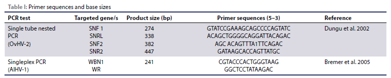
Single tube nested PCR for the detection of OvHV-2 was performed as previously described (Dungu et al. 2002). Oligonucleotide sequences and amplicon sizes are presented below (Table I).
• Detection of wildebeest alcelaphine herpesvirus 1 (AlHV-1)
Singleplex PCR was carried out for the detection of AIHV-1 as previously reported by Bremer et al. (2005). Oligonucleotide sequences and amplicon sizes are presented (Table I).
Gel electrophoresis
The PCR amplicons underwent electrophoresis at 120 volts for 30 minutes in 1.5% agarose gel containing 8 μl (0.8%) ethidium bromide. Eletrophoresed gels were subjected to UV in a gel documentation system (Vacutec, Pretoria, South Africa).
Statistical analysis
Descriptive statistics are presented in frequencies and percentages. Continuous data are described either using mean ± standard deviation or median and interquartile ranges. Results of tests are presented as proportions with the 95% confidence interval using the Wilson Score 95% confidence limit. Statistical analyses were performed in OpenEpi Open Source Epidemiological Statistics for Public Health, Version 3.01, CDC, USA.
Ethics
No ethical considerations were required for this study as the specimens used were diagnostic materials submitted by clients to the laboratories for diagnostic and surveillance purposes. Permission to use the laboratory data was granted by the Limpopo Department of Agriculture and Rural Development director of Veterinary Services.
Results
A total of 385 samples were submitted to the various laboratories for routine testing of MCF between 2001 and 2021. Of the 385 samples, 64.67% (n = 249) were tested using PCR while the remaining 35.32% (n = 136) were examined by histopathology. The overall number of positive samples for this study was 57.40% (n = 221/385), of which 86.43% (n = 191/221) were confirmed by PCR and 13.57% (n = 30/221) by histopathology (Table II). Table II shows that the period between 2001 and 2014 had the highest positivity rates of MCF while 2015 to 2021 reported the lowest number of cases.
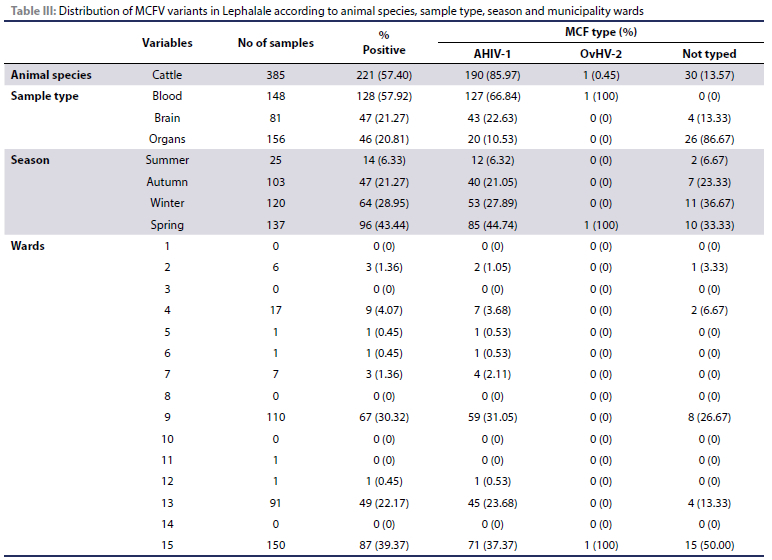
The distribution of the positive samples by season revealed that more cases were reported in spring (43.44%; n = 96) and winter (28.95%; n = 64), followed by autumn (21.27%; n = 47) and summer (6.33%; n = 14) (Table III). Based on sample types, the frequencies of detection were high in blood (83.12%; n = 128) followed by brain (57.83%; n = 48) and organs (29.30%; n = 46).
Of the 191 PCR-positive samples, 99.5% (n = 190) were genotyped as AIHV-1 and 0.5% (n = 1) as OvHV-2. AIHV-1 was detected throughout the two decades (2001-2022) under investigation, with the highest frequency observed in the years 2013, 2007, 2002, 2004, 2009, 2014, 2008, 2011, 2005, 2010, 2016 (Table II).
Based on sample type, AIHV-1, was detected in all specimens, with the highest frequency observed in blood, followed by brain and organs. AIHV-1, was detected in all seasons with spring having the highest frequency followed by autumn, winter and summer.
Our study also analysed incidences of MCF according to wards. The highest incidences in the Lephalale municipality were detected in wards 15, 9, and 13, with detection rates of 39.37%, 30.32%, and 22.17%, respectively, while frequencies of less than 10%, were observed in the remaining wards (Table III).
Discussion
MCF is an important disease in many countries of southern Africa due to the practice of farming of cattle in close proximity to wildebeest and sheep (Wambua et al. 2016; Cook et al. 2019; Sharma et al. 2019). Due to the absence of targeted surveillance of MCF in various parts of the world, the true burden of this disease is currently unknown.
The diagnosis of MCF is primarily based on a combination of clinical signs, necropsy findings, histopathology and detection of viral DNA in clinical samples by PCR (Pesca et al. 2019). However, PCR has become the method of choice for diagnoses of any forms of this virus. In this study, 86.43% of the samples were positive for MCF on PCR while only 13.57%, were detected on histopathology. Orono et al. (2019) also reported 94% positive samples from PCR. The high number of positive samples on PCR was expected since it is more sensitive compared to histopathology and should be a preferred method (Pesca et al. 2019; Riaz et al. 2021). It is further noted that the figures in this study could have been higher, but due to financial constraints and lack of compensation for confirmed cases, one animal from a group showing MCF clinical signs was sampled.
Annual incidences of WA-MCF are highly variable globally. However, annual incidences of this disease in South Africa remain unknown with losses of up to 34% previously reported in the North West province (Honiball et al. 2008). In the current study, rates of occurrence of MCF fluctuated through the years, with some years exhibiting high positivity compared to others. The higher frequencies in these years may be due to the meteorological factors, viral infective doses, availability of carrier and susceptible animals, newly introduced wildebeest in the area and also due to a higher reporting rate to laboratories.
The current study also investigated the occurrence of MCF in various seasons. During the period under review, there were higher incidents of the disease in spring and winter than autumn and summer. These results are in agreement with the South African literature, which documented high positive MCF cases in spring and late winter, respectively (Barnard et al. 1989; Honiball et al. 2008). Moreover, the results of the current study are in disagreement with those previously reported in Kenya (Orono et al. 2019) and Tanzania (Swai et al. 2013) which reported positive MCF cases in April, which constitute autumn season in their respective countries (Wambua et al. 2016). It has been established that occurrence of MCF in Kenya and Tanzania is due to close contact between cattle and wildebeest. Moreover, they coincide with the wildebeest calving season, with peaks observed when wildebeest calves are three to four months of age (Orono et al. 2019). However, this is in total disagreement with occurrence in South Africa, where transmission over long distances have been documented. Furthermore, high peaks are recorded during spring when wildebeest calves are eight to nine months old and no longer shedding highly significant virus (Barnard et al. 1989; Wambua et al. 2016). This supports the fact that these incidences are neither dependent on wildebeest calving season nor close contact.
It has been reported that wildebeest stress levels contribute to outbreaks of MCF (Rweyemamu et al. 1974). In South Africa, the winter months are associated with drought and scarcity of lush nutritious pastures, thus imposing nutritional stresses on veld-grazed production animals, including wildebeest (Lamega et al. 2021). Moreover, it is during this season, when wildebeest are hunted for trophy and meat (Hoffman et al. 2011). This exerts a lot of stress on the wildebeest and causes reactivation of latent virus, leading to transmission to cattle (Barnard et al. 1989; Honiball et al. 2008). Moreover, the calving season in beef cattle is usually synchronised to take place in spring in RSA and the period is associated with stress factors such as rising heat and humidity levels, escalating occurrences of infectious diseases, and increased demands for high quality feed in large quantities for physiological maintenance and milk production for the offspring (Lucy 2019). The cows' immune systems normally weaken as a result, and they become prone to diseases, including MCF. This may be the reason why more incidences are reported during spring and winter seasons.
The Lephalale municipality is divided into wards comprising residential areas (villages) as well as farms. The highest MCF cases were detected in wards 15, 9 and 13, which represent the majority of commercial game farms while the low detection rates were observed in other wards. These high differences in detection may be attributed to knowledge and reporting on the disease.
Conclusion
MCF is an economically important and notifiable disease of cattle in southern Africa. The study has highlighted that the disease in the Lephalale municipality conforms to the South African perspective in terms of season and transmission, as these animals (wildebeest and reservoirs) are not in close contact. The study has also highlighted the areas that require more attention in terms of control measures. However, there is currently no vaccine available against MCF. Furthermore, the transmission route of the disease is still being speculated. All wildebeest owners can pay into a collective insurance scheme and cattle farmers can claim for their confirmed MCF from this insurance. More research on vaccine production and transmission of the disease from wildebeest to cattle needs to be undertaken, so that we can have a better understanding of the disease in the country. Moreover, stricter measures on the movement of wildebeest, awareness campaigns and surveillance programmes should be put in place to help inform stakeholders, mitigate the problems and monitor the disease. Our study also shows the necessity for further research into the financial or economic impact of MCF on cattle farmers.
Acknowledgement
The authors would like to thank the Limpopo Department of Agriculture and Rural Development for granting them permission to use their laboratory data.
Conflict of interest
The author(s) declare no potential conflicts of interest with respect to the research, authorship, and/or publication of this article.
Funding
No funding was required for this study.
Ethics
No ethical considerations were required for this study as the specimens used were diagnostic materials submitted by clients to the laboratories for diagnostic and surveillance purposes.
ORCID
E Seakamela https://orcid.org/0000-0002-9212-1066
DD Lazarus https://orcid.org/0000-0001-5683-7887
D Malema https://orcid.org/0009-0008-6053-4262
A Lubisi https://orcid.org/0000-0003-4905-9516
I Matle https://orcid.org/0000-0002-1495-357X
References
Barnard, B.J., Van de Pypekamp, H.E., 1988, Wildebeest-derived malignant catarrhal fever: unusual epidemiology in South Africa. Onderstepoort J Vet Res 55, 69-71. [ Links ]
Barnard, B.J., Bengis, R.G., Griessel, M.D., et al., 1989, Excretion of alcelaphine herpesvirus-1 by captive and free-living wildebeest (Connochaetes taurinus). Onderstepoort J Vet Res 56, 131-134. [ Links ]
Bremer, C.W., Swart, H., Doboro, F., et al., 2005, Discrimination between sheep-associated and wildebeest-associated malignant catarrhal fever virus by means of a single-tube duplex nested PCR. Onderstepoort J Vet Res 72, 285-91. https://doi.org/10.4102/ojvr.v72i4.184. [ Links ]
Carruthers, J., 2008, Wilding the farm or farming the wild? The evolution of scientific game ranching in South Africa from the 1960s to the present. Transactions of the Royal Society of South Africa 63, 160-181. https://doi.org/10.1080/00359190809519220. [ Links ]
Chiyangwa, T., 2018, Financial implications of converting from livestock to game farming in the Karoo region, South Africa. (Doctoral dissertation, Stellenbosch: Stellenbosch University). 111. [ Links ]
Cloete, P.C., Taljaard, P.R., Grove, B., 2007, A comparative economic case study of switching from cattle farming to game ranching in the Northern Cape province: research article. S Afr J Wildlife Research 37, 71-78. https://doi.org/10.3957/0379-4369-37.1.71. [ Links ]
Cook, E., Russell, G., Grant, D. et al., 2019, A randomised vaccine field trial in Kenya demonstrates protection against wildebeest-associated malignant catarrhal fever in cattle. Vaccine 37, 5946-5953. https://doi.org/10.1016/j.vaccine.2019.08.040. [ Links ]
Decker, C., Hanley, N., Czajkowski, M. et al., 2021, Predicting uptake of a malignant catarrhal fever vaccine by pastoralists in northern Tanzania: Opportunities for improving livelihoods and ecosystem health. Ecological Economics 190, 107189. https://doi.org/10.1016/j.ecolecon.2021.107189. [ Links ]
Dungu, B., Bosman, A.M., Kachelhoffer, C., et al., 2002, Single-tube nested PCR for detection of the sheep-associated agent. The Veterinary Record 151(23):703-6. [ Links ]
Headley, S.A., Oliveira, T.E.S., Li, H., et al., 2020, Immunohistochemical detection of intralesional antigens of ovine gammaherpesvirus-2 in cattle with sheep-associated malignant catarrhal fever. Journal of Comparative Pathology 174, 86-98. https://doi.org/10.1016/j.jcpa.2019.11.002. [ Links ]
Hoffman, L.C., Van Schalkwyk, S., Muller, M., 2011, Quality characteristics of blue wildebeest (Connochaetes taurinus) meat: short communication. S Afr J Wildlife Research 41, 210-213. https://doi.org/10.3957/056.041.0208. [ Links ]
Hussain, I., Kashoo, Z.A., Wani, A.H., et al., 2017, Malignant catarrhal fever: recent update. Indian J Anim Sci 87, 260-269. https://doi.org/10.56093/ijans.v87i3.68792. [ Links ]
Honiball, E.J., van Essen, L.D., du Toit, J.G., 2008, A review of malignant catarrhal fever in the republic of South Africa. University of Pretoria, Centre for Wildlife Management, Faculty of Natural and Agricultural Sciences 96. Available from: chrome-extension://efaidnbmnnnibpcajpcglclefindmkaj/https://savf.org.za/wp-content/uploads/2018/09/A-Review-of-malignant-catarrhal-fever-in-the-republic-of-South-Africa-2010.pdf. [ Links ]
Lamega, S.A., Komainda, M., Hoffmann, M.P., et al., 2021, It depends on the rain: Smallholder farmers' perceptions on the seasonality of feed gaps and how it affects livestock in semi-arid and arid regions in Southern Africa. Climate Risk Management 34, 100362. https://doi.org/10.1016/j.crm.2021.100362. [ Links ]
Lankester, F., Lugelo, A., Kazwala, R., et al., 2015, The economic impact of malignant catarrhal fever on pastoralist livelihoods. PLOS ONE 10, e0116059. https://doi.org/10.1371/journal.pone.0116059. [ Links ]
Lankester, F., Russell, G.C., Lugelo, A., et al., 2016, A field vaccine trial in Tanzania demonstrates partial protection against malignant catarrhal fever in cattle. Vaccine 34, 831-838. https://doi.org/10.1016/j.vaccine.2015.12.009. [ Links ]
Li, H., Cunha, C.W., Taus, N.S., 2011, Malignant catarrhal fever: understanding molecular diagnostics in context of epidemiology. International Journal of Molecular Sciences 12, 6881-6893. https://doi.org/10.3390/ijms12106881. [ Links ]
Lucy, M.C., 2019, Stress, strain, and pregnancy outcome in postpartum cows. Animal Reproduction 16, 455-464. https://doi.org/10.21451/1984-3143-AR2019-0063. [ Links ]
Mangani, T., Coetzee, H., Kellner, K., et al., 2020, Socio-economic benefits stemming from bush clearing and restoration projects conducted in the D'Nyala Nature Reserve and Shongoane Village, Lephalale, South Africa. Sustainability 12, 5133. https://doi.org/10.3390/su12125133. [ Links ]
Meravi, P., Tiwari, A., Shukla, P.C., et al., 2019, Emerging threats of sheep-associated malignant catarrhal fever in India: A Review. Journal of Pharmacognosy and Phytochemistry 8, 1891-1895. [ Links ]
Myster, F., Gong, M.J., Javaux, J., et al., 2020, Alcelaphine herpesvirus 1 genes A7 and A8 regulate viral spread and are essential for malignant catarrhal fever. PLoS pathogens 16, e1008405. https://doi.org/10.1371/journal.ppat.1008405. [ Links ]
Orono, S.A., Gitao, G.C., Mpatswenumugabo, J.P., et al., 2019, Field validation of clinical and laboratory diagnosis of wildebeest-associated malignant catarrhal fever in cattle. BMC Veterinary Research 15, 69. https://doi.org/10.1186/s12917-019-1818-8. [ Links ]
Parameswaran, N., Dewals, B.G., Giles, T.C., et al., 2014, The A2 gene of alcelaphine herpesvirus-1 is a transcriptional regulator affecting cytotoxicity in virus-infected T cells but is not required for malignant catarrhal fever induction in rabbits. Virus Research 188, 68-80. https://doi.org/10.1016/j.virusres.2014.04.003. [ Links ]
Patel, J.R., Heldens, J.G.M., Bakonyi, T., et al., 2012, Important mammalian veterinary viral immunodiseases and their control. Vaccine 30, 1767-1781. https://doi.org/10.1016/j.vaccine.2012.01.014. [ Links ]
Pesca, C., Gobbi, M., Palombi, C., et al., 2019, Bovine malignant catarrhal fever: case reporting in central Italy. Veterinaria Italiana 55, 279-283. https://doi.org/10.12834/VetIt.1708.9037.4. [ Links ]
Reid, H.W., Van Vuuren, M., 2004, Malignant catarrhal fever, in: Coetzer, J.A.W., Tustin, R.C. (Eds.), Infectious Diseases of Livestock. Oxford University Press, Cape Town. [ Links ]
Riaz, A., Dry, I., Dalziel, R., et al., 2021, Molecular detection and characterization of ovine herpesvirus-2 using heminested PCR in Pakistan. J Vet Sci 22, e51. https://doi.org/10.4142/jvs.2021.22.e51. [ Links ]
Rweyemamu, M.M., Karstad, L., Mushi, E.Z., et al., 1974, Malignant catarrhal fever virus in nasal secretions of wildebeest: a probable mechanism for virus transmission. J Wildl Dis 10, 478-487. https://doi.org/10.7589/0090-3558-10.4.478. [ Links ]
Sharma, B., Parul, S., Basak, G., et al., 2019, Malignant catarrhal fever (MCF): An emerging threat. Journal of Entomology and Zoology Studies 7, 26-32. [ Links ]
Swai, E.S., Kapaga, A.M., Sudi, F., et al., 2013, Malignant catarrhal fever in pastoral Maasai herds caused by wildebeest-associated alcelaphine herpesvirus-1: An outbreak report. Vet Res Forum 4, 133-136. [ Links ]
Taylor, A., Lindsey, P.A., Davies-Mostert, H., et al., 2016, An assessment of the economic, social and conservation value of the wildlife ranching industry and its potential to support the green economy in South Africa. The Endangered Wildlife Trust 96-109. [ Links ]
Turan, T., Isidan, H., Atasoy, M.O., et al., 2020, Genetic diversity of ovine herpesvirus 2 strains obtained from malignant catarrhal fever cases in eastern Turkey. Virus Research 276, 197801. https://doi.org/10.1016/j.virusres.2019.197801. [ Links ]
Wambua, L., Wambua, P.N., Ramogo, A.M., et al., 2016, Wildebeest-associated malignant catarrhal fever: perspectives for integrated control of a lymphoproliferative disease of cattle in sub-Saharan Africa. Arch Virol 161, 1-10. https://doi.org/10.1007/s00705-015-2617-6. [ Links ]
Corresponding author: Email: bio4slim@gmail.com

