


Servicios Personalizados
Articulo
 Inglés (pdf)
 Articulo en XML
 Referencias del artículo
Indicadores

Links relacionados
-
 Citado por Google
 -
 Similares en Google
Compartir

 Permalink
Journal of the South African Veterinary Association
versión On-line ISSN 2224-9435
versión impresa ISSN 1019-9128
J. S. Afr. Vet. Assoc. vol.94 no.1 Pretoria 2023
http://dx.doi.org/10.36303/JSAVA.547
ORIGINAL RESEARCH
Poikilocytosis of Angora goats is associated with erythrocyte density and reticulocytosis
SDC ParsonsI, II; D BecksI; A VermeulenI; M HobsonIII; RM WarrenI; EH HooijbergIV
IDSI-NRF Centre of Excellence for Biomedical Tuberculosis Research, SAMRC Centre for Tuberculosis Research, Division of Molecular Biology and Human Genetics, Faculty of Medicine and Health Sciences, Stellenbosch University, South Africa
IIAfrivet Business Management, South Africa
IIIMohair SA, Camdeboo Veterinary Clinic, South Africa
IVDepartment of Companion Animal Clinical Studies, Faculty of Veterinary Science, University of Pretoria, South Africa
ABSTRACT
Angora goats in South Africa experience several syndromes that result in notable morbidity and mortality in juveniles and adults, but not kids. Insight into their causes is hampered by the lack of normal reference values for this breed, and the present study therefore aimed to characterise (1) differences in the haematology of healthy kids at birth and weaning, and (2) the haematology of apparently healthy yearlings. Selected variables were measured by blood smear analysis, and complete blood counts were performed using an ADVIA 2120i. Variables at 1, 11, and 20 weeks of age were compared using the Friedman test and associations between variables of yearlings were determined by correlation analysis. In kids, red blood cell count, mean corpuscular haemoglobin concentration (MCHC), and poikilocytosis increased over time, while mean corpuscular haemoglobin (MCH) and mean corpuscular volume (MCV) decreased. Yearlings displayed a lower MCHC, and higher haemoglobin distribution width than previously reported for goats, and these were positively correlated with poikilocytosis, as were reticulocyte counts. White cell counts of yearlings exceeded normal values previously reported for goats, with some individuals displaying remarkably high mature neutrophil counts. Changes in haemoglobin variant expression or cation and water fluxes are possible explanations for the findings in kids, while in yearlings, the associations between MCHC, HDW, poikilocytosis, and reticulocytosis suggest alterations in red cell hydration in adulthood that are associated with increased red cell turnover. These findings may prove informative in the further investigation of various clinical syndromes in this population.
Keywords: Angora goat, mean corpuscular haemoglobin concentration (MCHC), mean corpuscular volume (MCV), poikilocytosis, reticulocytosis
Introduction
South Africa produces over half of the world's mohair, a fibre that is highly valued for its lustre and sheen, making Angora goat farming a vital part of the rural economy of semi-arid parts of this country (Pienaar et al. 2018). Notably, juvenile and adult Angora goats of South African (SA) origin, but not kids, experience a remarkable array of poorly understood clinical syndromes, suggesting an age-related aetiology. These syndromes include "weaning shock" (Hobson 2019; Snyman 2007), severe sensitivity to cold (Wentzel et al. 1979), "habitual abortion" (Van Rensburg 1971), and "swelling disease", which presents as the sudden onset of subcutaneous oedema (Bath & Vermeulen 2011). Despite numerous investigations, their causes have not been elucidated and insight into the pathophysiology of these syndromes requires a better understanding of the normal haematology and serum chemistry of healthy individuals.
In the present study we aimed to characterise differences in the haematology of kids at birth and weaning as well as the haematology of apparently healthy SA Angora yearlings that were maintained under standard farming practices. As part of this exercise, we characterised red cell poikilocytosis, as this phenomenon has been described in Angora goats in the United States of America (Jain & Kono 1977) but not formally recorded in SA. This phenomenon has not been associated with clinical disease in Angora goats, although it has been reported to share some characteristics with red cell sickling of humans (Jain & Kono 1977).
Materials and methods
Animals and sampling
Animals were from a single herd from the Graaff-Reinet region of the Eastern Cape Province, South Africa, and sampled with the owner's consent. Newborn kids (n = 8, numbered K1 to K8) and yearlings (n = 28, numbered Y1 to Y28) were clinically healthy for the duration of the study, although faecal flotation tests performed three weeks earlier revealed that yearlings were infested with roundworms (100-2 000 eggs/g of faeces, median = 500 eggs/g) and coccidia (800-408 000 oocysts/g of faeces, median = 6 400 oocysts/g). All animals were maintained under free-range conditions and kids had additional access to lucerne pasture. On each sampling occasion, 4 ml of blood was collected from the jugular vein into Vacutainer tubes containing dipotassium ethylenediaminetetraacetic acid (EDTA) (BD, Franklin Lakes, NJ, USA). Samples were collected from the kids during the first, 11th, and 20th week after birth, and from yearlings at approximately one year old. On each occasion, air-dried blood smears were made within 30 min of sampling.
Haematological analysis
Whole blood samples were transported in a cooler box containing ice packs to the Clinical Pathology Laboratory, Faculty of Veterinary Science, University of Pretoria. Automated haematology analysis was performed circa 20 h after blood collection using an ADVIA 2120i (Siemens Healthcare, South Africa), with multispecies software set to the goat program. In goats, such a delay in processing has been shown to have a negligible effect on the measurement of erythrocyte variables and WBC (Püsch 2002). Quality control for this analyser included daily measurement of three levels of commercial quality control material; control results were within laboratory performance goals over the study period. Directly measured variables included whole blood haemoglobin (Hb) concentration (HGB), red blood cell count (RBC), mean corpuscular volume (MCV), red cell distribution width (RCDW), corpuscular Hb concentration mean (CHCM), Hb concentration distribution width (HDW), reticulocyte count, white blood cell count (WBC), platelet count (PLT), and mean platelet volume (MPV). Additionally, mean corpuscular Hb (MCH), mean corpuscular Hb concentration (MCHC), and haematocrit (HCT) were automatically calculated from HGB, RBC and MCV, cellular Hb content (CH) from CHCM and MCV, cellular Hb concentration (cell. HGB) from CH and RBC, and plateletcrit (PCT) from PLT and MPV. Blood smear evaluation and manual differential white cell counts (100 nucleated cells) were performed by experienced and registered veterinary technologists and verified by a board-certified veterinary clinical pathologist (EH).
Erythrocyte morphology
Blood smears were stained with Wright-Giemsa using an automated stainer (Hematek, Siemens Healthcare, South Africa). Using an Axiocam 305 colour camera attached to an Axio Observer 7 microscope with a 63x oil immersion objective lens (Carl Zeiss AG, Jena, Germany), five digital photomicrographs were taken of fields in the monolayer area that had few touching or deformed cells. Photomicrographs were viewed using ZEN (blue edition) imaging software (Carl Zeiss AG) and two representative images were exported in jpg format for measurement of cell roundness using ImageJ software (Schneider et al. 2012). To exclude debris, artifacts, overlapping cells, and platelets, the ImageJ particle analysis tool was set to exclude particles that touched the image edge, those with a circularity of less than 0.6, and those outside a range of 4 to 35 μm2 for one week old kids, and 4 to 18 μm2 for animals of 11 weeks and older. For each sampling occasion, the red cell roundness (4 x area/(π x [major axis]2) of 400 cells was measured. Poikilocytosis was defined as the coefficient of variation (CV) of red cell roundness and recorded as a percentage (CV x 100).
Statistical analysis
Goats have particularly small erythrocytes, and as an indicator of the accuracy of the measurement of RBC, MCV and CHCM, the mean absolute difference between the two independent measures of cellular HGB concentration, i.e. MCHC and CHCM, was calculated for the yearlings. A difference of more than 1.9 g/ dL can be indicative of inaccuracies in the measurement of these variables (Bauer & Moritz 2008a).
Results for kids at one, 11 and 20 weeks after birth, respectively, and for yearlings, were summarised using descriptive statistics. To identify differences in selected variables of kids at one, 11 and 20 weeks old, these were compared using the Friedman test and Dunn's post-hoc multiple comparisons test. For the yearlings, association between variables was investigated using Spearman's rank-order correlation analysis. Statistical analysis was done using Graphpad Prism 7 (GraphPad Software, San Diego, CA) and a p-value of < 0.05 was considered significant.
Results
Blood from one yearling (Y23) underwent marked visible in vitro haemolysis and haematology results for this individual were excluded from further analysis (Supplementary Table I). Notably, the CHCM of this animal (32.7 g/dL) was the lowest of all the yearlings and its red cells were highly rounded (11.6% poikilocytosis), a phenomenon that has previously been associated with osmotic fragility in Angora goats (Jain et al. 1980). No other samples were visibly haemolysed and the mean absolute difference between MCHC and CHCM of the remaining yearlings (n = 27) was 1.2 g/dL, indicating that in vitro haemolysis was negligible in these samples and that automated haematology results for this population were valid. In many instances, mild to severe platelet aggregation was observed on blood smears and data on platelet count, MPV and PCT are recorded in Supplementary Tables II and III but not included in further analysis.
Two kids were lost to follow-up and complete data sets were available for six animals. The red cell morphology of Angora goat kids at one, 11, and 20 weeks after birth is illustrated in Figure 1. The haematology results for these animals are given in Supplementary Table II and selected variables are summarised in Table I and Figure 2.
Between the ages of one and 20 weeks, kids showed a statistically significant increase in RBC, MCHC, and CHCM, and a significant decrease in HCT, MCV, MCH, CH, and RCDW. At 11 weeks old, HDW, WBC, as well as lymphocyte and monocyte counts, were significantly elevated compared to Week one values. Poikilocytosis increased over time, however these changes were not statistically significant (p = 0.14).
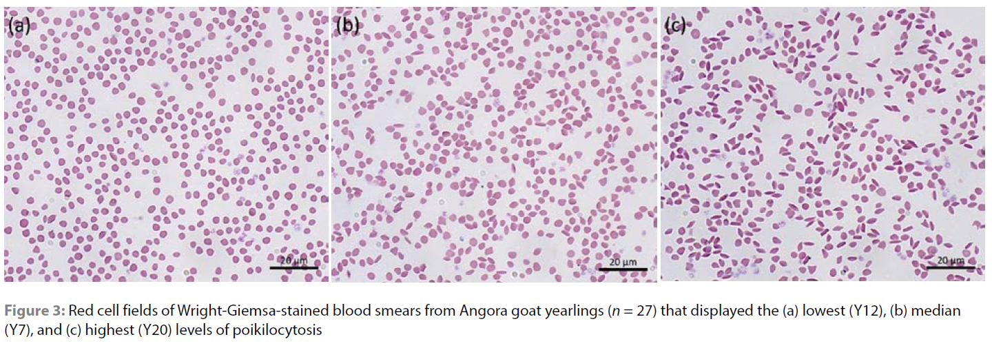
The red cell morphology of the Angora goat yearlings is illustrated in Figure 3, and the haematology data is given in Supplementary Table III and summarised in Table II. Correlation analysis between erythrocyte variables revealed 23 pairs that were significantly correlated (Supplementary Tables IV and V). Three pairs comprised equivalent variables (e.g. HGB and cell HGB). Six pairs included a composite variable that was directly dependent on the correlated variable (e.g. HCT and RBC). Seven pairs were complementary in red cell and HGB homeostasis (e.g. RBC and MCV). In addition, poikilocytosis was correlated with five variables: positively with MCHC, CHCM, HDW, and reticulocytosis (Figure 4), and negatively with HCT (r = 0.39, p < 0.05). Similarly, HDW was positively correlated with reticulocytosis (r = 0.51, p < 0.01) and negatively correlated with HCT (r = 0.39, p < 0.05).
Discussion
Angora kids displayed a significant decrease in MCH, MCV, and HCT, and a significant increase in RBC and MCHC during the first 20 weeks after birth. These changes were complementary, resulting in a relatively constant HGB level over time, and are similar to neonatal changes seen in other goat breeds (Holman & Dew 1964, 1965; Mbassa & Poulsen 1991b). In humans, foetal Hb (HbF) is associated with a relatively high MCH and MCV (Lim et al. 2015) and the changes in MCH of Angora kids may be similarly associated with a switch in Hb expression from foetal to adult variants (Huisman et al. 1969). A strong correlation between MCH and MCV is highly conserved across adult vertebrates and explains, in part, the concomitant decrease in MCV (Hawkey et al. 1991).
Despite the close association between MCH and MCV, the MCHC of Angora kids rose significantly over time. This phenomenon has previously been described in goats and calves (Brun-Hansen et al. 2006; Holman & Dew 1965), and again, may be associated with changes in the expression of Hb variants (Brun-Hansen et al. 2006; Olivieri et al. 1992). In addition, changes in cation flux, including that of potassium (K+), especially via the cation chloride family of cotransporters, directly affects the volume and density of erythrocytes (Browning et al. 2006; Quarmyne et al. 2011). This may indeed be an important regulator in ruminants, as foetal goat red cells have a far greater [K+] than do those of their dams (Blechner 1960), and red cells of neonatal lambs show a shift from high [K+] (HK) to low [K+] (LK) that is associated with changes in cation pump activity (Wolowyk & Ellory 1985). The SA Angora goat yearlings also displayed a lower MCH and MCV, and a higher MCHC and HDW, than reported for Turkish Angora goats and other goat breeds (Bauer & Moritz 2008b; Byers & Kramer 2010; Polat et al. 2018). Given that Angora goats display a red cell [K+] that is both markedly lower than other goat breeds and highly variable between individuals (Blechner 1960; Erkoç et al. 1987), it is plausible that variations in cation fluxes also account for the red cell volume and density characteristics of adult animals.
Poikilocytosis was mild at birth and became more pronounced with age. This change was not statistically significant, although it is probable that this results from the small sample size following the loss of two young kids from the study. Such losses are well described in the SA literature (Collins 2021). A similar increase in neonatal poikilocytosis is seen in white-tailed deer (Odocoilus virginianus), a species in which red cell sickling is associated with adult Hb (HbA) (Esin et al. 2018; Kitchen et al. 1964), and a similar phenomenon has been proposed as the cause of poikilocytosis of Angora goats (Jain et al. 1980). This conclusion was inferred from the observation that, during severe anaemia, poikilocytosis resolved with a concurrent change in Hb variant expression (Huisman et al. 1969; Jain et al. 1980). However, a causal relationship between these phenomena has not been conclusively shown and alternative explanations are therefore possible. In humans, a prerequisite for red cell sickling is cellular dehydration caused by changes in cation fluxes and reduced intracellular [K+] (Lew & Bookchin 2005), and notably, during periods of anaemia, LK sheep produce increased numbers of larger HK cells with higher K+ pump and leak fluxes (Kim et al. 1980). If the same is true for LK Angora goats, which also show increased MCV and decreased MCHC during anaemia (Jain et al. 1980), then changes in cation fluxes might be the primary cause of changes in red cell morphology during anaemia, rather than Hb switching. Red cell osmotic fragility also increases markedly in Angora goats during anaemia (Jain et al. 1980), further suggesting that changes in red cell cation and water fluxes occur during this time. In the present study, poikilocytosis was positively correlated with MCHC and HDW, supporting the notion that the phenomenon is closely associated with cell hydration.
Notably, all yearlings displayed a mild reticulocytosis, which is always a significant finding in goats (Jones & Allison 2007). A ready explanation for this phenomenon is that it reflects a response to blood loss caused by the intestinal parasites present at the time of sampling. However, the SA yearlings were not obviously anaemic given that their HGB was very similar to that reported for a Turkish population, and within the range of normal values reported for other goat breeds (Byers & Kramer 2010; Polat et al. 2018). Furthermore, although the correlation between reticulocytosis and both poikilocytosis and HDW was only moderate, this finding is nonetheless intriguing given the independence of the analytical methods, and raises the possibility that red cell dehydration and deformation are associated with increased red cell turnover in this breed.
Lastly, Angora kids displayed a significant increase in lymphocyte counts between birth and 11 weeks of age, a change that in humans has been attributed to the sudden exposure to novel antigens after birth (Comans-Bitter et al. 1997). Kids also displayed an increase in circulating neutrophils over time, and both phenomena have been documented in other goat breeds (Mbassa & Poulsen 1991a). In yearlings, the WBC exceeded the range of normal values reported for a Turkish Angora goat population as well as other goat breeds (Byers & Kramer 2010; Polat et al. 2018), with some individuals displaying remarkably high mature neutrophil counts, but no evidence of an inflammatory response. Similarly, SA Angora goats displayed a notably large variation in WBC in a study investigating vaccination against Ehlichia ruminantium (Haw 2013), and individuals presenting with non-infectious "habitual abortion" (Morgenthal 1966; Van Rensburg 1971) and "swelling disease" (Hobson 2016) displayed significantly higher WBC than did unaffected individuals. It is currently unknown if the leukocytosis associated with these syndromes predates the onset of clinical signs, however the present findings suggest that this possibility should be investigated.
Study limitations
Limitations to the present study include the fact that only a single, closely related goat population was investigated, and findings may not be reflective of the national herd; that intestinal parasitism was not controlled and may have affected red cell variables of heavily infested individuals; and, given the remarkable findings, that the delay between sample collection and analysis may have affected some measurements in this breed. It is also not clear from the present study if some of the variables, such as MCHC and poikilocytosis, reflect solely in vitro changes, as must be the case for the sample that underwent severe haemolysis, or also reflect differences in in vivo physiology.
Conclusion
This study provides reference haematology data for a population of SA Angora goats, evidence that in vitro poikilocytosis might be associated with changes in red cell hydration as well as red cell turnover, and insight into the WBC of apparently healthy Angora goats. These findings may prove informative in the further investigation of poorly understood syndromes in this population.
Acknowledgements
The authors thank Mr Francois Theron for kindly providing the study animals, the technologists of the Clinical Pathology Laboratory, Faculty of Veterinary Science, University of Pretoria, for assistance with sample analysis, and Dr Dalene de Swardt of the Central Analytical Facilities, Stellenbosch University, for her assistance with the microscopy. Financial support for this research was provided by the South African Medical Research Council (SAMRC) Centre for Tuberculosis Research, the Harry Crossley Foundation, and the Faculty of Medicine and Health Sciences Undergraduate Research Project Fund of Stellenbosch University.
Conflict of interest
The authors declare they have no conflicts of interest that are directly or indirectly related to the research.
Funding source
This work was undertaken with financial support from:
• The South African Medical Research Council (SAMRC) Centre for Tuberculosis Research
• The Harry Crossley Foundation
• The Faculty of Medicine and Health Sciences Undergraduate Research Project Fund of Stellenbosch University.
Study sponsors were not involved in any way in the study design, collection, analysis and interpretation of data; the writing of the manuscript; and the decision to submit the manuscript for publication.
Ethical approval
The author/s declare that this submission is in accordance with the principles laid down by the Responsible Research Publication Position Statements as developed at the 2nd World Conference on Research Integrity in Singapore, 2010.
This article does not contain any studies with human subjects. Prior to the commencement of the study, ethical approval was obtained from the following ethical review board: Animal Care and Use Committee of Stellenbosch University (protocol ACU-2019-10760).
All institutional and national guidelines for the care and use of laboratory animals were followed.
ORCID
SDC Parsons https://orcid.org/0000-0002-9033-9686
RM Warren https://orcid.org/0000-0001-5741-7358
EH Hooijberg https://orcid.org/0000-0002-4367-799X
References
Bath, G., Vermeulen, S., 2011, Swelling disease, Available from: http://gadi.agric.za/articles/Agric/swellingdisease. Accessed 18 Oct 2022. [ Links ]
Bauer, N., Moritz, A., 2008a, Evaluation of three methods for measurement of hemoglobin and calculated hemoglobin parameters with the ADVIA 2120 and ADVIA 120 in dogs, cats, and horses, Vet Clin Pathol 37, 173-179. https://doi.org/10.1111/j.1939-165X.2008.00039.x. [ Links ]
Bauer, N., Moritz, A., 2008b, Evaluation of three methods for measurement of hemoglobin and calculated hemoglobin variables with the ADVIA 120® and ADVIA 2120® systems in goats, J Vet Diagn Invest 20, 593-597. https://doi.org/10.1177/104063870802000509. [ Links ]
Blechner, J.N., 1960, Sodium and potassium in erythrocytes of adult and fetal goats, Am J Physiol 199(6), 1 174-1176. https://doi.org/10.1152/ajplegacy.1960.199.6.1174. [ Links ]
Browning, J.A., Ellory, J.C., Gibson, J.S., 2006, Pathophysiology of red cell volume, in F. Lang (ed.) Mechanisms and Significance of Cell Volume Regulation. Karger Publishers, pp. 241-268. https://doi.org/10.1159/000096327. [ Links ]
Brun-Hansen, H.C., Kampen, A.H., Lund, A., 2006, Hematologic values in calves during the first 6 months of life, Vet Clin Pathol 35, 182-187. https://doi.org/10.1111/j.1939-165X.2006.tb00111.x. [ Links ]
Byers, S.R., Kramer, J.W., 2010, Normal hematology of sheep and goats, in D.J. Weiss, K.J. Wardrop, O.W. Schalm (eds) Schalm's Veterinary Hematology. Wiley-Blackwell, Ames, Iowa, USA, pp. 836-842. [ Links ]
Collins, T., 2021, Don't count your lambs, in The Scapegoat. Reach Publishers, Westville, pp. 31-33. [ Links ]
Comans-Bitter, W.M., De Groot, R., Van den Beemd, R., et al., 1997, Immunophenotyping of blood lymphocytes in childhood. Reference values for lymphocyte subpopulations, J Pediatr 130, 388-393. https://doi.org/10.1016/S0022-3476(97)70200-2. [ Links ]
Erkoç, F.Ü., Alparslan, Z.N., Uğrar, E., 1987, Red blood cell potassium types of Angora goats (Capra hircus), Comp Biochem Physiol A Physiol 87, 9-11. https://doi.org/10.1016/0300-9629(87)90416-6. [ Links ]
Esin, A., Bergendahl, L.T., Savolainen, V., et al., 2018, The genetic basis and evolution of red blood cell sickling in deer, Nat Ecol Evol 2, 367. https://doi.org/10.1038/s41559-017-0420-3. [ Links ]
Haw, A., 2013, Attenuated heartwater vaccine (Ehrlichia ruminantium Welgevonden): immunization of Angora goats using the intra-muscular route of administration, MSc thesis, University of Pretoria, Pretoria. [ Links ]
Hawkey, C.M., Bennett, P.M., Gascoyne, S.C., et al., 1991, Erythrocyte size, number and haemoglobin content in vertebrates, Br J Haematol 77, 392-397. https://doi.org/10.1111/j.1365-2141.1991.tb08590.x. [ Links ]
Hobson, M., 2016, Swelsiekte - possible role of the inflammatory process. Available from: https://www.angoras.co.za/article/swelsiekte-possible-role-of-the-inflammatory-process. Accessed 18 Oct 2022. [ Links ]
Hobson, M., 2019, Weaning shock in Angora goat kids. Available from: https://www.angoras.co.za/article/weaning-shock-in-angora-goat-kids. Accessed 16 Mar 2023. [ Links ]
Holman, H.H., Dew, S.M., 1964, The blood picture of the goat: II. - Changes in erythrocytic shape, size and number associated with age, Res Vet Sci 5, 274-287. https://doi.org/10.1016/S0034-5288(18)34792-1. [ Links ]
Holman, H.H., Dew, S.M., 1965, The blood picture of the goat: III. Changes in haemoglobin concentration and physical measurements occurring with age, Res Vet Sci 6, 245-255. https://doi.org/10.1016/S0034-5288(18)35064-1. [ Links ]
Huisman, T.H., Lewis, J.P., Blunt, M.H., et al., 1969, Hemoglobin C in newborn sheep and goats: a possible explanation for its function and biosynthesis, Pediatr Res 3, 189-198. https://doi.org/10.1203/00006450-196905000-00001. [ Links ]
Jain, N.C., Kono, C.S., 1977, Fusiform erythrocytes resembling sickle cells in Angora goats: light and electron microscopic observations, Res Vet Sci 22, 169-180. https://doi.org/10.1016/S0034-5288(18)33282-X. [ Links ]
Jain, N.C., Kono, C.S., Myers, A., et al., 1980, Fusiform erythrocytes resembling sickle cells in Angora goats: observations on osmotic and mechanical fragilities and reversal of cell shape during anaemia, Res Vet Sci 28, 25-35. https://doi.org/10.1016/S0034-5288(18)32767-X. [ Links ]
Jones, M.L., Allison, R.W., 2007, Evaluation of the ruminant complete blood cell count, Vet Clin North Am Food Anim Pract 23, 377-402, v. https://doi.org/10.1016/j.cvfa.2007.07.002. [ Links ]
Kim, H.D., Theg, B.E., Lauf, P.K., 1980, LK sheep reticulocytosis: effect of anti-L on K influx and in vitro maturation, J Gen Physiol 76, 109-121. https://doi.org/10.1085/jgp.76.1.109. [ Links ]
Kitchen, H., Putnam, F.W., Taylor, W.J., 1964, Hemoglobin polymorphism: its relation to sickling of erythrocytes in white-tailed deer, Science 144, 1237-1239. https://doi.org/10.1126/science.144.3623.1237. [ Links ]
Lew, V.L., Bookchin, R.M., 2005, Ion transport pathology in the mechanism of sickle cell dehydration, Physiol Rev 85, 179-200. https://doi.org/10.1152/physrev.00052.2003. [ Links ]
Lim, W.F., Muniandi, L., George, E., et al., 2015, HbF in HbE/ß-thalassemia: a clinical and laboratory correlation, Hematology 20, 349-353. https://doi.org/10.1179/1607845414Y.0000000203. [ Links ]
Mbassa, G.K., Poulsen, J.S.D., 1991a, Leukocyte profile in growing dwarf and landrace kids, Zentralbl Veterinarmed A 38, 389-397. https://doi.org/10.1111/j.1439-0442.1991.tb01026.x. [ Links ]
Mbassa, G.K., Poulsen, J.S.D., 1991b, Haematological profile in neonatal dwarf and landrace kids, J Vet Med Ser A 38, 510-522. https://doi.org/10.1111/j.1439-0442.1991.tb01042.x. [ Links ]
Morgenthal, J.C., 1966, The haematology of the Angora goat with special reference to the habitual aborter. I. The pregnant doe, Onderstepoort J Vet Res 33, 363-378. [ Links ]
Olivieri, O., Vitoux, D., Galacteros, F., et al., 1992, Hemoglobin variants and activity of the (K+C1-) cotransport system in human erythrocytes, Blood 79, 793-797. https://doi.org/10.1182/blood.V79.3J93.793. [ Links ]
Pienaar, L., Partridge, A., Morokong, T., 2018, The mohair industry: economic impact of possible market closure. Western Cape Department of Agriculture. [ Links ]
Polat, H., Pehlivan, E., Dellal, G., 2018, Annual change of haematological parameters in Angora goats, Biol Rhythm Res 0, 1-11. https://doi.org/10.1080/09291016.2018.1557850. [ Links ]
Püsch, M., 2002, Hämatologiesystem ADVIA 120, Softwareadaptation und Evaluation bei den Tierarten Schaf und Ziege [Hematology analyzer ADVIA 120, software adaptation and evaluation for the species sheep and goat], PhD thesis, Justus-Liebig-Universität, Giessen. Available from: http://bibd.uni-giessen.de/gdoc/2002/uni/d020109.pdf. [ Links ]
Quarmyne, M.-O., Risinger, M., Linkugel, A., et al., 2011. Defining a phenotype for red cell volume regulation and potassium chloride cotransport, Blood Cells Mol Dis 47, 95-99. https://doi.org/10.1016/j.bcmd.2011.04.007. [ Links ]
Schneider, C.A., Rasband, W.S., Eliceiri, K.W., 2012, NIH Image to ImageJ: 25 years of image analysis, Nat Methods 9, 671-675. https://doi.org/10.1038/nmeth.2089. [ Links ]
Snyman, M.A., 2007, Body weight and growth rate of South African Angora goat kids under different pre- and post-weaning management systems. S Afr J Anim Sci 37, 132-141. https://doi.org/10.4314/sajas.v37i2.4037. [ Links ]
Van Rensburg, S.J., 1971, Reproductive physiology and endocrinology of normal and habitually aborting Angora goats, Onderstepoort J Vet Res 38, 1-62. [ Links ]
Wentzel, D., Viljoen, K.S., Botha L.J.J., 1979, Physiological and endocrinological reactions to cold stress in the Angora goat. Agroanimalia 11, 19-22. [ Links ]
Wolowyk, M.W., Ellory, J.C., 1985, Changes in the cation composition and active K+ transport in the red cells of fetal sheep prepartum, Can J Physiol Pharmacol 63, 1454-1459. https://doi.org/10.1139/y85-238. [ Links ]
 Correspondence:
email: svenparsons@hotmail.com
Supplementary Data
The supplementary data is available in pdf: [Supplementary data]

