


Servicios Personalizados
Articulo
 Inglés (pdf)
 Articulo en XML
 Referencias del artículo
Indicadores

Links relacionados
-
 Citado por Google
 -
 Similares en Google
Compartir

 Permalink
Journal of the South African Veterinary Association
versión On-line ISSN 2224-9435
versión impresa ISSN 1019-9128
J. S. Afr. Vet. Assoc. vol.93 no.2 Pretoria 2022
http://dx.doi.org/10.36303/jsava.490
ORIGINAL RESEARCH
Prevalence of radiographic changes in forelimb digits and metacarpophalangeal joints of South African endurance racehorses
E HollenbachI; MP RobertII; C le RouxI; Y SmitI
IDepartment of Companion Animal Clinical Studies, Faculty of Veterinary Science, University of Pretoria, South Africa
IICentre Hospitalier Vétérinaire Equin de Livet, France
ABSTRACT
If the number of events alone is considered, endurance riding is the fastest growing and the second-most popular Federation Equestre Internationale (FEI) discipline. Lameness is the most common cause of elimination from endurance races worldwide. To the authors' knowledge, no studies have been published investigating the prevalence of radiographic changes in the forelimb digits and metacarpophalangeal joints (MCP) of endurance racehorses in South Africa.
The study aimed to investigate the prevalence of radiographic changes in the forelimb digits and MCP joints of South African endurance racehorses.
One hundred endurance racehorses registered with ERASA were volunteered by their owners to partake in the current study. Radiographs were obtained from horses competing in endurance races during the 2018-2019 endurance racing season. Radiographs included seven standard views of each distal forelimb. Radiographic images were independently evaluated by three observers, point prevalence and inter-rater reliability (IRR) was calculated.
Data analysis of the forelimb digits revealed a large proportion of horses with bilateral signs of dorsopalmar hoof imbalance (95%); a diversion from a straight digital axis (91%), with an extended (broken back) proximal interphalangeal joint (67%) being the most common abnormality. Osteoarthritis of the proximal (16%) and distal (7%) interphalangeal joints was only observed in a low percentage of horses. Interestingly, the hoof-distal-phalanx-ratio of the majority (86%) of horses was more than 25% but none of these horses showed any other signs of chronic laminitis, indicating that hoof-distal-phalanx-ratio might not be a reliable indicator of chronic laminitis in this population of horses. Ossification of the ungular cartilages was observed in the majority (69%) of horses, either affecting one or both distal phalanges. Descriptive data analysis of the MCP joints showed that a large proportion of horses displayed radiological signs of MCP joint osteoarthritis (28%), with 10% being bilateral.
The current study provides insight into radiographic changes and their prevalence in the distal front limbs of South African endurance racehorses. Knowledge about the prevalence of specific radiographic changes would enable equine practitioners to better evaluate and manage horses that are affected. Although no correlations were made with age, speed or number of competitive kilometres competed, the current study may serve as a basis for future research.
Keywords: endurance riding, endurance racing, radiographs, prevalence
Introduction
The number of Fédération Equestre Internationale (FEI) endurance events has grown immensely from 16 worldwide events in 1994, 276 events in 2011 to 922 events in 2018. (Nagy, Dyson & Murray 2012) Endurance riding is the fastest-growing FEI discipline and the second-most popular FEI discipline after show-jumping (Nagy, Dyson & Murray 2012).
Lameness is considered to be the most common cause of elimination from endurance races worldwide (Bennet & Parkin 2018; Marlin, McEwen & Sluyter 2008; Nagy, Murray & Dyson 2010, 2014; Robert et al. 2012; Younes et al. 2016). The prevalence of lameness in endurance racehorses seems to be higher than in dressage horses or elite showjumpers (Nagy, Dyson & Murray 2017). Despite this, a surprisingly low proportion of lameness episodes are investigated by veterinarians and as a consequence limited information exists on orthopaedic injuries in endurance racehorses to date (Nagy, Dyson & Murray 2012, 2017; Robert et al. 2012).
Musculoskeletal injuries that were previously thought to be more common in Thoroughbred flat-racing horses are now seen in endurance racehorses (Misheff, Alexander & Hirst 2010). Although endurance racehorses race and train at lower speeds than Thoroughbred flat-racehorses, the former are at risk of athletically-induced bone pathology because of the long distances covered, at ever-increasing speeds (Misheff, Alexander & Hirst 2010).
Many studies previously investigated the prevalence of radiographic changes in Thoroughbred racehorses internationally (Contino, Park & McIlwraith 2012; Furniss, Carstens & Van den Berg 2011; Kane et al. 2003b; Smit 2014). Two studies have been performed on South African Thoroughbred racehorses (Furniss, Carstens & Van den Berg 2011; Smit 2014). Similar studies have also been carried out in Standardbred trotters and Quarter horses intended for cutting (Contino, Park & McIlwraith 2012; Grondahl & Engeland 1995). One study has compared high-level endurance horses to showjumpers (Robert et al. 2012).
To date, no studies have been published investigating the point prevalence of radiographic changes in the distal forelimb, i.e. digits and metacarpophalangeal (MCP) joints of endurance racehorses in South Africa. The objective of the study was to describe the point prevalence and distribution of radiographic changes in the digits and MCP joints of the forelimbs of 100 competing endurance racehorses in South Africa from 2018 to 2019. The authors hypothesised that radiographic changes would be present in the distal forelimb (digits and MCP joints) of endurance racehorses competing in South Africa in the 20182019 season.
Materials and methods
Study design
The current study followed an observational study design. Radiographs were obtained from 100 horses competing in endurance races within a two-and-a-half-hour (250 km) radius of the University of Pretoria during the 2018-2019 endurance racing season.
All horses competing in the 2018-2019 endurance season whose owners gave informed consent were eligible to be radiographed. Other inclusion criteria required that horses be microchipped, have a valid passport, up to date vaccinations (African horse sickness and equine influenza) and be registered with Endurance Riding Association of South Africa (ERASA).
The first 100 horses, where a complete series of radiographic images were obtained, were included in the study (n = 100). No additional selection was made. Radiographic examinations were comprised of seven views of each distal forelimb (Lateral to medial [LM] and dorsal to palmar [DPa] views of the front digits and an LM, flexed LM, dorsoproximal-palmarodistal oblique view [angled at 15° from the standing surface] [D15°PrPaDO], dorsolateral-palmaromedial oblique view [angled at 45° from the standing surface] [D45°LPaMO] and dorsomedial-palmarolateral oblique view [angled at 45° from the standing surface] [D45°MPaLO] views of the MCP joints). Radiographs were obtained using a digital radiography system and a mobile x-ray generator. Radiographs were evaluated using standard DICOM viewer software. All radiographs were taken either at completion of the race or immediately after a horse was eliminated. No radiographs were taken prior to the start of the race. Where it was difficult to take the radiographs in a safe manner, chemical restraint by means of intravenous detomidine hydrochloride (Dosage: 0.02-0.04 mg/kg IV, once off; Equidine, Virbac) was used. Sedation was preceded by a clinical examination proving the horse to be clinically stable, not fatigued or metabolically compromised. All radiographic images were independently evaluated by three observers who were either qualified surgeons (MR and YS) or an imaging specialist (CLR) as a set in a randomised order.
Categorisation of radiographic changes
The digit
Medial and lateral solar thickness were evaluated on horizontal dorsopalmar projections. The distance was measured from the medial and lateral distal border of distal phalanx (P3) to the ipsilateral solar surface and measurements were divided into five categories (< 5 mm, 5-9 mm, 10-14 mm, 15-19 mm, > 20 mm).
The angle of the solar margin of P3 to the ground is the angle created between the distal (solar) surface of the hoof capsule and the solar margin of P3. (Sherlock & Parks 2013) This angle was measured and divided into four categories (< 0°, 0-2°, 3-10°, > 10°).
Dorsopalmar hoof balance was evaluated on horizontal lateromedial projections. A vertical line from the centre of the condyles of middle phalanx (P2) was categorised as either bisecting the solar surface of the hoof capsule or being more dorsal or more palmar. Furthermore, the angle of the heel to the ground was measured and this angle was categorised as either being smaller, equal, or larger than the angle between the dorsal hoof wall and the ground.
Dorsal hoof wall length was measured and was divided into three categories (< 7.6 cm, 7.6-8.9 cm, > 8. 9 cm) (O'Grady 2014).
The digital axis was evaluated on lateromedial projections and each joint (distal interphalangeal [DIP] and proximal interphalangeal [PIP]) was examined for diversion from the straight axis (flexion and extension).
The dorsal hoof wall thickness and the palmar length of P3 were measured and the ratio between these two distances was calculated (DHW:PCL) (Parks & Belknap 2016). The hoof capsule was examined for signs of chronic laminitis (the presence or absence of dorsal hoof wall separation, capsular and phalangeal rotation, a depression immediately proximal to coronary band and the coronary band to P3 extensor process distance [CE distance] was measured).
Signs of osteoarthritis were recorded as the presence of bony remodelling, osteophytes or enthesophytes at the insertion of the joint capsule of the PIP and DIP or at the insertion of the common digital extensor tendon on the extensor process of P3.
The ungular cartilages were examined for ossification and categorised (grade 0 to 5) (Ruohoniemi, Tulamo & Hackzell 1993). Fractures of P3 were categorised according to type (Type 0 to 6) (Butler et al. 2017; Kidd 2011).
The metacarpophalangeal joint
Radiographs of the MCP joint were evaluated for the presence or absence of soft tissue swelling, changes consistent with MCP osteoarthritis (enthesophytes at the insertion of the joint capsule, osteophytes) and presence of osteochondral fragments or subchondral bone cysts. Dorsal fragments were categorised as articular or non-articular based on their location relative to the MCP joint capsule insertion.
Changes to the proximal sagittal ridge on the dorsal distal third metacarpal bone (MCIII) (none, notch, lucency, fragment/loose body, flattening), palmar MCIII (none, flat, lucency, sclerosis) and the distal sagittal ridge (none, flat, lucency) were examined and categorised accordingly. Palmar supracondylar lysis in distal MCIII was categorised as absent, slight, or moderate and severe.
The proximal sesamoid bone (PSB) were assessed for the presence or absence of elongation, irregular shape, fractures (Type 0 to 5) (Schnabel & Redding 2018), osteophytes (proximally and distally), the number of well-defined circular lucencies (0 to 3) (Kane et al. 2003a) and the number of regular as well as irregular vascular channels. Regular vascular channels were defined as linear lucencies that had parallel sides for their entire length and were < 2 mm in width (Kane et al. 2003a).
Data analysis
Each observer completed a standardised radiographic datasheet form once for each radiographic series. Discrepancies were settled by consensus where possible. If no consensus could be reached, the mathematical mode was recorded as the judgement. The frequency of radiographic changes was obtained for each structure evaluated as listed above. From the frequency, the point prevalence of each variable was calculated.
Inter-rater reliability was investigated by calculating Fleiss' kappa (adaptation of Cohen's kappa for three or more raters) for each variable after converting each variable into binominal categories. Fleiss' kappa results were interpreted according to McHugh (McHugh 2012; Wise et al. 2009). The p-value for each kappa was calculated and variables where Fleiss' kappa had a p-value < 0.05 were interpreted as having no statistically significant agreement (McHugh 2012). Intra-rater repeatability was not assessed as each observer evaluated the radiographs once.
Results
The median age of the study population (n = 100) was eight years old (5-17 yo, mean 9 yo) and consisted of geldings (59%), mares (36%) and stallions (5%). Most of the population was Arab (46%) or Arab cross (48%), other breeds (6%) included South African Boerperd and other crossbreeds.
The digit
Due to non-diagnostic quality radiographs (i.e. poor quality, incorrect positioning, etc.), only 93% (186/200 forelimbs) of foot radiographs could be evaluated for medial and lateral solar thickness. All other evaluations were made on all 200 forelimbs.
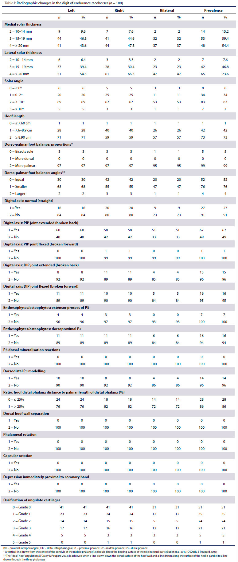
The results of the digit evaluations are summarised in Table I.
No horse had a medial or lateral solar thickness less than 10 mm. The prevalence of thin soles (10-14 mm) was higher on the medial aspect (15.2%) when compared to the lateral aspect (7.6%). The mean lateral solar thickness (20.7 mm [standard deviation (SD) = 4.4]) was higher than the mean medial solar thickness (18.7 mm [SD = 4.7]).
The prevalence of a negative solar angle was 8%. The prevalence of horses with a solar angle < 2° but above horizontal was 34%. Normal solar angles (3°-10°) had a prevalence of 83%.
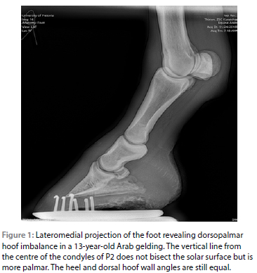
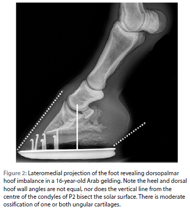
The majority of horses in the current study had evidence of "long toe low heel"syndrome as defined by O'Grady and Poupard (2003). In 95% of horses, the centre of rotation was more palmar bilaterally (Figure 1). The angle of heel to the ground was smaller than the angle of the dorsal hoof capsule to the ground (Figure 2) in 47% of horses bilaterally. Fifty-seven per cent of horses had bilateral dorsal hoof wall length larger than that expected for their size (O'Grady 2014).
The digital axis of 91% of horses diverged from the straight axis, with 73% of horses having an abnormal digital axis bilaterally. Extension of the PIP joint was the most common abnormality (67%).
The prevalence of enthesophytes or osteophytes on the extensor process of P3 where the common digital extensor tendon inserts was 7% (left [4%] and right [3%]).
The prevalence of enthesophytes or osteophytes at the insertion of the joint capsule on the dorsoproximal P2 was 11% in left front digits, 11% in right front digits and 6% of horses bilaterally.
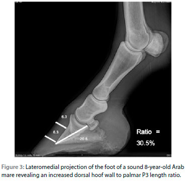
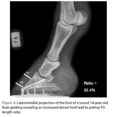
During the current study the DHW:PCL ratio ranged from 22.6% to 33.3% and had a mean of 27.1% (SD = 2.1). The prevalence of a ratio > 25% was 86% (Figures 3 and 4). The prevalence of other indicators of chronic laminitis was low (modelling of the dorsodistal margin [14%], no dorsal hoof wall separation, phalangeal or capsular rotation or a depression immediately proximal to the coronary band).
The CE distance (distance between the coronary band and extensor process) ranged from 5.0 mm to 17.0 mm (median 11 mm; mean 10.7 mm; SD = 1.8 mm).
The prevalence of grade 1 to 5 ungular cartilage ossification was 69% and is summarised in Table I. More severe ungular cartilage ossification had a low prevalence. Grade 4 ungular cartilage ossification had a prevalence of 5% and was present bilaterally in 3% of horses, while grade 5 ungular cartilage ossification had a prevalence of 1%.
No fractures of P3 were observed during the current study.
The metacarpophalangeal joint
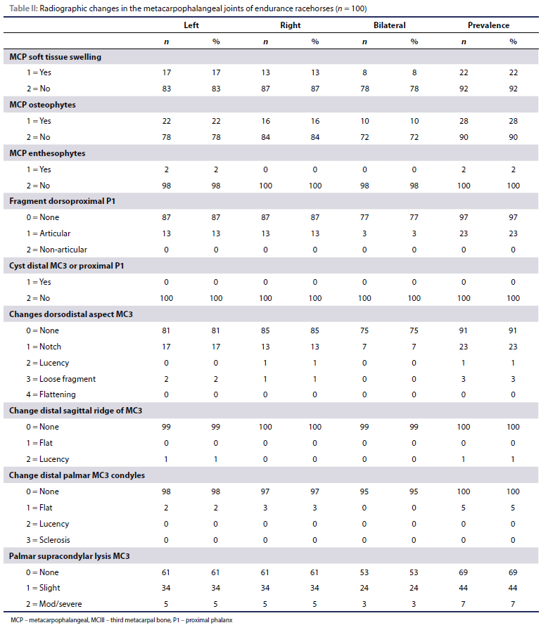
The results of the evaluation of the MCP joint are summarised in Table II. The prevalence of MCP osteophytes was 28%, with 10% of horses having bilateral MCP osteophytes. The prevalence of MCP enthesophytes was 2% and was unilateral.
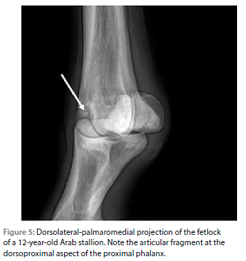
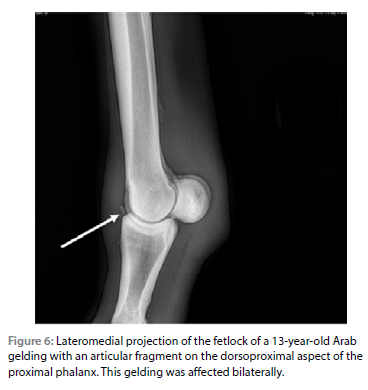
The prevalence of articular dorsoproximal fragments of proximal phalanx (P1) (Figure 5) was 23% and occurred bilaterally in 3% of horses (Figure 6). No non-articular fragments on the dorsoproximal P1 were observed.
Changes to the dorsodistal aspect of MCIII were present in 25% of horses overall. A notch was the most common change with a prevalence of 23%, followed by the presence of a fragment (3%) and lucency (1%). Three horses (3%) were affected with more than one type of dorsodistal MCIII change. No flattening was recorded.
The prevalence of flattening of the distal palmar MCIII condyles was 5%. No lucency or sclerosis was observed.
The overall prevalence of palmar supracondylar lysis was 47%. The prevalence of mild supracondylar lysis was 34% in both left and right front limbs while 24% of horses had mild lysis bilaterally. Moderate to severe lysis had a prevalence of 5% in both left and right front limbs and 3% horses had severe lysis bilaterally.
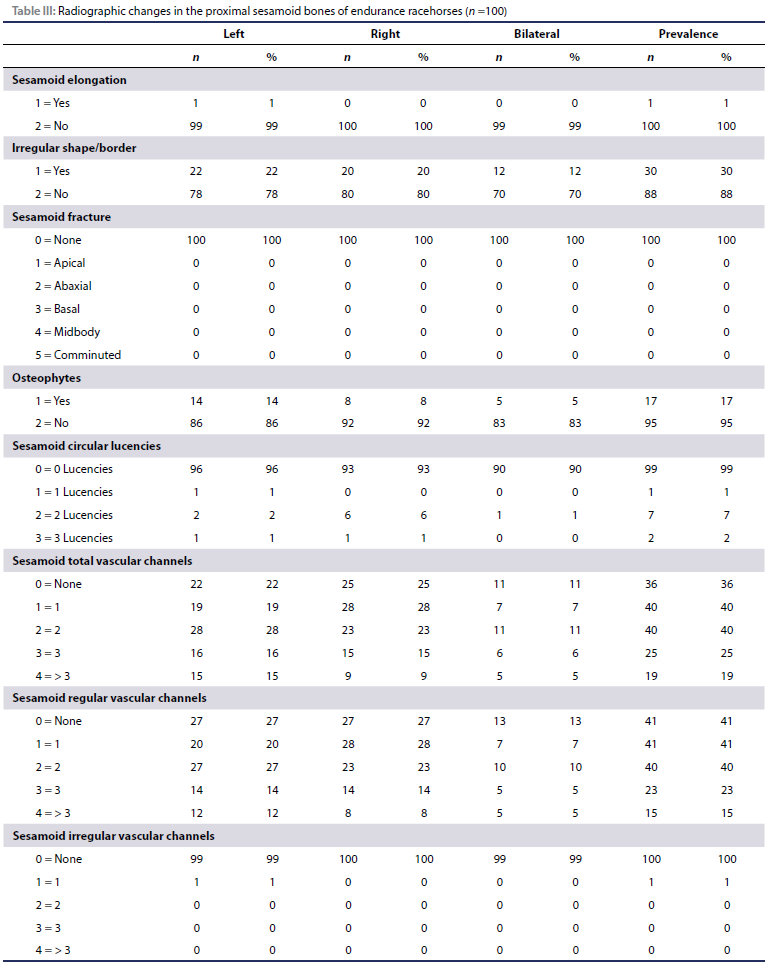
The results of the evaluation of the proximal sesamoid bones are summarised in Table III. Proximal sesamoid bone elongation was observed in only one left forelimb. No PSB fractures were observed. The prevalence of PSB with an abnormal shape or irregular border was 30%. Osteophytes of the PSB were observed in 14% of left fore [LF] limbs and 8% of right fore [RF] limbs with 5% of horses having osteophytes bilaterally. The prevalence of circular PSB lucencies was 10%. Only one irregular vascular channel was reported.
Inter-rater reliability
Raters agreed on dorsopalmar foot balance (0.2), and most measurements of digital axis normality (0.19-0.59) although they did not agree on digital axis normality itself (0.01). They also agreed on the presence of enthesophytes or osteophytes on the extensor process of P3 (0.32) and the dorsoproximal aspect of P2 (0.42) as well as modelling on the dorsodistal aspect of P3 (0.25).
Agreement was calculated for ungular cartilage ossification when absent (grade 0) (0.25), grade three (0.47), grade four (0.55), but no agreement existed for grade one or grade five ossification.
No statistically significant agreement was calculated for toe and heel angle measurements, digital axis normality or dorsal mineralisation reactions on P3.
No agreement existed for indications of laminitis (dorsal hoof wall separation, phalangeal rotation and capsular rotation), the presence of MCP cysts or changes to the distal palmar MC3 (flattening, lucency or sclerosis). Raters reached better agreement for changes to the distal dorsal aspect of MC3 (notch [0.31] and fragmentation [0.32]).
Raters agreed on MCP soft tissue swelling (0.18), osteophytes (0.41) and enthesophytes (0.17) as well as the absence of a dorsoproximal P1 fragment (0.81). They also agreed on the severity of palmar supracondylar lysis (0.32) and irregularities on the sesamoid bones (0.54). No agreement existed for sesamoid elongation, changes to the distal palmar MC3 condyles (flattening, lucency, sclerosis) and circular sesamoid lucencies. Sesamoid vascular channels were difficult to assess according to the raters and very little agreement was recorded.
Due to the low prevalence of certain variables, kappa and thus agreement, IRR could not be calculated for phalangeal rotation, coronary depression, P3 fractures, sclerosis of the distal palmar MC3 and sesamoid fractures.
Discussion
As hypothesised, radiographic changes were observed in the population of South African endurance horses. The current study population was similar to international endurance populations with regard to breed, age and sex distribution (Nagy, Dyson & Murray 2017; Robert et al. 2012).
Given the paucity of reports on radiographic changes in endurance horses (Robert et al. 2012), comparisons were made with Thoroughbreds (Contino, Park & McIlwraith 2012; Furniss, Carstens & Van den Berg 2011; Kane et al. 2003b; Smit 2014). It must be noted that horses in such studies are significantly younger (Contino, Park & McIlwraith 2012; Furniss, Carstens & Van den Berg 2011; Kane et al. 2003b; Smit 2014). Furthermore, one should keep in mind that horses entered in an endurance race are generally free from apparent lameness or overt clinical signs of orthopaedic disease. The prevalence of radiographic changes recorded in the current study may thus not be a true reflection of the larger endurance racehorse population.
Lateral solar length appears to exceed medial solar length in the majority of horses in the current study and is similar to a report of a mixed population of horses (Grundmann et al. 2015). This could be due to uneven weight distribution. Abnormal forces or weight distribution on a specific portion of the foot will cause decreased growth and distortion in the hoof capsule over time (O'Grady 2014). This can be related to lameness in the front limb (O'Grady 2014) as it is associated with conditions like sheared heels, distorted hoof walls and hoof cracks (O'Grady & Poupard 2003).
The prevalence of a negative solar angle (8%) was similar to findings in Thoroughbred yearlings (8.7%) (Smit 2014).
The majority (95%) of horses in the current study had evidence of "long toe low heel" syndrome. This could be due to breed conformation, general poor hoof care and farriery or the belief that horses with longer toes also have thicker soles and may be less likely to concussion injuries and solar bruising. Radiographic measurements of dorsopalmar hoof balance can also be influenced by the stage of the shoeing cycle, which was not recorded in the current study. Poor dorsopalmar balance contributes remarkably to the long toe/underrun heel conformation and the altered biomechanics that it causes (O'Grady & Poupard 2003). In this syndrome, the long toe delays break over causing an excessive pull on the deep digital flexor tendon [DDFT] and associated structures and has been associated with lameness (O'Grady & Poupard 2003).
Extension of the PIP joint was the most common digital axis abnormality (67%) in 60% of left front digits, 58% of right front digits. This is higher than in a study that looked at Thoroughbred yearlings (Furniss, Carstens & Van den Berg 2011) (15.1% and 18.9% respectively). Again, this could be due to breed conformation or poor farriery but warrants further investigation. Extension of the DIP joint (15%) was similar to Thoroughbred yearlings (Furniss, Carstens & Van den Berg 2011). DIP flexion (16%) occurred in 11% of left front digits and 10% of right front digits which was higher than in Thoroughbred yearlings (Furniss, Carstens & Van den Berg 2011) (0.4-0.8% respectively). The same study (Furniss, Carstens & Van den Berg 2011) reported similar findings on the prevalence of enthesophytes or osteophytes on the extensor process of P3.
The prevalence of enthesophytes or osteophytes on the dorsoproximal P2 (15%) was four times higher than findings reported in young horses (Contino, Park & McIlwraith 2012; Furniss, Carstens & Van den Berg 2011). Caution should be applied when comparing young horses with older horses as in the current study. Older horses have most likely undergone more training and riding and are thus more likely to show radiographic evidence of degenerative changes associated with athletic wear. The clinical significance of these changes warrants further investigation. Entheseous new bone does not necessarily imply osteoarthritis, but often occurs when osteoarthritis is present and must thus be interpreted with caution (Dyson 2003).
No dorsal mineralisation reactions were reported on P3 during the current study, similar to South African Thoroughbreds (Furniss, Carstens & Van den Berg 2011) (0.4-0.8%).
According to the reference range reported by Grundmann et al. (2015) the DHW:PCL ratio calculated in this study classified 86% of horses as having chronic laminitis. This raises the question on the reliability and accuracy of this ratio when evaluating this population for laminitis. When a cut-off ratio of 28% is used as an indicator for chronic laminitis, 32% of feet would still be classified as having chronic laminitis. When 30% is used, 7% of front feet would be classified as laminitic. It is possible that other radiographic signs of laminitis could have been underreported in the current study as the coronary band and dorsal hoof wall was not marked. The possibility exists that dorsal hoof wall thickening has an alternative aetiology in endurance horses and that laminitis is not the only cause of an increased DHW:PCL ratio in this population.
Coronary extensor distance in the current study (5-17 mm) was similar to some previous assessments (Redden 2003; Sherlock & Parks 2013), but higher than most published means of 3.36.9 mm (Grundmann et al. 2015; Parks & Belknap 2016). Care must be taken when interpreting these values as there is marked individual (breed and size) variability (Parks & Belknap 2016).
The prevalence of ungular cartilage ossification (grade 1-5) was 69% and although the clinical significance thereof remains controversial it is generally reported that extensive (grade 4 and 5) ossification or severe lateromedial asymmetrical ossification may be predisposing factors for fracture or contribute to foot-related pain and is thus of clinical significance (Butler et al. 2017; Jones & Dyson 2015). The prevalence of grade 4 and 5 ungular cartilage ossification was 5% and 1% respectively.
The prevalence of MCP osteophytes (28%) was high. This raises the question whether these horses suffered previous lameness episodes or have undiagnosed subclinical osteoarthritis. Elite endurance horses had a higher osteoarthritis score compared to showjumpers when pre-purchase radiographs were evaluated (Robert et al. 2012).
The prevalence of articular dorsoproximal fragments of P1 (23%) was higher than previously reported (0.6-2%) (Furniss, Carstens & Van den Berg 2011; Smit 2014) in 566 Thoroughbred yearlings. The exact aetiology of these fragments are unknown but they may be a manifestation of developmental orthopaedic disease (Butler et al. 2017; Declercq et al. 2009) or traumatic chip fractures. Although they are rarely associated with joint effusion, the majority of horses show arthroscopic evidence of synovitis and cartilage fibrillation (Butler et al. 2017). The presence of more than one fragment or fragments in horses older than seven years of age (such as in the current study) were risk factors associated with lameness (Declercq et al. 2009). The lower prevalence in young Thoroughbreds may be explained by pre-sale arthroscopic removal of fragments.
No extra-articular fragments on the dorsoproximal P1 were observed during the current study, similar to the prevalence in Thoroughbreds reported (Furniss, Carstens & Van den Berg 2011; Smit 2014).
No cysts were observed in the dorsodistal MCIII or P1 during the current study. This is similar to findings in Quarter horses (Contino, Park & McIlwraith 2012) and Thoroughbreds (Miyakoshi et al. 2017; Smit 2014). Affected horses usually show obvious lameness exacerbated by distal limb flexion (Dyson 2003).
Changes to the dorsodistal aspect of MCIII were present in 25% of horses which is higher than in previous studies (Contino, Park & McIlwraith 2012; Furniss, Carstens & Van den Berg 2011; Miyakoshi et al. 2017; Smit 2014). This could be explained by pre-sale arthroscopic removal of fragments.
The prevalence of changes of the distal palmar MCIII condyles (flattening 5%; no lucency or sclerosis) in this study was similar to that reported in Thoroughbred yearlings (6.0%) (Smit 2014).
Overall mild supracondylar lysis is more prevalent in the current study of endurance racehorses when compared to younger horses while moderate to severe palmar supracondylar lysis is most prevalent in young Thoroughbreds (Smit 2014). This is most likely due to the repetitive and speed-related trauma young Thoroughbreds are subjected to, while horses in the current study are subjected to a larger amount of lower-speed exercise. When one considers that the prevalence of other indicators of osteoarthritis, such as MCP osteophytes, is also high in the current study, mild supracondylar lysis could be another indication of subclinical or early osteoarthritis in this population. This could also be due to the higher mean horse age of the sample population, the higher number of kilometres these horses have completed both in competition and training, the higher weight of endurance riders when compared to flat racing jockeys, the nature of the discipline itself or the terrain these horses are expected to compete on. The possibility does exist that supracondylar lysis is not caused by synovial effusion due to osteoarthritis but has a different aetiology in endurance horses. This change may be adaptive or protective and not necessarily pathological.
The prevalence of PSB elongation (1%) and PSB fractures (0%) were similar to findings in Quarter horses and Thoroughbreds (Contino, Park & McIlwraith 2012; Smit 2014).
The prevalence of PSB with an abnormal shape or irregular border (30%) was three times that reported in Thoroughbred yearlings (10.8%) (Smit 2014). No PSB with an abnormal shape was reported in young Quarter horses (Contino, Park & McIlwraith 2012).
The prevalence of PSB osteophytes (17%) was higher than in young Quarter horses (1.5% in yearlings and 3.7% in two-year-olds) and Thoroughbred yearlings (0.4%) (Contino, Park & McIlwraith 2012; Smit 2014).
The prevalence of circular PSB lucencies (10%) is similar to that reported in Thoroughbred yearlings (Smit 2014).
More vascular channels (both regular and irregular) were reported in Thoroughbred yearlings compared to the current study (irregular: 1%) (Smit 2014). Two or more irregular vascular channels are associated with poor performance (Kane et al. 2003b; Spike-Pierce & Bramlage 2010).
A limiting factor of the current study was the poor inter-rater reliability and the lack of evaluation of intra-rater repeatability.
A further limiting factor one must keep in mind is the small radius (250 km) in which data was collected when compared to the country as a whole. The sample population consisted of 100 horses (1.7%) out of 5 922 registered horses (ERASA 2019). The prevalence of lesions may be biased due to the relatively small sample population evaluated and the very specific geographical location. This might warrant future investigation and comparison to other regions in South Africa.
This is the first study to describe radiographic findings in the distal aspect of the limb in competing endurance horses in South Africa. The clinical relevance of these radiographic changes warrants further investigation as the current study makes no correlations with factors such as horse age, conformation, lameness, kilometres competed, breed, distance, terrain, speed, rider weight or training regimens.
Comparisons to earlier studies on the prevalence of radiographic changes in horses should be done with caution. Most of these studies have been performed on yearling Thoroughbreds intended for flat racing and yearling Quarter horses intended for cutting. The mean age was nine-years-old (5-17 years old). Most of the population was Arab (48%) or Arab cross (46%). The difference in prevalence of changes compared with previous studies could thus be due to age, breed, or differences between disciplines and the injuries associated with them.
This information about prevalence and distribution of radiographic changes will enable equine veterinarians and riders to better manage horses showing these changes regarding shoeing and training. It may also serve as comparison for future similar studies in the endurance discipline in South Africa and possibly internationally.
Footnotes
• Cuattro Slate 6 EQ Digital Radiography System (Cuattro, Golden, CO USA).
• Mobile x-ray generator (PXP-40HF, Poskom Co., Gyeonggi-do, Republic of Korea).
• RadiAnt DICOM viewer (Medixant, Poznari, Poland).
• Horos (4.0.0 RC3) DICOM viewer software (Nimble Co LLC d/b/a Purview, Annapolis, MD USA).
Conflict of interest
The authors declare that there were no conflicts of interest.
Funding source
Submitted in fulfilment of the requirements for the degree of MSc in the Department of Companion Animal Clinical Studies at the Faculty of Veterinary Science, University of Pretoria from which all funding was obtained.
Radiographs were obtained using a digital radiography system sponsored by IVM Imaging South Africa, and a mobile x-ray generator.
Ethical approval
This study protocol was approved by both the Animal Ethics Committee (Project nr V041-18, approved 28 May 2018) and the Faculty of Veterinary Science Ethics Committee of the University of Pretoria.
ORCID
E Hollenbach: https://orcid.org/0000-0001-7771-8027
MP Robert: https://orcid.org/0000-0002-9497-3852
C le Roux: https://orcid.org/0000-0001-8333-5372
Y Smit: https://orcid.org/0000-0002-4298-4496
References
Bennet, E.D. & Parkin, T.D.H., 2018, Fédération Equestre Internationale endurance events: Risk factors for failure to qualify outcomes at the level of the horse, ride and rider (2010-2015), Vet J 236, 44-48. https://doi.org/10.1016/j.tvjl.2018.04.011. [ Links ]
Butler, J., Colles, C.M., Dyson, S.J., et al., 2017, Clinical radiology of the horse. 4th edn, West Sussex: Wiley Blackwell. [ Links ]
Contino, E.K., Park, R.D., Mcllwraith, C.W., 2012, Prevalence of radiographic changes in yearling and 2-year-old Quarter Horses intended for cutting, Equine Vet J 44(2), 185-195. https://doi.org/10.1111/j.2042-3306.2011.00432.x. [ Links ]
Declercq, J., Martens, A., Maes, D., et al., 2009, Dorsoproximal proximal phalanx osteochondral fragmentation in 117 warmblood horses, Vet Comp Orthop Traumatol 22(1), 1-6. https://doi.org/10.3415/VCOT-08-02-0016. [ Links ]
Dyson, SJ., 2003, Radiography and radiology, in Dyson, Sue J. and Ross, M.W. (eds) Diagnosis and Management of Lameness in the Horse 2nd edn, pp. 153-166. https://doi.org/10.1016/B978-0-7216-8342-3.50022-X. [ Links ]
ERASA (2019) ERASA Data Base - Horses, Endurance Ride Association of South Africa. Available from: https://www.erasa.co.za/index.php/content/Horses/2001/lib_defaults/content.php/2603. Accessed 18 Aug 2019. [ Links ]
Furniss, C., Carstens, A., Van den Berg, S.S., 2011, Radiographic changes in Thoroughbred yearlings in South Africa, J S Afr Vet Assoc 82(4), 194-204. https://doi.org/10.4102/jsava.v82i4.74. [ Links ]
Grondahl, A.M. & Engeland, A., 1995, Influence of radiographically detectable orthopedic changes on racing performance in standardbred trotters, J Am Vet Med Assoc 206(7), 1013-1017. [ Links ]
Grundmann, I.N.M., Drost, W.T., Zekas, L.J., et al., 2015, Quantitative assessment of the equine hoof using digital radiography and magnetic resonance imaging, Equine Vet J 47(5), 542-547. https://doi.org/10.1111/evj.12340. [ Links ]
Jones, L.E. & Dyson, S.J., 2015, Radiographic characterization of ossification of the ungular cartilages in horses: 271 cases (2005-2012), J Am Vet Med Assoc 247(7), 801-811. https://doi.org/10.2460/javma.247J.801. [ Links ]
Kane, A.J., Park, R.D., McIllwraith, C.W., et al., 2003a, Radiographic changes in Thoroughbred yearlings. Part 1: Prevalence at the time of the yearling sales, Equine Vet J 35(4), 354-365. https://doi.org/10.2746/042516403776014280. [ Links ]
Kane, A.J., McIlwraith, C.W., Park, R.D., et al., 2003b, Radiographic changes in Thoroughbred yearlings. Part 2: Associations with racing performance, Equine Vet J 35(4), 366-374. https://doi.org/10.2746/042516403776014307. [ Links ]
Kidd, J., 2011. Pedal bone fractures, Equine Veterinary Education 23(6), 314-323. https://doi.org/10.1111/j.2042-3292.2011.00227.x. [ Links ]
Marlin, D.J., McEwen, J., Sluyter, F., 2008, Completion and treatment rates in modern endurance racing, Proceedings of 4th International Equitation Science Conference, p. 67. [ Links ]
McHugh, M.L., 2012, Interrater reliability: the kappa statistic, Biochemia Med (Zagreb) 22(3), 276-282. https://doi.org/10.11613/BM.2012.031. [ Links ]
Misheff, M.M., Alexander, G.R., Hirst, G.R., 2010, Management of fractures in endurance horses, Equine Veterinary Education 22(12), 623-630. https://doi.org/10.1111/j.2042-3292.2010.00150.x. [ Links ]
Miyakoshi, D., Senba, H., Shikichi, M., et al., 2017, A retrospective study of radiographic abnormalities in the repositories for Thoroughbreds at yearling sales in Japan, J Vet Med Sci 79(11), 1807-1814. https://doi.org/10.1292/jvms.16-0425. [ Links ]
Nagy, A., Dyson, S.J., Murray, J.K., 2012, A veterinary review of endurance riding as an international competitive sport, Vet J 194(3), 288-293. https://doi.org/10.1016/j.tvjl.2012.06.022. [ Links ]
Nagy, A., Dyson, S.J., Murray, J.K., 2017, Veterinary problems of endurance horses in England and Wales, Prev Vet Med 140, 45-52. https://doi.org/10.1016/j.prevetmed.2017.02.018. [ Links ]
Nagy, A., Murray, J.K., Dyson, S.J., 2010, Elimination from elite endurance rides in nine countries: A preliminary study, Equine Vet J 42, 637-643. https://doi.org/10.1111/j.2042-3306.2010.00220.x. [ Links ]
Nagy, A., Murray, J.K., Dyson, S.J., 2014, Horse-, rider-, venue- and environment-related risk factors for elimination from Federation Equestre Internationale endurance rides due to lameness and metabolic reasons, Equine Vet J 46(3), 294-299. https://doi.org/10.1111/evj.12170. [ Links ]
O'Grady, S.E., 2014, How to evaluate the equine hoof capsule, American Farriers Journal 59, 54-61 [ Links ]
O'Grady, S.E. & Poupard, D.A., 2003, Proper physiologic horseshoeing, Vet Clin North Am Equine Pract, 19(2), 333-351. https://doi.org/10.1016/S0749-0739(03)00020-8. [ Links ]
Parks, A.H. & Belknap, J.K., 2016, Diagnostic Imaging, in Belknap, James K. and Geor, R.J. (eds) Equine Laminitis. Hoboken, NJ, USA: John Wiley & Sons, Inc., pp. 226-239. https://doi.org/10.1002/9781119169239.ch27. [ Links ]
Redden, R.F., 2003, Radiographic imaging of the equine foot, Vet Clin North Am Equine Pract 19, 379-392. https://doi.org/10.1016/S0749-0739(03)00026-9. [ Links ]
Robert, C., Coussediére, M., Pélissier, C., et al., 2012, Chevaux d'endurance et de saut d'obstacles: quelles differences á la radiographie en visite d'achat? Pratique Vétérinaire Equine 44, 25-29. [ Links ]
Ruohoniemi, M., Tulamo, R., Hackzell, M., 1993, Radiographic evaluation of ossification of the collateral cartilages of the third phalanx in Finnhorses, Equine Vet J 25(5), 453-455. https://doi.org/10.1111/j.2042-3306.1993.tb02989x [ Links ]
Schnabel, L.V. & Redding, W.R., 2018, Diagnosis and management of proximal sesamoid bone fractures in the horse, Equine Veterinary Education 30(8), 450-455. https://doi.org/10.1111/eve.12615. [ Links ]
Sherlock, C. & Parks, A.H., 2013, Radiographic and radiological assessment of laminitis, Equine Veterinary Education 25(10), 524-535. https://doi.org/10.1111/eve.12065. [ Links ]
Smit, Y., 2014, MSc Dissertation: Prevalence of radiographic changes in South African Thoroughbred racehorses at the yearling sales, 2008-2010. University of Pretoria. [ Links ]
Spike-Pierce, D.L. and Bramlage, L.R, 2010, Correlation of racing performance with radiographic changes in the proximal sesamoid bones of 487 Thoroughbred yearlings, Equine Vet J 35(4), 350-353. https://doi.org/10.2746/042516403776014262. [ Links ]
Wise, L.N., Bryan J.N., Sellon, D.C., et al., 2009, A retrospective analysis of renal carcinoma in the horse, J Vet Intern Med 23(4), 913-918. https://doi.org/10.1111/j.1939-1676.2009.0326.x. [ Links ]
Younes, M., Barrey, E., Cottin, F., et al., 2016, Elimination in long-distance endurance rides: insights from the analysis of 7,032 starts in 80 to 160 km competitions, Comparative Exercise Physiology 12(4), 157-167. https://doi.org/10.3920/CEP160022. [ Links ]
 Correspondence:
E Hollenbach
Email: elza.hollenbach@up.ac.za

