


Serviços Personalizados
Artigo
 Inglês (pdf)
 Artigo em XML
 Referências do artigo
Indicadores

Links relacionados
-
 Citado por Google
 -
 Similares em Google
Compartilhar

 Permalink
Journal of the South African Veterinary Association
versão On-line ISSN 2224-9435
versão impressa ISSN 1019-9128
J. S. Afr. Vet. Assoc. vol.93 no.2 Pretoria 2022
http://dx.doi.org/10.36303/JSAVA.515
CASE REPORT
Lung lobe torsion in association with a pulmonary papillary carcinoma in a dog
E Ciriano; M Marrington; J Grant
Northwest Veterinary Specialists, United Kingdom
ABSTRACT
Lung lobe torsion (LLT) is an uncommon condition in dogs reported to be most commonly idiopathic or secondary to trauma, pleural effusion, lung lobectomy or thoracic neoplasia. Carcinomas are the most common primary lung tumours in dogs, but only a few cases have been reported in association with LLT in veterinary medicine.
This case describes an adult male neutered Labrador, which presented with lethargy, weight loss and pleural effusion. Computed tomography (CT), cytology of the lung, thoracocentesis and fluid analysis were performed. CT revealed pleural effusion and torsion of the left cranial lung lobe with no evidence of a pulmonary mass or metastatic disease. Thoracotomy and left cranial lung lobectomy were performed. Intraoperatively there was no macroscopic evidence of pulmonary neoplasia. Histopathology of the lobar tissue confirmed grade 2 pulmonary papillary carcinoma.
It is possible that early detection and surgical management might help to prevent the morbidity and mortality associated with LLT. However, as in this case, the underlying cause for the LLT will ultimately determine the patient's prognosis. The final diagnosis of papillary carcinoma in this case, was only made via histopathological assessment of the pulmonary tissue as it was unclear on the advanced imaging and macroscopic intraoperative evaluation of the lungs. This case highlights the importance of considering pulmonary neoplasia as a differential for LLT even in the absence of a macroscopic mass, and therefore the value of performing histopathology on the excised lung tissue.
Keywords: pulmonary papillary carcinoma, lung lobe torsion, oncology, surgery, thoracotomy
Introduction
Lung lobe torsion (LLT) is an uncommon and potentially life-threatening condition in dogs. Torsion results from rotation of a lobe around the broncho-vascular pedicle compromising the airways and pulmonary vasculature at the hilus, leading to congestion and consolidation of the affected lobar tissue. In dogs, the most common reported cause of LLT is spontaneous, although any condition that interferes with lung lobe mobility could lead to LLT, such as thoracic neoplasia, pleural effusion, and trauma (Benavides et al. 2019; Park et al. 2018).
Carcinomas are the most common primary lung tumours in dogs, followed by sarcoma, adenoma and pulmonary neuroendocrine tumours (McPhetridge et al. 2022). LLT associated with a primary pulmonary neoplasia is a rare finding with only a few cases reported in veterinary medicine. There is one study of 80 dogs assessing the cause of LLT. An idiopathic cause was considered in 77% of the population, while 21% had an underlying condition. Adenocarcinoma was diagnosed only in two of the patients (Rossanese et al. 2020).
Patient presentation
An 11-year-old male neutered Labrador was presented as an emergency for bilateral pleural effusion. A three-week history of lethargy, hyporexia, weight loss, coughing, gagging and intermittent episodes of haemoptysis were reported. Vaccination and parasite control were up to date and there was no history of trauma or thoracic surgery. The patient was on long-term meloxicam (0.1 mg/kg orally SID) for osteoarthritis.
On physical examination, the dog was quiet, alert and responsive, with a body condition score of 3/9. Tachycardia and mild increased inspiratory effort were detected, as well as bilateral reduction of the bronchovesicular lung sounds. Weak femoral pulses and pyrexia at 39.6 "Celsius were detected.
Complete blood count and biochemistry showed no abnormalities. Lung worm snap test was negative. Orthogonal radiographs of the thorax revealed moderate bilateral pleural effusion.
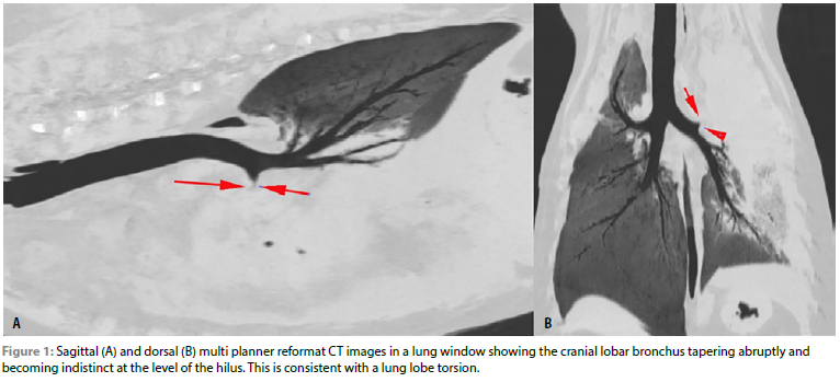
Subsequently, full body CT scan revealed a left cranial LLT with severe pulmonary congestion. The left cranial lung lobe was severely enlarged and caudo-dorsally displaced with a vesicular gas pattern detected in the mid-dorsal caudal thorax. There were no signs of contrast enhancing or focal mass lesions detectable within the affected lobar parenchyma. The left caudal lung lobe was medially and caudally displaced by the cranial lung lobe (Figure 1). The right lung appeared unremarkable. Moderate amount of bilateral pleural effusion was present. The remaining thoracic structures and abdominal imaging revealed no abnormalities.
Thoracocentesis was performed and 550 ml (20 ml/kg) of se-rosanguinous fluid was drained. Pleural effusion was consistent with an exudate with pyogranulomatous inflammation. Cytologically more than 50% of the inflammatory cells were neutrophils with no signs of degeneration, and 35% of the cell population were described to be large pleomorphic mononuclear cells and epithelioid-mesothelioid-type cells. Based on cytological findings, a chronic process secondary to inflammation or infection was suspected. There were no bacteria seen. The large pleomorphic mononuclear cells detected were highly consistent with reactive mesothelial cells. An underlying neoplastic process, such as mesothelioma or carcinoma was not excluded.
Left cranial lung lobe cytology revealed an abundance of red blood cells, neutrophils, vacuolated macrophages, as well as a background population of unidentifiable cells. These cells occasionally appeared discrete and macrophage-like but their overall appearance was more epithelioid. These cells were arranged in clusters with occasional cells exhibiting multiple criteria of malignancy. These findings were consistent with a pyogranulomatous inflammation and abundant necrosis of the lung tissue with suspected underlying epithelial neoplasia.
Management and outcome
Intercostal thoracotomy for left cranial lung lobectomy was performed. The left cranial lung lobe was rotated 180 degrees around the hilus and was excised by hilar ligation. There was no macroscopic evidence of neoplasia affecting the excised left cranial lung lobe, the remainder of the pulmonary parenchyma or the thoracic cavity. Thoracotomy drain was placed for postoperative management. Meloxicam (0.1 mg/kg orally SID), amoxicillin-clavulanic acid (20 mg/kg orally BID), paracetamol (10 mg/kg intravenously TID), gabapentin (10 mg/kg orally TID) and omeprazole (1 mg/kg orally BID) were also prescribed during hospitalisation.
Five days postoperatively, given the high production of sero-sanguinous fluid (10 mg/kg/day), meloxicam was discontinued and anti-inflammatory dose of prednisolone (0.5 mg/kg orally SID) was prescribed 48 hours later. After a couple of days, reduction of the production of the pleural fluid was noted and the thoracostomy drain was removed. The patient was discharged nine days post-surgery.
Histopathology from the left cranial lung lobe revealed neoplas-tic pulmonary epithelial cells arranged in a papillary pattern, consistent with a grade 2 pulmonary papillary carcinoma (Figure 2). There were no neoplastic cells detected on the submitted tissue borders.

Adjuvant systemic and intracavitary chemotherapy options were discussed with the owners but were declined.
Three weeks from surgery, persistent pleural effusion was noted. Cytology was most consistent with a neoplastic effusion secondary to an underlying epithelial tumour. Culture and sensitivity analysis was negative. Further diagnostic imaging (CT or thoracic radiography) was advised, and potential adjunctive chemotherapy was again discussed. Both were declined by the owners. Two weeks later, the dog developed episodes of regurgitation which were thought to be due to progression of the disease and increasing volume of pleural effusion. Further investigations and thoracocentesis were declined. The patient was euthanised 83 days from diagnosis.
Discussion
The initial clinical signs of the patient (weight lost, coughing, gagging and haemoptysis) were explained by the presence of pleural effusion which could lead to tracheal compression, and the intrapulmonary neoplastic disease (Sherding & Birchard 2006). Thoracic radiographs can be diagnostic or suggestive of LLT. Specific radiographic patterns have been described in dogs with LLT, such as vesicular gas pattern, lobar consolidation, displaced or abrupt bronchus, mediastinal shift, displaced trachea, and axial rotation of the carina (D'Anjou et al. 2005). In this case, the presence of pleural effusion at the time of thoracic radiographs may have masked these radiographic features preventing radiographic diagnosis of LLT. CT scan is more sensitive at identifying LLT, pulmonary metastasis, thoracic neoplasia and thoracic lymphadenopathy in the presence of pleural effusion (Lamb et al. 2019; Seiler et al. 2008). In this case, no pulmonary mass was detected in the left cranial lung lobe on the post-contrast CT images. However, compromised vascularity in the rotated lung would have led to reduced blood flow and subsequently caused decreased contrast distribution into the lung preventing visualisation of a mass-like lesion within the lung parenchyma.
Pleural effusion is a common finding associated with LLT. In one study of 39 animals with LLT, 90% of the population had pleural effusion. Pleural effusion prior to the development of LLT was identified in three patients with underlying thoracic pathologies (Benavides et al. 2019). In this case, we cannot determine if the pleural effusion was secondary to the underlying neoplastic process and therefore a trigger factor for lobar torsion, or if pleural effusion was secondary to LLT.
Tumour stage, histological tumour type, tumour grade, lymph node metastasis and overexpression of epidermal growth factor receptor (EGFR) are prognostic factors associated with primary lung cancer (Sabattini et al. 2012). Dogs with stage T1 lung cancer have a better prognosis than dogs diagnosed with more advanced disease. In one study of 42 dogs, the median survival time (MST) of pulmonary papillary tumours stage T1 was 555 days, compared to 72 days for the more advanced stages. Only one case with papillary adenocarcinoma (T1N0M0) survived for less than six months (Polton et al. 2008). There are other studies assessing the histological tumour type concluding that pulmonary papillary adenocarcinoma has better outcomes than other pulmonary tumours (McNiel et al. 1997; Polton et al. 2018). Lymph node metastasis has also been described as a negative prognostic factor associated with MST of 26 days, compared to 452 days for patients with stage T1 (McNiel et al. 1997).
In this case report, the patient was diagnosed with a stage T1, grade 2, pulmonary papillary carcinoma with an MST of 83 days from diagnosis. Thoracic lymphadenopathy was not detected on CT images or exploratory thoracotomy. However, early nodal metastasis (HN1, HN2) could not be excluded without histopathological diagnosis of the regional lymph nodes. Carcinomatosis could also have been a cause of death in this patient.
Lung lobectomy is the treatment of choice for primary lung cancer in patients with no evidence of regional or distant metastasis. Chemotherapy can be used as palliative or adjuvant therapy in patients with stage T2 or more advanced pulmonary disease. Injectable chemotherapy agents such as cisplatin, carboplatin, mitoxantrone and vinorelbine could be considered but further studies are required to support their benefit (Polton et al. 2008; Wouda et al. 2015). There is a study showing clinical benefit of metronomic chemotherapy with low dose cyclophosphamide, thalidomide and piroxicam for advanced-stage pulmonary cancer in dogs (Polton et al. 2018). Another oral chemotherapy agent to consider is tyrosine kinase inhibitors, such as toceranib phosphate (London et al. 2003). Intracavitary chemotherapy with mitoxantrone and/or carboplatin is an effective treatment for dogs with carcinomatosis and other pleural tumours, despite the presence of malignant effusions (Charney 2005).
Conclusion
This case description proves the importance of considering pulmonary neoplasia as a differential in cases of LLT until excluded by histopathology. As in this case, there was no evidence of neoplasia on advanced imaging or macroscopic tissue evaluation to indicate neoplasia as the cause for the LLT. Histopathology will aid in formulating a prognosis and deciding whether adjuvant treatment is required.
Conflict of interest
The authors have declared that no competing interest exists.
Funding source
This case report received no specific grant from any funding agency in the public, commercial, or not-for-profit sectors.
Ethical approval
Owners' consent was obtained for the procedures undertaken and the use of the data for research purposes. Established internationally recognised high standards of veterinary clinical patient care were followed. Ethical approval from a committee was therefore not specifically required.
ORCID
E Ciriano https://orcid.org/0000-0001-8985-1642
M Marrington https://orcid.org/0000-0001-9218-2742
J Grant https://orcid.org/0000-0002-9010-6006
References
Benavides, K.L., Rozanski, E.A., Oura, T.J., 2019, Lung lobe torsion in 35 dogs and 4 cats, Can Vet J 60(1), 60-66. [ Links ]
Charney, S., Bergman, P., McKnight, J., et al., 2005, Evaluation of intracavitary mitoxantrone and carboplatin for treatment of carcinomatosis, sarcomatosis and mesothelioma, with or without malignant effusions: a retrospective analysis of 12 cases (1997-2002), Vet Comp Oncol 3(4), 171-181. https://doi.org/10.1111/j.1476-5810.2005.00075.x. [ Links ]
D'Anjou, M.A., Tidwell, A.S., Hecht, S., 2005, Radiographic diagnosis of lung lobe torsion, Vet Radiol Ultrasound 46, 478-484. https://doi.org/10.1111/j.1740-8261.2005.00087.x. [ Links ]
Lamb, C., Whitlock, J., Foster-Yeow, A., 2019, Prevalence of pulmonary nodules in dogs with malignant neoplasia as determined by CT, Vet Radiol Ultrasound 60(3), 300-305. https://doi.org/10.1111/vru.12723. [ Links ]
London, C.A., Hannah, A.L., Zadovoskaya, R., et al., 2003, Phase I dose-escalating study of SU11654, a small molecule receptor tyrosine kinase inhibitor, in dogs with spontaneous malignancies, Clin Cancer Res 9(7), 2755-2768. [ Links ]
McNiel, E.A., Ogilvie, G.K., Powers, B.E., 1997, Evaluation of prognostic factors for dogs with primary lung tumors: 67 cases (1985-1992), J Am Vet Med Assoc 211(11),1422-1427. [ Links ]
McPhetridge, J., Scharf, V., Regier, P., et al., 2022, Distribution of histopathologic types of primary pulmonary neoplasia in dogs and outcome of affected dogs: 340 cases (2010-2019), J Am Vet Med Assoc 260(2), 234-243. https://doi.org/10.2460/javma.20.12.0698. [ Links ]
Park, K., Grimes, J., Wallace, M., et al., 2018, Lung lobe torsion in dogs: 52 cases (2005-2017), Vet Surg 47(8), 1002-1008. https://doi.org/10.1111/vsu.13108. [ Links ]
Polton, G., Brearley, M., Powell, S., et al., 2008, Impact of primary tumour stage on survival in dogs with solitary lung tumours, J Small Anim Pract, 49(2), 66-71. https://doi.org/10.1111/j.1748-5827.2007.00403.x. [ Links ]
Polton, G., Finotello, R., Sabattini, S., et al., 2018, Survival analysis of dogs with advanced primary lung carcinoma treated by metronomic cyclophosphamide, piroxicam and thalidomide, Vet Comp Oncol 16(3), 399-408. https://doi.org/10.1111/vco.12393. [ Links ]
Rossanese, M., Wustefeld-Janssens, B., Price, C., et al., 2020, Long-term survival after treatment of idiopathic lung lobe torsion in 80 cases, Vet Surg 49(4), 659-667. https://doi.org/10.1111/vsu.13406. [ Links ]
Sabattini, S., Mancini, F., Marconato, L., et al., 2012, EGFR overexpression in canine primary lung cancer: pathogenetic implications and impact on survival, Vet Comp Oncol 12(3), 237-248. https://doi.org/10.1111/vco.12002. [ Links ]
Seiler, G., Schwarz, T., Vignoli, M., et al., 2008, Computed tomographic features of lung lobe torsion, Vet Radiol Ultrasound 49(6), 504-508. https://doi.org/10.1111/j.1740-8261.2008.00435.x. [ Links ]
Sherding, R. & Birchard, S., 2006, Saunders manual of small animal practice, St. Louis, Mo.: Saunders Elsevier, pp. 1696-1707. https://doi.org/10.1016/B0-72-160422-6/50166-2. [ Links ]
Wouda, R., Miller, M., Chon, E., et al., 2015, Clinical effects of vinorelbine administration in the management of various malignant tumor types in dogs: 58 cases (1997-2012), J Am Vet Med Assoc 246(11), 1230-1237. https://doi.org/10.2460/javma.246.11.1230. [ Links ]
 Correspondence:
E Ciriano
Email: estel.laciriano@gmail.com

