


Serviços Personalizados
Artigo
 Inglês (pdf)
 Artigo em XML
 Referências do artigo
Indicadores

Links relacionados
-
 Citado por Google
 -
 Similares em Google
Compartilhar

 Permalink
Journal of the South African Veterinary Association
versão On-line ISSN 2224-9435
versão impressa ISSN 1019-9128
J. S. Afr. Vet. Assoc. vol.93 no.2 Pretoria 2022
http://dx.doi.org/10.36303/JSAVA.83
ORIGINAL RESEARCH
Molecular detection of zoonotic pathogens causing gastroenteritis in humans: Salmonella spp., Shigella spp. and Escherichia coli isolated from Rattus species inhabiting chicken farms in North West Province, South Africa
TA RamatlaI, II; N MphuthiII; T RamailiII; M TaioeI, III; O ThekisoeI; M SyakalimaII, IV
IUnit for Environmental Sciences and Management, North-West University, South Africa
IIDepartment of Animal Health, Faculty of Natural and Agricultural Sciences, North-West University, South Africa
IIIEpidemiology, Parasites and Vectors, Agriculture Research Council, Onderstepoort Veterinary Research, South Africa
IVUniversity of Zambia, School of Veterinary Medicine, Department of Disease Control, Zambia
ABSTRACT
Rodents are key carriers and reservoirs of various pathogens of public health importance to both human and animal diseases. This research was carried out in order to identify the selected pathogens, namely, Shigella spp., Salmonella spp. and Escherichia coli from rats that inhabit the poultry houses. A total of 154 samples from captured rats were examined for the zoonotic bacterial pathogens, of which 3.3%, 29.9% and 20.7% were harbouring Shigella spp., Salmonella spp., and E. coli, respectively. A total of 14 Shigella isolates expressed presence of ipaH gene, of which eight and five were positive for S. sonnei and S. boydii, respectively. For Salmonella, 68 isolates were positive for invA and other genes including spy with 26 (38%), sdf\ 2 (18%), spvC 14 (20%), hilA 28 (41%), misL 43 (63%), orfL 31 (46%) and spiC 38 (56%). For E. coli, the aggR gene was the most prevalent (62 [42%]), followed by the eae gene, which was only detected in 21 (14%) isolates, while stx gene was not detected in any of the samples. This study shows that zoonotic pathogens with virulence genes are circulating in rodents from selected chicken farms in the North West Province of South Africa. Rodents must therefore be regarded as important carriers of zoonotic pathogens that can potentially infect both humans and animals.
Keywords: poultry farms, Rattus species, Salmonella, Shigella, Escherichia coli, zoonotic pathogens
Introduction
Rodents are the main carriers and hosts of many important zoonotic pathogens in humans and animals (El-Sharkawy et al. 2017). It is generally known that they play a role in the transmission and spread of human diseases. They mostly eat feed in storage places and leave their excreta behind leading to the spread of pathogenic organisms (Franssen et al. 2016; Jemilehin et al. 2016). Others carry ectoparasites that are vectors of important pathogens of animals and humans. The selected bacteria, that is, Escherichia coli, Shigella spp. and Salmonella spp. for this investigation are of particular public concern as they are known to cause serious diseases in human beings (Hong et al. 2018; Wang et al. 2015).
Salmonella has been isolated from captured rats and mice at poultry farms in several studies elsewhere and is linked to human and animal diseases (Nkogwe et al. 2011; Li et al. 2018; Umali et al. 2012). Rodents have been shown to play a vital role in spreading disease and thus are regarded as important indicator species for disease outbreaks (Jemilehin et al. 2016; Lapuz et al. 2008; López et al. 2012).
Shigellosis is an acute invasive human intestinal infection caused by the bacteria of the Shigella genus. (Mokhtari et al. 2012). There are four species in the Shigella genus, which include S. dysenteriae (serogroup A), S. flexneri (serogroup B), S. sonnei (serogroup C) and S. boydii (serogroup D) (Theron et al. 2001). Rattus spp. are known vectors and reservoirs of this pathogen and rabbits, calves, and monkeys have been reported as hosts (Jianjun 2005; Maurelli et al. 1998; Pan et al. 2006).
Escherichia coli is an enterobacterial zoonosis that can be transmitted by rodents, and rats have been implicated in the transmission of this zoonotic pathogen in previous studies (Caballero et al. 2015; Nkogwe et al. 2011). E. coli has also been isolated from several animals, birds, reptiles, bats, and wild deer (Adesiyun et al. 1999; Gopee et al. 2000). All these vectors or reservoirs of the pathogen have been linked to infections in human beings. Based on its virulence, this species is classified into five classes; Enteroaggregative E. coli (EAEC), Shiga toxin-producing E. coli (STEC), enterohaemorrhagic E. coli (EHEC), enterotoxigenic E. coli (ETEC), attaching and effacing E. coli (AEEC) and enteroinvasive E. coli (EIEC) (Vendramin et al. 2014).
This study was conducted in order to isolate and identify Shigella spp., Salmonella spp. and E. coli from rats that are found in poultry houses as a way of understanding the potential risk of infections.
Materials and methods
Study area, sampling and rodent identification
The study was conducted in the North West Province of South Africa, which is located between 25 and 28 degrees south of the equator and 22 to 28 degrees longitude east of the Greenwich Meridian. The temperature in the region ranges from 17 °C to 31 °C in summer and 3 °C to 21 °C in winter (Ramatla et al. 2017). A total of 154 rat samples were collected from six randomly selected commercial farms, transported to the laboratory and were euthanised. Rats were dissected following a procedure described previously (Lapuz et al. 2008; Ramatla 2019) whereby kidney and caecal samples were removed. For rodent identification, DNA was extracted from kidney tissue using QIAamp DNA Blood and Tissue Kit (Qiagen, Hilden, Germany) and then polymerase chain reaction (PCR) was conducted for amplification of cytochrome oxidase subunitI gene (COI) as reported by Ramatla et al. (2019).
Isolation and identification of bacterial species
Shigella spp.
Shigella was isolated according to the method described by Phiri et al. (2021). Tryptic soy broth (TSB) 10 ml (Oxoid, Basingstoke, UK), was added into the faecal samples collected from caecum which were homogenised by vortexing for two minutes followed by incubation at 37 °C ± 1 °C for 24 hours. Xylose lysine deoxycholate agar (XLD) (Merck, Wadeville, South Africa) was used to increase the chances of culturing and isolation of the Shigella spp. The plates were incubated at 37 °C ± 1 °C for 24 hours in an overturned position. Pinkish to reddish colonies were selected and subjected to Gram staining.
Salmonella spp.
Salmonella was isolated according to the modified version of the International Organization for Standardization (ISO-6579: 2002) method as described previously by Mainar-Jaime et al. (2013). Briefly, samples were first pre-enriched in buffered peptone water (BPW) (Oxoid, Biolab, South Africa), then transferred to Mueller-Kauffmann Tetrathionate Novobiocin (MKTTn) broth (Sigma-aldrich, S.A. Barcelona, Spain). Rappaport-Vassiliadis medium with soya (RVS) broth was used to complete the enrichment process (Sigma-Aldrich, S.A. India). Xylose lysine deoxycholate (XLD) agar (Merck, Wadeville, South Africa) and brilliant green agar (BGA) were used to streak loopfuls of the RVS broth-enriched cultures separately onto two selective agar plates, namely: BGA (Scharlau Chemie S.A. Barcelona, Spain) and XLD agar (Merck, Wadeville, South Africa), and they were incubated for 24 hours in an inverted posture at 37 °C ± 1 °C. On XLD agar, two to three red colonies with or without black centres, colourless or opaque-white colonies surrounded by pink or red zones, and red colonies on BGA were recognised as Salmonella suspects. These putative colonies were purified by incubation at 37 °C for 24 hours on nutrient agar (NA) (Merck, Wadeville, South Africa).
E. coli
Isolation of E. coli from the samples was done following a previously described enrichment method (Caballero et al. 2015) with some modifications. A weight of 5 g of samples was vortexed vigorously in 10 ml of TSB (Oxoid, UK) and incubated at 37 °C for 24 hours. The broth culture was subcultured onto MacConkey's agar (Biolab, supplied by Merck, Johannesburg, South Africa) then incubated at 37 °C for 24 hours. Following incubation, two to three rose pink colonies were selected and sub-cultured on NA (Merck, Wadeville, South Africa), which was then incubated at 37 °C for 24 hours.
Biochemical tests
Shigella spp.
The Gram-negative isolates were subjected to biochemical tests as described by Mokhtari et al. (2012). For the Indole test, the formation of a red ring indicated a positive reaction and were picked for further tests, while a yellow-brown ring indicated a negative reaction. Colonies of the presumptive Shigella were added into the urea broth (Biolab, Merck, South Africa) and incubated. The positive isolates of Shigella did not show any changes in colour. Furthermore, isolates were also subjected to triple sugar iron (TSI) agar (Biolab, Merck, South Africa) to test for glucose, lactose, and sucrose fermentation. Bergey's manual of systematic bacteriology was used as an identification aid.
Salmonella spp.
All colonies showing rod-shaped Gram-negative bacteria were subjected to biochemical testing as described by Samaxa et al. (2012). Salmonella spp. was subjected to catalase, hydrogen peroxide. The presence of enzyme in bacteria is evident with rapid production of gas (oxygen) bubbles and this is considered as indicating the presence of Salmonella spp. The isolates were also subjected to the Oxidase test; the isolates which did not show any colour change after smearing the colony over Microbact oxidase strips (MBO266A, Oxoid Ltd., Basingstoke, England) were suspected to be Salmonella. Suspected colonies were tested for Indole by using three to five drops of Kovac's reagent (Pro-LAB Diagnostics, IVD, Bromborough, Wirral, UK), whereby yellow or no colour was an indication of suspected Salmonella.
E. coli
The E. coli isolates were then subjected to Gram-staining. All colonies showing the Gram-negative rod-shape were subjected to biochemical tests, whereby the isolates which were catalase-positive, indole-positive and Voges-Proskauer-negative were considered as E. coli strains (Dobrowsky et al. 2014).
Haemolysis test for E. coli
Isolates that satisfied morphological structures and primary biochemical tests were plated on blood agar plates and incubated aerobically at 37 °C for 24 hours. E. coli specifically appears as mucoid with haemolysis on blood agar plates as described by March and Ratnam (1986).
Molecular characterisation of bacterial isolates
Pure colonies of suspected Shigella, Salmonella, and E. coli were used to obtain genomic DNA coli cultures using the Fungal/ Bacterial Soil Microbe DNA Mini Prep kit (Zymo-Research Fungal/ Bacterial Soil Microbe DNA Mini Prep kit, USA) according to the manufacturer's instructions. Extracted DNA was eluted in a clean 1.5 ml micro-centrifuge tube with 100 ml of DNA elution buffer. The existence of any contaminants was checked using the NanoDrop ND-1000 UV spectrophotometer (Thermo-Fisher Scientific Inc., USA) by measuring the optical density at 280 nm and 260 nm. The isolated DNA was kept at 20 °C until the PCR was done.
Polymerase chain reaction
The final reaction mixture for all PCR assays for E. coli, Shigella and Salmonella in this study was 25 ul and consisted of 8.5 ul double-distilled water, 2 µl of template DNA, the primer mix contained 1 µm of each primer, 2X Dream Taq Green PCR Master Mix (2X Dream Taq Green buffer, 4 mM MgCl2, 0.4 mM of each dNTP and 1 unit/µl of thermo stable Taq polymerase) (Thermo Scientific, USA). The amplification conditions were as follows: 94 °C for 30 sec, 30 cycles at 94 °C for 45 sec, 68 °C for 1 min, followed by 5 min final extension at 68 °C and specific annealing temperatures for each primer are shown in Tables I, II, and III. Standard reference strains were used as a positive control in this study (Shigella sonnei ATCC® 25931, Shigella boydii ATCC® 9207, and Shigella flexneri ATCC® 12022, Salmonella Typhimurium ATCC® 14028 and Escherichia coli ATCC® 25922) and nuclease-free water was used as a negative control. Amplified fragments of DNA were fractionated on a 2% w/v agarose gel in 0.5 x TAE buffer and visualised under UV light after staining with ethidium bromide (Ekwanzala et al. 2017; Ramatla 2019). To allow standardisation, the 1-kb and 100-bp reference markers (Sigma, D7058) were utilised.
Detection of the Shigella spp.
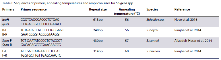
PCR was conducted for amplification of the invasion plasmid antigen H (ipaH) gene including three sets of primers to amplify the target genes of S. boydii (Conserved Hypothetical Protein), S. sonnei (Putative Restriction Endonuclease) and S. flexneri (rfc/ wzy). A method described by Nave et al. (2016) and Alizadeh-Hesar et al. (2014) was used to perform PCR, and primer sequences used are shown in Table I.
Detection of the Salmonella species
Eleven genes namely: fliC, spiC, misL, Ppb23, spy, spvC, orfL, invA, sdf I, hilA, and fliB were investigated by PCR. Salmonella was confirmed using the invA gene, and the virulence of the isolates was determined using the other genes (Table II). Five microlitres of sterile nuclease-free water was used as negative control.
Detection of E. coli
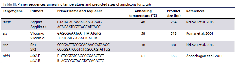
The PCR targeted the amplification of the uidA gene encoding for adhesin of E. coli, aggR gene encoding for antiaggregative protein (app) gene of Enteroaggregative E. coli, stx(Shiga toxins) encoding for lambdoid bacteriophages and eaeA gene encoding for enteropathogenic E. coli intimin. The primers and product sizes and annealing temperatures are shown in Table III.
Statistical analysis
Chi-square was used to test for significance of infection rate between male and female rats as well as between different rat species, whereas ANOVA was used to test for variance in observations made across the different sampling sites.
Results
Rodent identification
The PCR amplification and sequencing of the COI gene revealed that 55 (36%) and 99 (64%) of 154 rodent samples were R. tanezumi and R. rattus, respectively. Detailed information is provided in our previous publication (Ramatla et al. 2019).
Biochemical results
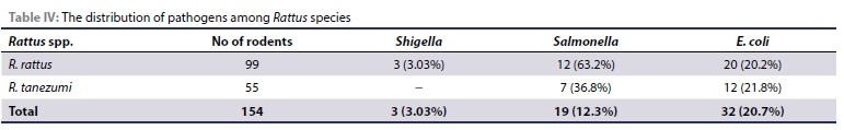
One hundred and fifty-four captured rat samples were investigated for the presence of zoonotic bacterial pathogens that they were possibly carrying, of which 3.3%, 20.7% and 12.3% were harbouring Shigella spp., E. coli and Salmonella spp., respectively as shown in Table IV. All chemical tests for Salmonella spp., Shigella spp. and E. coli isolates are shown in Table IV. A total of 33 presumptive isolates were suspected to be Shigella spp., 120 samples were presumptive Salmonella isolates and 162 were E. coli isolates.
Molecular results
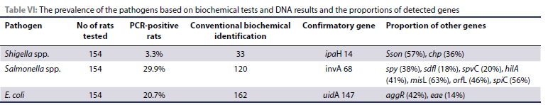
Out of five farms where samples were collected, Shigella spp. was not detected in two farms as shown in Table V. Based on biochemical tests, 33 presumptively positive Shigella isolates were identified. All 33 isolates were subjected to PCR of which 14 isolates were harbouring ipaH gene (613 bp) for Shigella spp., eight were positive for Sson (430bp), and five were positive for chp (248bp) genes for S. sonnei and S. boydii, respectively. One ipaH-positive isolate was negative for S. sonnei, S. boydii and S. flexneri, which we suspect might be S. dysenteriae.
The invA positive isolates (n = 68) were thus confirmed as Salmonella spp. All the other virulence genes detected are shown in Table VI. However, the genes: fliC, fliB, and Ppb23 were not amplified. Among the 162 presumptive E. coli isolates tested, a total of 147 showed amplification of the uidA E. coli housekeeping gene (Table V). After running the gel on electrophoresis for 65 minutes, the desired 556 bp fragments were obtained. Therefore, all 147 samples were confirmed as E. coli isolates. The aggR (42%), and eae (14%) genes were detected while none of the samples had the stx gene.
Discussion
Rodents can act as reservoirs of several pathogens, and controlling them effectively leads to control of these pathogens and the diseases they cause. They primarily transmit pathogens from the farm environment through their droppings and urine to food animals and subsequently to human beings. The poultry industry in South Africa does not yet fully appreciate the significant role played by rodents in the spread of E. coli, Shigella spp. and Salmonella spp. contamination in poultry farms and ultimately into the food chain. These pathogens are very important causes of serious diseases, colibacillosis, shigellosis and salmonellosis in human beings (Hong et al. 2018; Zhang et al. 2018). The current study, therefore, puts into perspective the important role that these rodents may play in possible circulation of these pathogens to poultry and ultimately humans.
In this study, the ipaH gene of Shigella spp. was detected from 3.3% of screened faecal rat samples. This gene, encoding the IpaH protein family (Lin et al. 2010), is found in four species of Shigella (S. dysenteriae, S. sonnei, S. boydii, and S. flexneri) (Lin et al. 2010; Mokhtari et al. 2012) and is therefore used as an indicator to detect the occurrence of Shigella spp. (Alizadeh-Hesar et al. 2014). Other studies have also found S. sonnei as the predominant species (Ranjbar et al. 2008; Tajbakhsh et al. 2012), compared to S. dysenterae which was rarely detected (Nave et al. 2016). It has also been reported that S. sonnei is the most isolated serotype in chickens (Zhang et al. 2013) and in human beings (Duran et al. 2013), thus supporting our findings in this case. S. sonnei invades human intestines, and then spreads, which is the critical process of its mechanism for its virulence. It has been found to be one of the most common species causing shigellosis (Ranjbar et al. 2008; Tajbakhsh et al. 2012). S. boydii was also detected in this study. These findings are in line with previous studies whereby S. boydii was detected (Ranjbar et al. 2014; Ud-Din et al. 2013). These findings, therefore, show that rats are a big public health risk factor for shigellosis.
The expression of invasion factor A (invA) gene has been confirmed in Salmonella spp. and is used for Salmonella spp. identification (Akinola et al. 2019; Fekry et al. 2018; Li et al. 2018; Refai et al. 2017). All the isolates which were invA positive (n = 68) in the current study were thus confirmed as Salmonella spp. The invA gene enables and shows the capacity of Salmonella spp. to infiltrate and cause gastroenteritis (Ekwanzala et al. 2017; Li et al. 2018) and is thus a marker of the pathogen's virulence. In our study, a proportion of 12.3% of Salmonella spp. was identified in rats, which is substantially lower than that which has been reported in other countries such as the USA with 16.2% (Henzler & Opitz 1992), Nigeria with 18.0% (Wakawa et al. 2015), as well as Japan with 31.8-41.2% (Lapuz et al. 2008). However, in this study the detection rate of Salmonella is higher than that found on poultry farms in other countries, including the UK with 10.0% (Hilton et al. 2002) and 2% in Trinidad and Tobago (Nkogwe et al., 2011). The proportional discrepancies could be owing to the analytical methodologies utilised, the number of samples analysed, and the various geographical/regional circumstances.
In 68 invA harbouring Salmonella isolates, the spyC, hilA, misL, orfL, sdfl and spy virulence genes were also detected. The spvC gene, which is translocated into the host cell cytoplasm by the SPI-2 TTSS (type three secretion system), was found in 21% of the isolates (Browne et al. 2008). The spvC gene is necessary for host cell survival and is important for systemic infections (Chaudhary et al. 2015). The hilA gene was carried by eight isolates (11%). This gene is significant in Salmonella pathogenesis because it is required for bacterial colonisation of the host intestine's extracellular luminal compartment (Peixoto et al. 2017). About 65% of the Salmonella isolates were carrying misL gene and the gene increases pathogenicity by allowing the infection to survive inside macrophages (Hughes et al. 2008). About 47% of the isolates were carrying the orfL gene which helps with adhesion and survival in macrophages, and it also possesses a secretion mechanism that regulates toxin secretion (Odjadjare & Olaniran 2015), hence promoting the pathogenicity of the Salmonella isolate engaged. About 12 (18%) of the isolates had the sdfI, for the detection of S. Enteritidis (Mohd Afendy & Son 2015), whereas the spy gene was found to be present in 26 (38%), and many researchers have used it to detect S. Typhimurium (De Freitas et al. 2010). The S. Enteritidis and S. Typhimurium strains have been identified as causes of human diseases (Collard et al. 2008) and also infecting some animals (Rodriguez et al. 2018). Therefore, this study confirms the presence of these Salmonella serovars from rats and further shows that these strains are virulent.
Most E. coli strains are known to be commensal bacteria in all warm-blooded animals' gastrointestinal tracts (Karmali et al. 2010; Madoshi et al. 2016). The aggR and eae genes were detected in this study, while the stx gene was not detected from all the rat faecal samples. The findings of this study concur with previous results obtained in Tunisia (Salem et al. 2011) and in South Africa (Ndlovu et al. 2015). However, the detection of aggR, stx and eae genes among the E. coli isolates is of great concern. The eae gene, on the other hand, is found in the enteropathogenic (EPEC) E. coli strains and used as a marker for LEE-positive STEC strains (Alonso et al. 2017; Phillips & Frankel 2000) and is responsible for attacking and destroying the intestinal epithelial cell of the host (Malik et al. 2017). Shiga toxin (Stx) is the virulent factor in Shiga toxin-producing E. coli (STEC) which causes human gastrointestinal diseases (Phillips & Frankel 2000). The aggR is described as a transcriptional regulator of enteroaggregative E. coli (EAEC). Enteroaggregative E. coli causes diarrhoea in humans (Morin et al. 2013). The presence of these E. coli strains in rats that are capable of contaminating both feed and poultry products is an important public health concern.
Conclusion
The detection of Shigella, Salmonella and E. coli bacteria in the faecal samples highlights the importance of these rodents in the spread of these pathogens among rats, chickens, and finally human beings. The fact that the strains detected also had the genes that signify virulence and are similar to species known to cause disease in humans and animals is of great significance. The rats provide the chance for environment-rat-chicken interaction during ingestion of Shigella spp., Salmonella spp. and E. coli-contaminated rodent faecal droppings by poultry, thus increasing the risk of circulation and re-introduction of the pathogens even after the flock has been evacuated and the environment cleansed and disinfected. Consequently, the risks posed by the presence of rodents (Rattus spp.) in the transmission cycle in the human environment and in chicken farms should not be overlooked. To limit the risk of Shigella, Salmonella, and E. coli transmission to chickens and, ultimately, humans, it is critical to manage rats on poultry farms. Control and prevention of these pathogens within these poultry farming settings requires a "One Health" approach.
Ethical approval
Prior to the commencement of the study, the research proposal was approved based on Animal Research Ethics Committee (NWU-00274-18-A5) guidelines by North-West University Research Ethics Regulatory Committee (NWU-RERC).
ORCID
TA Ramatla: https://orcid.org/0000-0002-0473-8075
N Mphuthi: https://orcid.org/0000-0001-6956-4152
T Ramaili: https://orcid.org/0000-0002-8025-0979
M Taioe: https://orcid.org/0000-0002-2905-4104
O Thekisoe: https://orcid.org/0000-0003-0700-4787
M Syakalima: https://orcid.org/0000-0002-0649-5409
References
Adesiyun, A.A., 1999, Absence of Escherichia coli O157 in a survey of wildlife from Trinidad and Tobago, J Wildl Dis 35(1), 1 15-120. https://doi.org/10.7589/0090-3558-35.1.115. [ Links ]
Akinola, S.A., Mwanza, M., Ateba, C.N., 2019, Occurrence, genetic diversities, and antibiotic resistance profiles of Salmonella serovars isolated from chickens, Infect Drug Resist 12, 3327-3342. https://doi.org/10.2147/IDR.S217421. [ Links ]
Alizadeh-Hesar, M., Bakhshi, B., Najar-Peerayeh, S., 2014, Molecular diagnosis of Salmonella enterica and Shigella spp. in stool sample of children with diarrhea in Tehran, International Journal of Enteric Pathogens 2, 1-5. https://doi.org/10.17795/ijep17002. [ Links ]
Alonso, C.A., Mora, A., Diaz, D., et al., 2017, Occurrence and characterization of stx and/or eae-positive Escherichia coli isolated from wildlife, including a typical EPEC strain from a wild boar, Vet Microbiol 207, 69-73. https://doi.org/10.1016/j.vetmic.2017.05.028. [ Links ]
Alvarez, J., Sota, M., Vivanco, A.B., 2004, Development of a multiplex PCR technique for detection and epidemiological typing of Salmonella in human clinical samples, J Clin Microbiol 42(4), 1734-1738. https://doi.org/10.1128/JCM.42.4.1734-1738.2004. [ Links ]
Anbazhagan, D., Mui, W.S., Manso, M., et al., 2011, Development of conventional and real-time multiplex PCR assays for the detection of nosocomial pathogens, Braz J Microbiol 42(2), 448-458. https://doi.org/10.1590/S1517-83822011000200006. [ Links ]
Ateba, C. & Mochaiwa, B., 2014, Use of invA gene specific PCR analysis for the detection of virulent Salmonella species in beef products in the North West province, South Africa, Journal of Food and Nutrition Research 2, 294-300. https://doi.org/10.12691/jfnr-2-6-5. [ Links ]
Browne, S.H., Hasegawa, P., Okamoto, S., et al., 2008, Identification of Salmonella SPI-2 secretion system components required for SpvB-mediated cytotoxicity in macrophages and virulence in mice, FEMS Immunol Med Microbiol 52(2), 194-201. https://doi.org/10.1111/j.1574-695X.2007.00364.x. [ Links ]
Caballero, M., Rivera, I., Jara, L.M., et al, 2015, Isolation and molecular identification of potentially pathogenic Escherichia coli and Campylobacter jejuni in feral pigeons from an urban area in the city of Lima, Peru, Rev Inst Med Trop Sao Paulo 57(5), 393-396. https://doi.org/10.1590/S0036-46652015000500004. [ Links ]
Chaudhary, J.H., Nayak, J.B., Brahmbhatt, M.N., et al., 2015, Virulence genes detection of Salmonella serovars isolated from pork and slaughterhouse environment in Ahmedabad, Gujarat, Vet World 8(1), 121-124. https://doi.org/10.14202/vetworld.2015.121-124. [ Links ]
Collard, J.M., Bertrand, S., Dierick, K., et al., 2008, Drastic decrease of Salmonella Enteritidis isolated from humans in Belgium in 2005, shift in phage types and influence on foodborne outbreaks, Epidemiol Infect 136(6), 771-781. https://doi.org/10.1017/S095026880700920X.d. [ Links ]
De Freitas, C.G., Santana, A.P., Da Silva, P.H.C., et al., 2010, PCR multiplex for detection of Salmonella Enteritidis, Typhi and Typhimurium and occurrence in poultry meat, Int J Food Microbiol 139(1-2), 15-22. https://doi.org/10.1016/j.ijfoodmicro.2010.02.007. [ Links ]
Dobrowsky, P.H., Van Deventer, A., De Kwaadsteniet, M., et al., 2014, Prevalence of virulence genes associated with pathogenic Escherichia coli strains isolated from domestically harvested rainwater during low- and high-rainfall periods, Appl Environ Microbiol 80(5), 1633-1638. https://doi.org/10.1128/AEM.03061-13. [ Links ]
Duran, C., Nato, F., Dartevelle, S., et al., 2013, Rapid diagnosis of diarrhea caused by Shigella sonnei using dipsticks; comparison of rectal swabs, direct stool and stool culture, PLoS One 8(11), e80267. https://doi.org/10.1371/journal.pone.0080267. [ Links ]
Ekwanzala, M.D., Abia, A.L.K., Keshri, J. et al., 2017, Genetic characterization of Salmonella and Shigella spp. isolates recovered from water and riverbed sediment of the Apies River, South Africa, Water SA 43, 387-397. [ Links ]
El-Sharkawy, H., Tahoun, A., El-Gohary A.E.G.A., et al., 2017, Epidemiological, molecular characterization and antibiotic resistance of Salmonella enterica serovars isolated from chicken farms in Egypt, Gut Pathog 9, 8. https://doi.org/10.1186/s13099-017-0157-1. [ Links ]
Fekry, E., Ammar, A.M., Hussien, A., 2018, Molecular detection of InvA, OmpA and Stn genes in Salmonella serovars from broilers in Egypt, Alexandria Journal of Veterinary Science 56, 69-74. https://doi.org/10.5455/ajvs.288089. [ Links ]
Franssen, F., Swart, A., Van Knapen, F., et al., 2016, Helminth parasites in black rats (Rattus rattus) and brown rats (Rattus norvegicus) from different environments in the Netherlands, Infect Ecol Epidemiol 6, 31413. https://doi.org/10.3402/iee.v6.31413. [ Links ]
Gopee, N.V., Adesiyun, A.A., Caesar, K., 2000, Retrospective and longitudinal study of salmonellosis in captive wildlife in Trinidad, J Wildl Dis 36(2), 284-293. https://doi.org/10.7589/0090-3558-36.2.284. [ Links ]
Henzler, D. & Opitz, H., 1992, The role of mice in the epizootiology of Salmonella enteritidis infection on chicken layer farms, Avian Dis 36(3), 625-631. https://doi.org/10.2307/1591757. [ Links ]
Hilton, A.C., Willis, R.J., Hickie, S.J., 2002, Isolation of Salmonella from urban wild brown rats (Rattus norvegicus) in the West Midlands, UK, Int J Environ Health Res 12(2), 163-168. https://doi.org/10.1080/09603120220129328. [ Links ]
Hong, Y.P.V., Wang, Y.W., Huang, I.H., 2018, Genetic relationships among multidrug-resistant Salmonella enterica serovar Typhimurium strains from humans and animals, Antimicrob Agents Chemother 62(5), 213-218. https://doi.org/10.1128/AAC.00213-18. [ Links ]
Hughes, L.A., Shopland, S., Wigley P., et al., 2008, Characterisation of Salmonella enterica serotype Typhimurium isolates from wild birds in northern England from 2005-2006, BMC Vet Res 4, 4. https://doi.org/10.1186/1746-6148-4-4. [ Links ]
Jemilehin, F., Ogunleye, A., Okunlade, A., et al., 2016, Isolation of Salmonella species and some other Gram negative bacteria from rats cohabitating with poultry in Ibadan, Oyo State, Nigeria, African Journal of Microbiology Research 10, 1104-1110. https://doi.org/10.5897/AJMR2015.7774. [ Links ]
Jianjun, J.I.A.N.G., 2005, Isolation and identification of rabbits Shigelladysenteriae in a large-scale warren, Journal of Anhui Agricultural Sciences 33, 1666. [ Links ]
Karmali, M.A., Gannon, V., Sargeant, J.M., 2010, Verocytotoxin-producing Escherichia coli (VTEC), Vet Microbiol 140(3-4), 360-370. https://doi.org/10.1016/j.vetmic.2009.04.011. [ Links ]
Kumar, J.K., Tabor, S., Richardson, C.C., 2004. Proteomic analysis of thioredoxin-targeted proteins in Escherichia coli, Proc Natl Acad Sci U S A 101(11), 3759-3764. https://doi.org/10.1073/pnas.0308701101. [ Links ]
Lapuz, R., Tani, H., Sasai, K., et al., 2008, The role of roof rats (Rattus rattus) in the spread of Salmonella Enteritidis and S. Infantis contamination in layer farms in eastern Japan, Epidemiol Infect 136(9), 1235-1243. https://doi.org/10.1017/S095026880700948X. [ Links ]
Li, X., Liu, L., Li, Q., et al., 2018, Salmonella contamination in layer farms in China: detection and genetic analysis, J Poult Sci 55(1), 1-9. https://doi.org/10.2141/jpsa.0160144. [ Links ]
Lin, W.S., Cheng, C.M., Van, K.T., 2010, A quantitative PCR assay for rapid detection of Shigella species in fresh produce, J Food Prot 73(2), 221-233. https://doi.org/10.4315/0362-028X-73.2.221. [ Links ]
López, F.E., De las Mercedes Pescaretti, M., Morero, R., et al., 2012, Salmonella Typhimurium general virulence factors: A battle of David against Goliath?, Food Res Int 45(2), 842-851. https://doi.org/10.10167j.foodres.2011.08.009. [ Links ]
Madoshi, B.P., Kudirkiene, E., Mtambo, M.M., et al., 2016, Characterisation of commensal Escherichia coli isolated from apparently healthy cattle and their attendants in Tanzania, PloS One 11(12), e0168160. https://doi.org/10.1371/journal.pone.0168160. [ Links ]
Mainar-Jaime, R.C., Andrés, S., Vico, J.P., et al., 2013, Sensitivity of the ISO 6579: 2002/ Amd 1: 2007 standard method for detection of Salmonella spp. on mesenteric lymph nodes from slaughter pigs, J Clin Microbiol 51(1), 89-94. https://doi.org/10.1128/JCM.02099-12. [ Links ]
Malik, A., Nagy, B., Kugler, R., et al., 2017, Pathogenic potential and virulence genotypes of intestinal and faecal isolates of porcine post-weaning enteropathogenic Escherichia coli, Res Vet Sci 115, 102-108. https://doi.org/10.1016/j.rvsc.2017.02.002. [ Links ]
March, S.B. & Ratnam, S., 1986, Sorbitol-MacConkey medium for detection of Escherichia coli O157: H7 associated with hemorrhagic colitis, J Clin Microbiol 23(5), 869-872. https://doi.org/10.1128/jcm.23.5.869-872.1986. [ Links ]
Maurelli, A.T., Routh, P.R., Dillman, R.C., et al., 1998, Shigella infection as observed in the experimentally inoculated domestic pig, Sus scrofa domestica, Microb Pathog 25(4), 189-196. https://doi.org/10.1006/mpat.1998.0230. [ Links ]
Mohd Afendy, A.T. & Son, R., 2015, Pre-enrichment effect on PCR detection of Salmonella Enteritidis in artificially contaminated raw chicken meat, International Food Research Journal 22, 2571-2576. [ Links ]
Mokhtari, W., Nsaibia, S., Majouri, D., et al., 2012, Detection and characterization of Shigella species isolated from food and human stool samples in Nabeul, Tunisia, by molecular methods and culture techniques, J Appl Microbiol 1 13(1), 209-222. https://doi.org/10.1111/j.1365-2672.2012.05324.x. [ Links ]
Morin, N., Santiago, A.E., Ernst, R.K., et al., 2013, Characterization of the AggR regulon in enteroaggregative Escherichia coli, Infect Immun, 81(1), 122-132. https://doi.org/10.1128/IAI.00676-12. [ Links ]
Nave, H.H., Mansouri, S., Sadeghi, A. et al., 2016, Molecular diagnosis and anti-microbial resistance patterns among Shigella spp. isolated from patients with diarrhea, Gastroenterol Hepatol Bed Bench 9(3), 205-210. [ Links ]
Ndlovu, T, Le Roux, M., Khan, W., et al., 2015, Co-detection of virulent Escherichia coli genes in surface water sources, PLoS One 10(2), e0116808. https://doi.org/10.1371/journal.pone.0116808. [ Links ]
Nkogwe, C., Raletobana, J., Stewart-Johnson, A., et al., 2011, Frequency of detection of Escherichia coli, Salmonella spp., and Campylobacter spp. in the faeces of wild rats (Rattus spp.) in Trinidad and Tobago, Vet Med Int 2011, 686923. https://doi.org/10.4061/2011/686923. [ Links ]
Odjadjare, E.C. & Olaniran, A.O., 2015, Prevalence of antimicrobial resistant and virulent Salmonella spp. in treated effluent and receiving aquatic milieu of wastewater treatment plants in Durban, South Africa, Int J Environ Res Public Health 12(8), 9692-9713. https://doi.org/10.3390/ijerph120809692. [ Links ]
Olobatoke, R.Y. & Mulugeta, S.D., 2015, Incidence of non-typhoidal Salmonella in poultry products in the North West Province, South Africa, South African Journal of Science 111(11/12), 7. https://doi.org/10.17159/sajs.2015/20140233. [ Links ]
Pan, B., Wang, W., Xie, Y.P., et al., 2006, Detection, serology classification and drug susceptibility of Shigella from experimental monkeys, Guangxi Agricultural Science 37, 331-332. [ Links ]
Peixoto, R.J., Alves, E.S., Wang, M., et al., 2017, Repression of Salmonella host cell invasion by aromatic small molecules from the human fecal metabolome, Appl Environ Microbiol 83(19), e01148-01117. https://doi.org/10.1128/AEM.01148-17. [ Links ]
Perera, K. & Murray, A., 2008, Development of a PCR assay for the identification of Salmonella enterica serovar Brandenburg, J Med Microbiol 57(Pt 10), 1223-1227. https://doi.org/10.1099/jmm.0.2008/002337-0. [ Links ]
Phillips, A.D. & Frankel, G., 2000. Intimin-mediated tissue specificity in enteropathogenic Escherichia coli interaction with human intestinal organ cultures, J Infect Dis, 181(4), 1496-1500. https://doi.org/10.1086/315404. [ Links ]
Phiri, A.F., Abia, A.L.K., Amoako, D.G., et al., 2021, Burden, antibiotic resistance, and clonality of Shigella spp. Implicated in community-acquired acute diarrhoea in Lilongwe, Malawi, Trop Med Infect Dis 6(2), 63. https://doi.org/10.3390/tropicalmed6020063 [ Links ]
Ramatla, T., Mphuthi, N., Gofaone, K., et al., 2019, Identification of rodent species that infest poultry houses in Mafikeng, North West Province, South Africa, International Journal of Zoology 2019, 1-8. https://doi.org/10.1155/2019/1280578. [ Links ]
Ramatla, T., Ngoma, L., Adetunji, M., et al., 2017, Evaluation of antibiotic residues in raw meat using different analytical methods, Antibiotics (Basel) 6(4), 34. https://doi.org/10.3390/antibiotics6040034. [ Links ]
Ramatla, T.A., 2019, The virulence and antimicrobial resistance of Salmonella spp. isolates from rodents inhabiting chicken farms in Mafikeng, South Africa (Doctoral dissertation, North-West University [South Africa]). Available from: http://repository.nwu.ac.za/handle/10394/35485. Accessed 23 Jan 2021. [ Links ]
Ramatla, T.A., Mphuthi, N., Ramaili, T., et al., 2020, Molecular detection of virulence genes in Salmonella spp. isolated from chicken faeces in Mafikeng, South Africa, J S Afr Vet Assoc 91(1), e1-e7. https://doi.org/10.4102/jsava.v91i0.1994. [ Links ]
Ranjbar, R., Afshar, D., Mehrabi Tavana, A., et al., 2014, Development of multiplex PCR for simultaneous detection of three pathogenic Shigella species, Iran J Public Health 43(12), 1657-1663. [ Links ]
Ranjbar, R., Dallal, M.M.S., Talebi, M., et al., 2008, Increased isolation and characterization of Shigella sonnei obtained from hospitalized children in Tehran, Iran, J Health Popul Nutr 26(4), 426-230. https://doi.org/10.3329/jhpn.v26i4.1884. [ Links ]
Refai, M., Hatem, M.E., Elhariri, M., et al., 2017, Using of molecular biology techniques compared with conventional detection methods for detection of Salmonella in cattle in Egypt, Journal of American Science 13, 46-50. [ Links ]
Rodriguez, F.I., Pascal, D.C., Pulido, D., et al., 2018, Prevalence, antimicrobial resistance profile and comparison of selective plating media for the isolation of Salmonella in backyard chickens from Entre Rios, Argentina, Zoonoses Public Health 65(1), 95-101. https://doi.org/10.1111/zph.12415. [ Links ]
Salem, I.B., Ouardani, I., Hassine, M., et al., 2011, Bacteriological and physico-chemical assessment of wastewater in different region of Tunisia: impact on human health, BMC Res Notes 4, 144. https://doi.org/10.1186/1756-0500-4-144. [ Links ]
Samaxa, R.G., Matsheka, M.I., Mpoloka, S.W., et al., 2012, Prevalence and antimicrobial susceptibility of Salmonella isolated from a variety of raw meat sausages in Gaborone (Botswana) retail stores, J Food Prot 75(4), 637-642. https://doi.org/10.4315/0362-028X.JFP-11-438. [ Links ]
Tajbakhsh, M., Garcia Migura, L., Rahbar, M., et al., 2012, Antimicrobial-resistant Shigella infections from Iran: an overlooked problem?, J Antimicrob Chemother 67(5), 1 128-1 133. https://doi.org/10.1093/jac/dks023. [ Links ]
Theron, J., Morar, D., Du Preez, M., et al., 2001, A sensitive seminested PCR method for the detection of Shigella in spiked environmental water samples, Water Res 35(4), 869-874. https://doi.org/10.1016/S0043-1354(00)00348-1. [ Links ]
Ud-Din, A.I., Wahid, S.U., Latif, H.A., et al., 2013, Changing trends in the prevalence of Shigella species: emergence of multi-drug resistant Shigella sonnei biotype g in Bangladesh, PloS One 8(12), e82601. https://doi.org/10.1371/journal.pone.0082601. [ Links ]
Umali, D.V., Lapuz, R.R.S.P., Suzuki, T., et al., 2012, Transmission and shedding patterns of Salmonella in naturally infected captive wild roof rats (Rattus rattus) from a Salmonella-contaminated layer farm, Avian Dis 56(2), 288-294. https://doi.org/10.1637/9911-090411-Reg.1. [ Links ]
Vendramin, T., Kich, D.M., Molina, R.D., et al., 2014, Molecular screening of bovine raw milk for the presence of Shiga toxin-producing Escherichia coli (STEC) on dairy farms, Food Sci Technol 34(3), 604-608. https://doi.org/10.1590/1678-457x.6422. [ Links ]
Wakawa, A., Mohammed, F., Mamman, H., 2015, Isolation and antibiotic susceptibility of Escherichia coli and Salmonella gallinarum isolated from rats in commercial poultry farms with recurrent Colibacillosis and Fowl typhoid cases in Zaria, Nigeria, Journal of Animal and Veterinary Advances 5, 1147-1152. [ Links ]
Wang, Y., Wang, Y., Lan, R., et al., 2015, Multiple endonuclease restriction real-time loop-mediated isothermal amplification: a novel analytically rapid, sensitive, multiplex loop-mediated isothermal amplification detection technique, J Mol Diagn 17(4), 392-401. https://doi.org/10.1016/j.jmoldx.2015.03.002. [ Links ]
Zhai L., Yu Q., Bie X., et al., 2014, Development of a PCR test system for specific detection of Salmonella Paratyphi B in foods, FEMS Microbiol Lett 355(1), 83-89. https://doi.org/10.1111/1574-6968.12443. [ Links ]
Zhang, H., Wang, R., Bao, H., 2013, Phage inactivation of foodborne Shigella on ready-to-eat spiced chicken, Poult Sci 92(1), 211-217. https://doi.org/10.3382/ps.2011-02037. [ Links ]
Zhang, L., Wei, Q., Han, Q., et al., 2018, Detection of Shigella in milk and clinical samples by magnetic immunocaptured-loop-mediated isothermal amplification assay, Front Microbiol 9, 94. https://doi.org/10.3389/fmicb.2018.00094. [ Links ]
Zishiri, O.T, Mkhize, N., Mukaratirwa, S., 2016, Prevalence of virulence and antimicrobial resistance genes in Salmonella spp. isolated from commercial chickens and human clinical isolates from South Africa and Brazil, Onderstepoort J Vet Res 83(1), a1067. https://doi.org/10.4102/ojvr.v83i1.1067. [ Links ]
 Correspondence:
TA Ramatla
Email: ra21205450@gmail.com

