


Serviços Personalizados
Artigo
 Inglês (pdf)
 Artigo em XML
 Referências do artigo
Indicadores

Links relacionados
-
 Citado por Google
 -
 Similares em Google
Compartilhar

 Permalink
Journal of the South African Veterinary Association
versão On-line ISSN 2224-9435
versão impressa ISSN 1019-9128
J. S. Afr. Vet. Assoc. vol.93 no.1 Pretoria 2022
http://dx.doi.org/10.36303/jsava.2022.93.1.505
ORIGINAL RESEARCH
https://doi.org/10.36303/jsava.2022.93.1.505
Prevalence of methicillin resistance in Staphylococcus pseudintermedius isolates from dogs with skin and ear infections in South Africa
CD PriorI; A MoodleyII; M KaramaIII; MN MalahlelaIV; A LeisewitzI
IDepartment of Companion Animal Clinical Studies, Faculty ofVeterinary Science Onderstepoort, University of Pretoria, South Africa
IIInternational Livestock Research Institute, Kenya
IIIVeterinary Public Health Section, Faculty ofVeterinary Science, University of Pretoria, South Africa
IVVeterinary Public Health Section, Department of Paraclinical Sciences, Faculty ofVeterinary Science Onderstepoort, University of Pretoria, South Africa
ABSTRACT
Staphylococcus pseudintermedius (SP) is an important opportunistic pathogen, frequently associated with pyoderma and otitis in dogs. The emergence and rapid expansion of methicillin-resistant Staphylococcus pseudintermedius (MRSP) is problematic due to multidrug resistance and reduced treatment options. The aim of this study was to determine i) the prevalence of MRSP in dogs with pyoderma or otitis externa, ii) the antimicrobial resistance patterns of MRSP from South African isolates, and iii) the risk factors for MRSP-associated pyoderma or otitis externa in dogs in South Africa (RSA).
Sixty-eight presumptive clinical SP isolates (collected from 65 dogs) from five geographically dispersed laboratories in RSA were collected over 2 years. Possible MRSP isolates were flagged when resistance to oxacillin was observed. Thereafter, all isolates were confirmed as SP by polymerase chain reaction (PCR) and further genotyped for the mecA gene.
Fifty-seven of 68 isolates were confirmed to be SP (83.8%), while 49/57 (85.9%) carried mecA. Our findings showed that preliminary phenotypic methods supplemented by genotypic methods increased the accuracy of correctly identifying SP. All isolates were resistant to at least one antimicrobial drug. There was a high incidence of amoxicillin (70.1%) and enrofloxacin (65%) resistance. Important risk factors for mecA positive carriage were previous hospital admission, pruritus, and previous antibacterial failure. This study demonstrates a high prevalence of mecA positive carriage (85.9% of samples) in MRSP pyoderma and otitis in dogs in RSA. There is an urgent need for better laboratory diagnosis of MRSP and surveillance of dogs presenting with pyoderma and otitis in South Africa.
Keywords: antimicrobial, disc diffusion, mecA, veterinary, pyoderma, otitis
Background
Staphylococcus pseudintermedius (SP) is the most frequent bacterial pathogen isolated from canine skin and ear infections, (Griffeth et al. 2008) and is a leading cause of pyoderma. SP accounts for 20% of ear infections in dogs presented to veterinarians (Cole et al. 2006). The worldwide spread of methicillin-resistant SP (MRSP) has become a significant animal health problem (Hensel et al. 2016). In South Africa alone, there are an estimated 9.2 million dogs living in households (Canine Zone 2019) and given the close interaction between humans and their pets, there is an increased risk of spread of multidrug-resistant bacteria (MDR), particularly in animals that are treated for life threatening diseases, which can become zoonotic (Hartantyo et al. 2018). Currently, the misuse and overuse of antimicrobials remains the key factor for selection of MRSP strains in healthy dogs, representing a huge challenge for effective veterinary treatment (Rota et al. 2013).
In the South African setting, the emergence of MRSP may have serious implications. Given the unreported prevalence of MRSP and mecA positive isolates, little is known about the impact of methicillin resistance in animal health and its effects on a One Health level. With human carriage of SP, certain mobile genetic elements from MRSP may promote the spread of resistant genes to commensal skin flora (Van Duijkeren et al. 2011).
Tuberculosis (TB) and human immunodeficiency virus (HIV) are common infections amongst impoverished South African communities. HIV is an important cause of immunosuppression. TB transmission is associated with low socio-economic status and frequently seen in HIV burdened communities (Tadokera et al. 2020). Clustering of domestic animals in these settings thus remains a proxy for transmission of MDR bacteria, which could pose a risk to already immunocompromised individuals.
Otitis externa and pyoderma are common diseases in dogs that have been associated with MRSP (Mathie et al. 2010). A number of antimicrobials are commonly used for the treatment of otitis including fusidic acid, aminoglycosides, polymyxin B, fluoroquinolones, silver sulfadiazine, and oxytetracycline (Jacobson 2002; Morris et al. 2017).
The World Association of Veterinary Dermatology (WAVD) provides guidelines regarding the management, therapeutic considerations and preventive measures for MRSP infections in dogs (Morris et al. 2017). Despite the availability of these guidelines, in South Africa the treatment and management of MRSP infections remains suboptimal or inappropriate, as seen by the high level of resistance encountered (Qekwana et al. 2019). This is likely due to a lack of awareness amongst practitioners. A study on Staphylococcus spp. isolates from South Africa reported a significant increase in S. aureus and SP isolates that were resistant to second- and third-line antibiotics including lincosamides, fluroquinolones and trimethoprim-sulphamethoxazole (Qekwana et al. 2019). Furthermore, Blunt et al. (2013) illustrated high levels of resistance to ampicillin and doxycycline in SP isolates associated with pyoderma in dogs in South Africa (Blunt et al. 2013; Qekwana et al. 2019).
Current phenotypic and biochemical lab methods used in the diagnosis of Staphylococcus isolates are fallible. Discrepancies in colour change and variation in the expression of key characteristics in biochemical testing result in the misidentification of bacterial species (Kasela & Malm 2018; Schissler et al. 2009; Speers et al. 1998). Whilst current phenotypic biochemical tests are rapid and user-friendly, the reproducibility of the individual substrate reactions ranges varies and has a low level of accuracy (Schissler et al. 2009). Furthermore, the ability of current biochemical methods to distinguish SP from other Staphylococcus spp. is inadequate as these species can be interchangeably misidentified and most tests for SP are heavily biased towards S. aureus (Bond & Loeffler 2012; Stull et al. 2014; Vrbovská et al. 2020). As a result, these tests cannot adequately distinguish between S. aureus and SP veterinary strains (Couto et al. 2001).
Currently, information on antimicrobial resistance patterns of pyoderma or otitis externa-associated SP isolates and risk factors for carriage in dogs is not available in South Africa. The aim of this study was to determine i) the prevalence of MRSP in dogs with pyoderma or otitis externa, ii) the antimicrobial resistance patterns of MRSP from South African isolates, and iii) the risk factors for MRSP-associated pyoderma or otitis externa in dogs in South Africa.
Research methods and design
Sampling and testing
A total of 49 skin and 16 ear samples were collected by veterinarians from 64 dogs that presented with pyoderma or otitis externa at 28 private veterinary clinics, six private veterinary specialist centres and one academic veterinary hospital in South Africa. Samples were collected over 25 months from November 2017 to December 2019 and submitted to five participating diagnostic laboratories located in five provinces of South Africa. In the clinic, skin and ear samples were collected with Amies transport swabs and transported on Amies Transport medium (Thermofisher Scientific, Oxoid Limited, Basingstoke, UK) before being sent to the appropriate laboratories. Individual labs then cultured the samples and identified the presence of SP using routine microbiological techniques (Clinical and Laboratory Standards Institute 2015).
The disk diffusion method was used in the lab to test presumptive SP isolates for antimicrobial resistance against a panel of 16 antimicrobials (Oxoid Thermo Scientifc, UK), as described by the Clinical Laboratory Standards Institute guidelines (Clinical and Laboratory Standards Institute 2015). The five bacteriology laboratories participating in this study followed the Clinical Laboratory Standards Institute guidelines (Clinical Laboratory Standards Institute 2007, 2008, 2009, 2010, 2011, 2012, 2013, 2014, 2015) to isolate and conduct antimicrobial susceptibility testing.
The panel of 16 antimicrobials (Oxoid Thermo Scientifc, UK), included penicillin (10 IU), oxacillin (1pg), amoxicillin/clavulanic acid (20/10 μg), cephalothin (30 μg), cefoxitin (1 μg), cefazidime (30 μg), ceftriaxone (30 μg), doxycycline (30 μg), enrofloxacin μg), erythromycin (15 μg), clindamycin/lincomycin (2 μg), gentamicin (10 μg), amikacin (30 μg), kanamycin (30 μg), chloramphenicol (30 μg) and trimethoprim/sulfamethoxazole (25 μg). Isolates displaying oxacillin (methicillin) resistance were forwarded to the researcher after undergoing phenotypic and biochemical screening at the referring lab.
Data collection
Participating laboratories submitted a total of 68 presumptive (not PCR confirmed) clinical SP isolates. In addition, antimicrobial susceptibility results and patient information data for every dog from which each of the 68 SP was recovered were collected from all the veterinarians who consented to participate in this study. Patient data consisted of age, gender, breed, sterilisation status, hospital visits, referral centre visits, hospital admission or not, surgery, wounds, pruritus, antimicrobial treatment route (systemic, topical or eardrops), use of systemic glucocorticoids, use of 1st and 2nd tier antimicrobial use, and antimicrobial failure resulting in culture.
Molecular detection of methicillin-resistant S. pseudintermedius
All 68 presumptive MRSP isolates were evaluated using PCR (Sasaki et al. 2010) and PCR-Restriction Fragment Length Polymorphism (RFLP) (Bannoehr et al. 2009) to confirm their SP status. All isolates including confirmed SP and other Staphylococci isolates were screened for the presence of the mecA gene by PCR amplification (Haenni et al. 2014).
All isolates that were PCR negative for SP were tested by multiplex PCR to verify their Staphylococcus status (Morot-Bizotet al. 2004). Multiplex PCR was carried out as described by Morot-Bizot et al., with primers that targeted the four staphylococcal species, namely S. aureus, S. saprophyticus, S. xylosus and S. epidermidis (Morot-Bizot et al. 2004). The conditions for all PCR reactions used were as described in the various references cited.
Data management and analysis
Data collected for 14 variables (age, gender, hospital visits, referral centre visits, admission to hospital, surgery, wounds, pruritus, antimicrobial route, systemic glucocorticoids, 1st and 2nd tier antimicrobial use, antimicrobial failure resulting in culture and eardrops) were captured into Microsoft Excel spreadsheets (Mac 2019, Version 16.3.5). All descriptive statistical analyses were performed using SPSS 17.0 software for Windows, whereas exact logistic regression analyses were done using Stata/IC 11.2. Variables were analysed by exact logistic regression model for risk factors from animals with mecA positive and mecA negative isolates. Statistical analysis was performed using Fisher's exact test to determine non-random associations between categorical variables. The variables were placed into a univariate analysis in the form of 2 x 2 tables with Fisher's exact p-values. Those with p < 0.25 were entered into a multiple exact logistic regression model and non-significant variables (p > 0.05) were eliminated until only significant ones remained.
Results
Of the 68 presumptive MRSP isolates received from participating laboratories, 83.8% (57/68) of the isolates were confirmed as SP. Only the 57 isolates confirmed to be true SP by PCR (Sasaki et al. 2010) and PCR-RFLP (Bannoehr et al. 2009) were evaluated further. The remaining 11 isolates were tested using the multiplex-PCR described by Morot-Bizot et al. (2004). The assay confirmed that only seven of the isolates were Staphylococcus spp. with four of the isolates testing negative with the species-specific primers included in the multiplex-PCR assay. Since the 68 samples were collected from 65 dogs, it is calculated that 83% (54/65) of the dogs sampled were infected with SP.
Of the 57 SP positive samples, a number of them were found to be positive for other bacterial species in addition to SP: Streptococcus canis was detected in 17.5% (10/57), Enterococcus spp. was found in 15.8% (9/57), Pseudomonas aeruginosa in 8.8% (5/57), Enterobacter spp. in 5.2% (3/57), Neisseria animaloris, Staphylococcus epidermidis, Proteus mirabilis and Staphylococcus aureus were present in 2/57 (3.5%) and Escherichia coli was in 1/57 (1.8%) of samples.
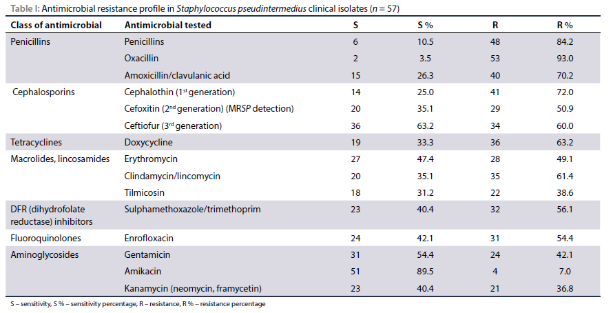
Of the 57 SP isolates that were tested for antimicrobial resistance on disc diffusion (Table I): 93.0% (53/57) were resistant to oxacillin, 84.2% (48/57) to penicillin, 72% (41/57) to cephalothin, 70.2% (40/57) to amoxicillin/clavulanic acid, 60.0% (34/57) to ceftiofur and 50.9% (29/57) to cefoxitin. Resistance to non-B-lactams included: 63.2% (36/57) to doxycycline, 61.4% (35/57) to clindamycin and lincomycin, 56.1% (32/57) to sulphamethoxazole/trimethoprim, and 55.4% (31/57) to enrofloxacin, 49.1% (28/57) to erythromycin, 42.1% (24/57) to gentamycin and 38.6% (22/57) to tilmicosin, 36.8% (21/57) to kanamycin, 12.3% (7/57) to amikacin and 3.5% (2/57) to chloramphenicol.
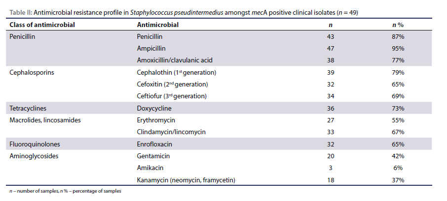
The mecA gene was found in 86.0% (49/57) of SP isolates, 100% (2/2) of S. aureus and 100% (2/2) in other staphylococcal isolates. Multi-resistance was defined as resistance to at least one antimicrobial in three or more antimicrobial categories (Magiorakos et al. 2012). MDR was observed in 49.1% (28/57) of SP that were mecA positive (Table II).
Demographics of samples
The highest number of clinical SP isolates were obtained from Gauteng province (47.4%; 27/57), followed by KwaZulu-Natal (28.1%; 16/57) and then the Western Cape (21%; 12/57).
The average age of the dogs in the study was 65 months (SD = 36 months). Of the 57 SP isolates, 18.5% (10/54) were detected in samples from the Africanus breed and 18.5% (10/54) from Bull Terrier dogs, while 12.97% (7/54) were from unknown breeds. A total of 5.5% (3/54) were detected in samples from German Shepherds, 7.4% (4/54) from Boerboel, 5.5% (3/54) from Jack Russell Terrier, 3.7% (2/54) from Border Collie, 3.7% (2/54) from Dachshund, 3.7% (2/54) from Rottweiler, 3.7% (2/54) Weimaraner, 3.7% (2/54) from Yorkshire Terrier, 1.8% (1/54) from Bulldog, 1.8% (1/54) from Staffordshire Bull Terrier, 1.8% (1/54) from Beagle, Miniature Pinscher 1.8% (1/54), Spaniel 1.8% (1/54), Chow Chow 1.8% (1/54), Doberman 1.8% (1/54), Great Dane 1.8% (1/54), Pekingese 1.8% (1/54), Poodle 1.8% (1/54) and 1.8% (1/54) were from Rhodesian Ridgeback.
Risk factors
Of all risk factors investigated, only hospital admission, pruritus and antibiotic failure increased the odds of recovering a mecA positive SP isolate from a dog skin or ear sample (p < 0.1). On univariate analysis, risk factors with p < 0.25 were entered into a multiple exact logistic regression model and non-significant variables (p > 0.05) were eliminated.
Discussion
This project was a prospective, descriptive study conducted from five geographically dispersed laboratories in RSA from November 2017 to December 2019.
S. pseudintermedius in dogs with pyoderma and otitis
Primary phenotypic identification of SP by diagnostic laboratories concurred with PCR (Sasaki et al. 2010) and PCR-RFLP (Bannoehr et al. 2009) in 83.8% (57/68) and 82.35% (56/68) of cases, respectively. These results reinforce previous studies in regard to the genera and species of bacterial isolates from cases of canine otitis and pyoderma where SP was the most frequently isolated organism (Bajwa 2016; Bajwa 2019; Grõnthal et al. 2017; Loeffler & Lloyd 2018; Maluping et al. 2014; Paul et al. 2011; Perreten et al. 2010).
Both PCR methods used in this study for the detection of SP have been shown to yield reliable results with a small margin of error (Bannoehr et al. 2009; Sasaki et al. 2007 ). The margin of erroneous identification when used together is less and substantially better than phenotypic methods of identification. This is the first molecular diagnostic test characterises the pta gene by the PCR-RFLP protocol according to Bannoehr et al. (2009). A small percentage of the SP population has been shown to be incorrectly identified because of heterogeneity in the Mbol restriction site (Slettemeâs et al. 2010). This necessitated the use of a second PCR methodology by Sasaki et al. (2007) that targets the 926-bp nuc gene. The second PCR method has been determined not to be specific for the final identification of SP as this gene has less variation for the specific species detection of SP. There was an acceptable level of concordance between phenotypic and molecular methods used to identify SP isolates. However, our findings showed that the use of a protocol whereby preliminary phenotypic methods are supplemented by molecular techniques increased the accuracy of identifying SP and eliminated false positive SP detected by phenotypic methods, thus enabling accurate treatment and surveillance of the SP.
It is clinically necessary to recognise methicillin resistance in Staphylococcus spp. isolates, as all methicillin-resistant staphylococci are considered resistant to all β-lactam antibiotics in vivo, irrespective of the results of disc diffusion (Stapleton & Taylor 2002). The most common form of methicillin resistance is conferred by the penicillin-binding protein 2a (PBP2a) encoded by the mecA gene (Matsuhashi et al. 1986). Detection of mecA via PCR is the gold standard for the diagnosis of methicillin resistance (Schissler et al. 2009). The disc diffusion method identified 77.9% (53/68) isolates to be methicillin resistant, while mecA was identified in 72.1% (49/68) of the methicillin-resistant isolates identified as MRSP on disc diffusion. Similarly to Graham et al. (2000) and Saptura,et al. (2017), the results of this study comparing mecA detection by PCR with resistant isolates by oxacillin disc test demonstrated that current phenotypic methods are suboptimal when compared to genotypic detection of MRSP. Methicillin resistance may also occur through hetero-resistance, alternative mechanisms or be observed in mecA-negative isolates that produce high levels of β-lactamase (Schissler et al. 2009). Hetero-resistance represents a minor subpopulation of antibiotic-resistant bacteria, which may go undetected in phenotypic testing (Band & Weiss 2019). Consequently, organisms that show sensitivity on antibiograms, may clinically exhibit resistance with subsequent treatment failures.
Other commonly used phenotypic tests for the detection of methicillin resistance include oxacillin resistance agar screening base and ORSAB selective supplement (Oxoid, Nepean, Canada), or a chromogenic MRSA agar (Brilliance MRSA Agar, Oxoid, Basingstoke, UK). While these tests are fallible and are typically used for MRSA screening, when combined with mecA PCR to determine phenotype and genotype, antimicrobial selection may be more clinically effective (Schissler et al. 2009).
Mixed infections - implications of methicillin-resistant S. pseudintermedius and other staphylococci in dogs with pyoderma and otitis externa
Recent studies of canine ear canal microbiota in dogs with otitis externa have been published (Bradley et al. 2020; Kasai et al. 2020; Korbelik et al. 2019; Tang et al. 2020). These studies demonstrate a decline in bacterial diversity in otitis externa and pyoderma compared to healthy dogs. Endemic canine ear and skin organisms, which are generally not the primary cause of the disease process, may become opportunists when primary pathological changes occur (Pye 2018). Otitis infections are commonly polymicrobial in nature with secondary opportunistic invaders (Lamm et al. 2010). The most common primary aetiological pathogens are members of the genus Staphylococcus (Penna et al. 2010).
The coagulase-positive species were the most common species of Staphylococcus isolated. Several staphylococcal species were identified, including coagulase-positive SP 83.8% (57/68), S. aureus and coagulase-negative S. epidermidis 7% (4/57). The high prevalence of coagulase-positive species in otitis and pyoderma is consistent with previous studies (Bajwa 2019; Bourély et al. 2019; Morris et al. 2006). The elevated prevalence of SP over S. aureus is expected, as this species is documented to be the dominant staphylococcal species in canine infections (Lyskova et al. 2007; Penna et al. 2010).
While staphylococci are a common resident organism on the dermis and mucosa of dogs, changes in the microenvironment on the surface of the skin can disrupt the equilibrium of the cutaneous ecosystem, allowing staphylococci to become pathogenic. As a result, other staphylococci are often cultured from dogs, suggesting that they play an important perpetuating role in the pathogenesis of otitis and pyoderma respectively (Lilenbaum et al. 2000; Loeffler & Lloyd 2018; Ma et al. 2020; Penna et al. 2010). Both S. aureus and S. epidermidis isolated in this study have been reported to develop methicillin resistance (Ma et al. 2020; Penna et al. 2010; Xu et al. 2020). Resultant antimicrobial resistance occurs due to the high frequency of conjugation and exchange of plasmids between members of the Staphylococcus species. Thus, knowledge of species members and their respective pathological properties in otitis and pyoderma provides practical information for the appropriate management of canine otitis externa (Kasai et al. 2020).
Mixed infections and implications in dogs with pyoderma and otitis externa
A smaller portion of samples had mixed infections that included MRSP and other bacteria, of which Enterococcus spp. was the most frequent (15.8%) followed by Pseudomonas aeruginosa (8.8%). The concomitant presence of MRSP with Enterococcus spp. and P. aeruginosa in pyoderma and/or otitis externa is consistent with previous studies (Kasai et al. 2020; Ngo et al. 2018).
The presence of P. aeruginosa in the clinical samples of canine otitis is clinically significant for the treatment of otitis among dogs (Pye 2018). P. aeruginosa is not typically a canine ear inhabitant but an opportunistic bacteria commonly found in soil, water, and decaying organic matter in the environment (Pye 2018). When present, it could potentially become reliant upon host factors to support its growth. P. aeruginosa produces biofilms protecting itself from antibiotics administered topically (Mekic et al. 2011). Due to the bacterium's resistance to several classes of antibiotics, selection of antibiotics for treatment can be challenging making it difficult to eradicate P. aeruginosa-associated canine otitis infection. Treatment is further complicated by the increasing number of multidrug-resistant strains with concurrent MRSP infection (Mekic et al. 2011).
In addition, Streptococcus canis, Enterococcus spp., P. aeruginosa, Enterobacter spp., N. animaloris, Proteus mirabilis and E. coli were also found in mixed infections. These organisms are normally located in the canine ear and skin and do not have a primary role in initiating the disease process, but rather become opportunists when pathological changes occur (Miller et al. 2013). Otitis infections are commonly polymicrobial in nature; some of these organisms are considered an incidental finding, however. most are secondary opportunistic invaders in dogs with otitis (Lamm et al. 2010).
Whilst non-staphylococci bacteria are members of normal ear and skin flora, their significance as opportunistic bacteria in canine otitis and pyoderma infections and predisposing factors are not fully established. Further research into the link between altered microbiota and disease severity are necessary, as this information is important for the successful treatment of otitis among dogs.
Antimicrobial resistance
All isolates were resistant to at least one antimicrobial drug. The degree of multidrug resistance in this study, 49.1% (28/57), was far higher than in previous studies (Blunt et al. 2013; Qekwana et al. 2019). This may be because the antimicrobials tested in this study are widely used in key formulations available for the management of otitis externa and pyoderma. This is a cause for concern as antimicrobial overuse may contribute to the selection of resistant strains of canine staphylococci (Penna et al. 2010).
The high incidence of amoxicillin resistance (70.1%; 40/57) found in this study was not unexpected given the preferential use of narrow-spectrum antimicrobials as a first-line as recommended by current antimicrobial guidelines (Buckland et al. 2016). In addition, methicillin-resistant isolates frequently displayed resistance to non-p-lactams during disc diffusion testing, especially to doxycycline, clindamycin, sulphamethoxazole/ trimethoprim and enrofloxacin.
The high levels of resistance of SP to enrofloxacin are in agreement with findings from other studies (Feng et al. 2012; Grõnthal et al. 2017; Kadlec & Schwarz 2012). This is of great concern considering its use in both human and veterinary medicine as an antimicrobial of last resort in the treatment of complicated infections. In agreement with previous studies evaluating resistance amongst isolates on disc diffusion, (Hanselman et al. 2009; Qekwana et al. 2017; Qekwana et al. 2019), levels of resistance to enrofloxacin in this study were especially high amongst the mecA positive isolates at 65% (32/49) (Table II).
Despite the high levels of resistance to enrofloxacin in this study, there was no statistically significant association with the use of other second-tier antibiotics such as cefalexin or cefpodoxime and mecA positive isolates in this study. This is contrary to Eckholm et al. (2013) and Fungwithaya et al. (2017) who demonstrated that the initiation of second-tier antibiotics, subsequently resulted in culture-positive methicillin resistance isolates, which were mecA positive. Only 51% (25/49) of the isolates in this study had a history of second tier antibiotic use (p > 0.1) compared to Eckholm et al. (2013) and Fungwithaya et al. (2017) in which 62.5% (n = 5) and 100% (n = 10) respectively.
Risk factors for mecA carriage in dogs with otitis externa and pyoderma
Regarding the different risk factors that were investigated in this study, findings showed that mecA occurrence rates in dog MRSP were associated with a number of factors, namely, hospital admission, pruritis and antibiotic failure. Univariable analysis of risk factor variables from animals were based on mecA positive isolates from PCR as this is considered the gold standard for the defining MRSP.
Based on the history provided by the referring veterinarians in this study, 32.7% (16/49) of mecA positive isolates reported a previous admission to hospital. Pruritis was found to have a significant association to mecA positive SP carriage - 57.1% (28/49) of mecA positive isolates had a history of pruritis, whereas none of the dogs with mecA negative isolates (0/8) displayed this clinical sign (p = 0.004). The majority of clinical isolates reported chronic pruritis as the main clinical sign. The association of MRSP with pruritis may be ascribed to the fact that pruritis alters the normal physiological barrier on the skin, which often requires antibacterial therapy for concurrent infections. This may be further exacerbated by inappropriate antimicrobial therapy and failure to recognise persistent underlying disease such as hypersensitivity (Bajwa 2016). Failure to address the underlying causes of pruritus will result in the recurrence of staphylococcal pyoderma and likely lead to repeated antimicrobial therapy. Repeated antimicrobial therapy is thus likely to become ineffectual, which will intensify the acquisition of mecA. Interventions including systemic and topical therapy to reduce inflammation and pruritus in dogs, such as the use of Oclacitinib, have demonstrated a decrease in antimicrobial use (Rynhoud et al. 2021). The overall reduction in antimicrobial use may thus decrease the prevalence of mecA amongst dogs with pyoderma and/or otitis. This further underscores the necessity of steering away from systemic antibiotic use to manage the surface and superficial pyoderma associated with hypersensitivity. It is crucial that antiseptic medical shampoos be the first treatment choice to manage these infections (Morris et al. 2017).
Of the 26 clinical isolates in this study that reported antimicrobial failure by referring clinicians, 25 were mecA positive. This highlights the importance of antimicrobial susceptibility testing of bacterial cultures associated with canine pyoderma and otitis after failure of empirical treatment with first-tier antibiotics.
The present study found that gender, sterilisation and age were not good predictors of mecA gene presence. The lack of a significant association between mecA and these variables could be attributed to the study's small sample size. Thus, further research is needed to investigate these factors.
Limitations of the study
A limitation of this study is that clinical isolates were not recruited randomly and the true prevalence of mecA carriage may therefore not be reflected. Additional limitations include its retrospective nature, the small number of isolates and the lack of a control group of methicillin-sensitive organisms. Whilst the methodology in this study touches on some of the molecular aspects in organism identification and the identification of mecA from isolates, the researcher did not assess the divergence between different MRSP strains. Additional molecular studies are required to further characterise the SP population from dogs in South Africa. Defining the behaviour and polymorphism of resistance genes in South Africa has both molecular and clinical value, as it would assist with antibiotic use guidelines and infection control strategies.
Conclusion
This study provides evidence of the high prevalence of mecA gene in clinical MRSP isolates from dogs with pyoderma and otitis in South Africa. Important risk factors for mecA positive carriage include hospital admission, pruritus and antimicrobial failure. Methicillin-resistant isolates were significantly more likely to exhibit non-B-lactam resistance, especially to doxycycline, clindamycin, sulphamethoxazole/trimethoprim and enrofloxa-cin. The findings of this study have provided additional baseline data on this important canine pathogen. Further molecular epidemiological investigations will prove useful to better characterise MRSP. Our findings suggest that, while standard phenotypic and susceptibility testing is a reasonable first step, optimal resistance detection may necessitate a combination of phenotypic and genotypic protocols. Moreover, there is a need for improved lab identification protocols to increase the accuracy of identifying SP, thus facilitating treatment choices and enabling accurate surveillance of the pathogen.
Acknowledgements
Dr Maryke Henton, Dr Annelize Jonker and Prof. Leonard Flemming.
Conflict of interest
The authors declare no conflict of interest.
Author contributions
CP carried out the experiment. CP wrote the manuscript with support from AL, AM and MK. AL and MK helped supervise the project. AL conceived the original idea. AL supervised the project.
Funding source
This work was supported by an ad hoc donation by Zoetis Animal
Health.
Data availability statement
The dataset that supports the findings of this study is available from Dr CD Prior and all the documentations have been approved and are in line with the regulations of the University of Pretoria
Disclaimer
The views expressed in the submitted article are the authors' own and not an official position of the institution or funder.
Ethical approval
The author has obtained, for research described in this work, the applicable research ethics approval from the Animal Ethics Committee. The author declares that he has observed the ethical standards required in terms of the University of Pretoria's Code of Ethics for Researchers and the Policy guidelines for responsible research. Ethical clearance number: S4285-12.
The author declares that the relevant Department of Agriculture, Forestry and Fisheries approval was obtained for permission to do research in terms of section 20 of the Animal Diseases Act, 1984 (Act no.35 of 1984). Project research number: V094-18
ORCID
CD Prior https://orcid.org/0000-0002-5254-3205
A Moodley https://orcid.org/0000-0002-6469-3948
Μ Ka rama https://orcid.org/0000-0002-6197-5309
MN Malahlela https://orcid.org/0000-0001-9789-9558
A Leisewitz https://orcid.org/0000-0001 -8432-9425
References
Bajwa, J., 2016, Canine superficial pyoderma and therapeutic considerations, Can Vet J 57, 204-206. [ Links ]
Bajwa, J., 2019, Canine otitis externa - treatment and complications, Can Vet J 60, 97-99. [ Links ]
Band, V.I. & Weiss, D.S., 2019, Heteroresistance - a cause of unexplained antibiotic treatment failure? Plos Pathog 15, e1007726. https://doi.org/10.1371/journal.ppat.1007726. [ Links ]
Bannoehr, J., Franco, A., Lurescia, M., et al., 2009, Molecular diagnostic identification of Staphylococcus pseudintermedius, J Clin Microbiol 47, 469-471. https://doi.org/10.1128/JCM.01915-08. [ Links ]
Blunt, C.A., Van Vuuren, M. & Picard, J., 2013, Antimicrobial susceptibility profiles of Staphylococcus intermedius isolates from clinical cases of canine pyoderma in South Africa, J S Afr Vet Assoc 84, E1-6. https://doi.org/10.4102/jsava.v84i1.276. [ Links ]
Bourély, C., Cazeau, G., Jarrige, N., et al., 2019, Antimicrobial resistance patterns of bacteria isolated from dogs with otitis, Epidemiol Infect 147, e121. https://doi.org/10.1017/S0950268818003278. [ Links ]
Bradley, C.W., Lee, F.F., Rankin, S.C., et al., 2020, The otic microbiota and mycobiota in a referral population of dogs in eastern USA with otitis externa, Vet Dermatol 31, 225-249. https://doi.org/10.1111/vde.12826. [ Links ]
Buckland, E.L., O'neill, D., Summers, J., et al., 2016, Characterisation of antimicrobial usage in cats and dogs attending UK primary care companion animal veterinary practices, Vet Rec 179, 489. https://doi.org/10.1136/vr.103830. [ Links ]
Canine Zone, 2019, Pet insurance has become a "must-have" for all pet owners. [Online]. Available from: https://www.magzter.com/stories/Animals-and-Pets/Canine-Zone/Pet-Insurance-Has-Become-A-Must-have-For-All-Pet-Owners. Accessed 20 Jul 2020. [ Links ]
Cole, L.K., Kwochka, K.W., Hillier, A., et al., 2006, Identification of oxacillin-resistant staphylococci in dogs with end-stage otitis, Vet Rec 159, 418-419. https://doi.org/10.1136/vr.159.13.418. [ Links ]
Couto, I., Pereire, S., Miragaia, M., et al., 2001. Identification of clinical staphylococcal isolates from humans by internal transcribed spacer PCR, J Clin Microbiol 39, 3099-3103. https://doi.org/10.1128/jcm.39.9.3099-3103.2001. [ Links ]
Clinical and Laboratory Standards Institute, 2007, Performance standards for antimicrobial susceptibility testing - seventeenth informational supplement, CLSI document M100-S17, Clinical and Laboratory Standards Institute. Wayne, PA. [ Links ]
Clinical and Laboratory Standards Institute, 2008, Performance standards for antimicrobial disk and dilution susceptibility tests for bacteria isolated from animals: Approved standard, 3rd edn., ClSI document M31-A3. Wayne, PA. [ Links ]
Clinical and Laboratory Standards Institute, Clinical and Laboratory Standards Institute, 2009, Performance standards for antimicrobial susceptibility testing - nineteenth informational supplement M100-S19, Clinical and Laboratory Standards Institute. Wayne, PA. [ Links ]
Clinical and Laboratory Standards Institute, 2010, Performance standards for antimicrobial susceptibility testing - twentieth informational supplement M100-S20, Clinical and Laboratory Standards Institute. Wayne, PA. [ Links ]
Clinical and Laboratory Standards Institute, 2011, Performance standards for antimicrobial susceptibility testing: Twenty-first informational supplement -approved standard, Clinical and Laboratory Standards Institute. Wayne, PA. [ Links ]
Clinical and Laboratory Standards Institute, 2012, Performance standards for antimicrobial susceptibility testing: Twenty-second informational supplement. This document provides updated tables for the Clinical and Laboratory Standards Institute antimicrobial susceptibility testing standards M02-A11 and M07, vol. 32. Clinical and Laboratory Standards Institute, Wayne, PA. [ Links ]
Clinical and Laboratory Standards Institute, 2015, Performance standards for antimicrobial disk and dilution susceptibility tests for bacteria isolated from animals; 3rd edn. CLSI supplement VET01S. [ Links ]
Eckholm, N.G., Outerbridge, C.A., White, S.D., et al., 2013, Prevalence of and risk factors for isolation of meticillin-resistant Staphylococcus spp. from dogs with pyoderma in northern California, USA, Vet Dermatol 24, 154-161.e34. https://doi.org/10.1111/j.1365-3164.2012.01051.x. [ Links ]
Feng, Y., Tian, W., Lin, D., et al., 2012, Prevalence and characterisation of methicillin-resistant Staphylococcus pseudintermedius in pets from South China, Vet Microbiol 160, 517-524. https://doi.org/10.1016/j.vetmic.2012.06.015. [ Links ]
Fungwithaya, P., Chanchaithong, P., Phumthanakorn, N., et al., 2017, Nasal carriage of methicillin-resistant staphylococcus pseudintermedius in dogs treated with cephalexin monohydrate, Can Vet J 58, 73-77. [ Links ]
Graham, J.C., Murphy, O.M., Stewart, D., et al., 2000, Comparison of PCR detection of mecA with methicillin and oxacillin disc susceptibility testing in coagulase-negative staphylococci, J Antimicrob Chemother 45, 111-113. https://doi.org/10.1093/jac/45.1.111. [ Links ]
Griffeth, G.C., Morris, D.O., Abraham, J.L., et al., 2008, Screening for skin carriage of methicillin-resistant coagulase-positive staphylococci and Staphylococcus schleiferi in dogs with healthy and inflamed skin, Veterinary Dermatology 19, 142-149. https://doi.org/10.1111/j.1365-3164.2008.00663.x. [ Links ]
Grònthal, T., Eklund, M., Thomson, K., et al., 2017, Antimicrobial resistance in Staphylococcus pseudintermedius and the molecular epidemiology of methicillin-resistant S. pseudintermedius in small animals in Finland, J Antimicrob Chemother 72, 1021-1030. https://doi.org/10.1093/jac/dkx086. [ Links ]
Haenni, M., De Moraes, N.A., Châtre, P., et al., 2014, Characterisation of clinical canine meticillin-resistant and meticillin-susceptible Staphylococcus pseudintermedius in France, J Glob Antimicrob Resist 2, 119-123. https://doi.org/10.1016/j.jgar.2014.02.002. [ Links ]
Hanselman, B.A., Kruth, S.A., Rousseau, J., et al., 2009, Coagulase positive staphylococcal colonisation of humans and their household pets, Can Vet J 50, 954-958. [ Links ]
Hartantyo, S.H.P., Chau, M.L., Fillon, L., et al., 2018, Sick pets as potential reservoirs of antibiotic-resistant bacteria in Singapore, Antimicrobial Resistance and Infection Control 7. https://doi.org/10.1186/s13756-018-0399-9. [ Links ]
Hensel, N., Zabel, S. & Hensel, P., 2016, Prior antibacterial drug exposure in dogs with meticillin-resistant Staphylococcus pseudintermedius (MRSP) pyoderma, Vet Dermatol 27, 72-8e20. https://doi.org/10.1111/vde.12292. [ Links ]
Jacobson, L.S., 2002, Diagnosis and medical treatment of otitis externa in the dog and cat, J S Afr Vet Assoc 73, 162-170. https://doi.org/10.4102/jsava.v73i4.581. [ Links ]
Kadlec, K. & Schwarz, S., 2012, Antimicrobial resistance of Staphylococcuspseudintermedius, Vet Dermatol 23, 276-282, e55. https://doi.org/10.1111/j.1365-3164.2012.01056.x. [ Links ]
Kasai, T., Fukui, Y., Aoki, K., et al., 2020, Changes in the ear canal microbiota of dogs with otitis externa, J Appl Microbiol 130(4), 1084-1091. https://doi.org/10.1111/jam.14868. [ Links ]
Kasela, M. & Malm, A., 2018, Overview of phenotypic methods used for differentiation of Staphylococcus aureus, Current Issues in Pharmacy and Medical Sciences 31, 117-121. https://doi.org/10.1515/cipms-2018-0023. [ Links ]
Korbelik, J., Singh, A., Rousseau, J., et al., 2019, Characterisation of the otic bacterial microbiota in dogs with otitis externa compared to healthy individuals, Vet Dermatol 30, 228-e70. https://doi.org/10.1111/vde.12734. [ Links ]
Lamm, C.G., Ferguson, A.C., Lehenbauer, T.W., et al., 2010, Streptococcal infection in dogs - a retrospective study of 393 cases, Vet Pathol 47, 387-395. https://doi.org/10.1177/0300985809359601. [ Links ]
Lilenbaum, W., Veras, M., Blum, E., et al., 2000, Antimicrobial susceptibility of staphylococci isolated from otitis externa in dogs, Lett Appl Microbio 31, 42-45. https://doi.org/10.1046/j.1472-765x.2000.00759.x. [ Links ]
Loeffler, A. & Lloyd, D. H., 2018, What has changed in canine pyoderma? A narrative review, Vet J 235, 73-82. https://doi.org/10.1016/j.tvjl.2018.04.002. [ Links ]
Lyskova, P., Vydrzalova, M. & Mazurova, J., 2007, Identification and antimicrobial susceptibility of bacteria and yeasts isolated from healthy dogs and dogs with otitis externa, J Vet Med A Physiol Pathol Clin Med 54, 559-563. https://doi.org/10.1111/j.1439-0442.2007.00996.x. [ Links ]
Ma, G.C., Worthing, K.A., Ward, M.P., et al., 2020, Commensal staphylococci including methicillin-resistant Staphylococcus aureus from dogs and cats in remote New South Wales, Australia, Microb Ecol 79, 164-174. https://doi.org/10.1007/s00248-019-01382-y. [ Links ]
Magiorakos, A.P., Srinivasan, A., Carey, R.B., et al., 2012, Multidrug-resistant, extensively drug-resistant and pandrug-resistant bacteria - an international expert proposal for interim standard definitions for acquired resistance, Clin Microbiol Infect 18, 268-281. https://doi.org/10.1111/j.1469-0691.2011.03570x [ Links ]
Maluping, R.P., Paul, N.C. & Moodley, A., 2014, Antimicrobial susceptibility of methicillin-resistant Staphylococcus pseudintermedius isolated from veterinary clinical cases in the UK, Br J Biomed Sci 71, 55-57. https://doi.org/10.1080/09674845.2014.11669965. [ Links ]
Mathie, R.T., Baitson, E.S., Hansen, L., et al., 2010, Homeopathic prescribing for chronic conditions in feline and canine veterinary practice, Homeopathy 99, 243-248. https://doi.org/10.1016/j.homp.2010.05.010. [ Links ]
Matsuhashi, M., Song, M.D., Ishino, F., et al., 1986, Molecular cloning of the gene of a penicillin-binding protein supposed to cause high resistance to beta-lactam antibiotics in Staphylococcus aureus, J Bacteriol 167, 975-980. https://doi.org/10.1128/jb.167.3.975-980.1986. [ Links ]
Mekic, S., Matanovic, K. & Seol, B., 2011, Antimicrobial susceptibility of Pseudomonas aeruginosa isolates from dogs with otitis externa, Vet Rec 169, 125. https://doi.org/10.1136/vr.d2393. [ Links ]
Miller, W.H., Griffin, C.E., Campbell, K.L., et al., 2013, Muller & Kirk's small animal dermatology. St. Louis, Mo, Elsevier. [ Links ]
Morot-Bizot, S.C., Talon, R. & Leroy, S., 2004, Development of a multiplex PCR for the identification of Staphylococcus genus and four staphylococcal species isolated from food, J Appl Microbiol 97, 1087-1094. https://doi.org/10.1111/j.1365-2672.2004.02399.x. [ Links ]
Morris, D.O., Loeffler, A., Davis, M.F., et al., 2017, Recommendations for approaches to meticillin-resistant staphylococcal infections of small animals - diagnosis, therapeutic considerations and preventative measures: Clinical Consensus Guidelines of the World Association for Veterinary Dermatology, Vet Dermatol 28, 304-e69. https://doi.org/10.1111/vde.12444. [ Links ]
Morris, D.O., Rook, K.A., Shofer, F.S., et al., 2006, Screening of Staphylococcus aureus, Staphylococcus intermedius, and Staphylococcus schleiferi isolates obtained from small companion animals for antimicrobial resistance - a retrospective review of 749 isolates (2003-04), Vet Dermatol 17, 332-337. https://doi. org/10.1111/j.1365-3164.2006.00536.x. [ Links ]
Ngo, J., Taminiau, B., Fall, P.A., et al., 2018, Ear canal microbiota - a comparison between healthy dogs and atopic dogs without clinical signs of otitis externa, Vet Dermatol 29, 425-e140. https://doi.org/10.1111/vde.12674. [ Links ]
Paul, N.C., Moodley, A., Ghibaudo, G., et al., 2011, Carriage of methicillin-resistant Staphylococcus pseudintermedius in small animal veterinarians - indirect evidence of zoonotic transmission, Zoonoses Public Health 58, 533-539. https://doi.org/10.111/j.1863-2378.2011.01398.x. [ Links ]
Penna, B., Varges, R., Medeiros, L., et al., 2010, Species distribution and antimicrobial susceptibility of staphylococci isolated from canine otitis externa, Vet Dermatol 21, 292-296. https://doi.org/10.1111/j.1365-3164.2009.00842.x. [ Links ]
Perreten, V., Kadlec, K., Schwarz, S., et al., 2010, Clonal spread of methicillin-resistant Staphylococcus pseudintermedius in Europe and North America - an international multicentre study, J Antimicrob Chemother 65, 1145-1154. https://doi.org/10.1093/jac/dkq078. [ Links ]
Pye, C., 2018, Otitis externa in dogs, Can Vet J 59, 1231-1234. [ Links ]
Qekwana, D.N., Oguttu, J.W. & Odoi, A., 2019, Geographic distribution of staphylococcus spp. Infections and antimicrobial resistance among dogs from Gauteng Province presented at a veterinary teaching hospital in South Africa, Spat Spatiotemporal Epidemiol 28, 14-23. https://doi.org/10.1016/j.sste.2018.11.004. [ Links ]
Qekwana, D.N., Oguttu, J.W., Sithole, F., et al., 2017, Patterns and predictors of antimicrobial resistance among Staphylococcus spp. from canine clinical cases presented at a veterinary academic hospital in South Africa, BMC Vet Res 13, 116. https://doi.org/10.1186/s12917-017-1034-3. [ Links ]
Rota, A., Milani, C., Corrò, M., et al., 2013, Misuse of antimicrobials and selection of methicillin-resistant Staphylococcus pseudintermedius strains in breeding kennels - genetic characterisation of bacteria after a two-year interval, Reprod Domest Anim 48, 1-6. https://doi.org/10.1111/j.1439-0531.2012.02012.x. [ Links ]
Rynhoud, H., Gibson, J.S., Meler, E., et al., 2021, The association between the use of oclacitinib and antibacterial therapy in dogs with allergic dermatitis: a retrospective case-control study, Front VetSci 8, 631443. https://doi.org/10.3389/fvets.2021.631443. [ Links ]
Saputra, S., Jordan, D., Worthing, K.A., et al., 2017, Antimicrobial resistance in coagulase-positive staphylococci isolated from companion animals in Australia - a one year study, PLoS One 12, e0176379. https://doi.org/10.1371/journal.pone.0176379. [ Links ]
Sasaki, T., Tsubakishita, S., Tanaka, Y., et al., 2010, Multiplex-PCR method for species identification of coagulase-positive staphylococci, J Clin Microbiol 48, 765-769. https://doi.org/10.1128/JCM.01232-09. [ Links ]
Schissler, J.R., Hillier, A., Daniels, J.B., et al., 2009, Evaluation of Clinical Laboratory Standards Institute interpretive criteria for methicillin-resistant Staphylococcus pseudintermedius isolated from dogs, J Vet Diagn Invest 21, 684-688. https://doi.org/10.1177/104063870902100514. [ Links ]
Slettemeâs, J. S., Mikalsen, J. & Sunde, M. 2010. Further diversity of the Staphylococcus intermedius group and heterogeneity in the MboI restriction site used for Staphylococcus pseudintermedius species identification, J Vet Diagn Invest 22, 756-759. https://doi.org/10.1177/104063871002200517. [ Links ]
Speers, D.J., Olma, T.R. & Gilbert, G.L., 1998, Evaluation of four methods for rapid identification of Staphylococcus aureus from blood cultures, J Clin Microbiol 36, 1032-1034. https://doi.org/10.1128/JCM.36.4.1032-1034.1998. [ Links ]
Stapleton, P.D. & Taylor, P.W., 2002, Methicillin resistance in Staphylococcus aureus: mechanisms and modulation, Sci Prog 85, 57-72. https://doi.org/10.3184/003685002783238870. [ Links ]
Tadokera, R., Bekker, L.G., Kreiswirth, B.N., et al., 2020, TB transmission is associated with prolonged stay in a low socio-economic, high burdened TB and HIV community in Cape Town, South Africa, BMC Infect Dis 20, 120. https://doi.org/10.1186/s12879-020-4828-z. [ Links ]
Tang, S., Prem, A., Tjokrosurjo, J., et al., 2020, The canine skin and ear microbiome - a comprehensive survey of pathogens implicated in canine skin and ear infections using a novel next-generation-sequencing-based assay, Vet Microbiol 247, 108764. https://doi.org/10.1016/j.vetmic.2020.108764. [ Links ]
Van Duijkeren, E., Catry, B., Greko, C., et al., 2011, Review on methicillin-resistant Staphylococcus pseudintermedius, J Antimicrob Chemother 66, 2705-2714. https://doi.org/10.1093/jac/dkr367. [ Links ]
Xu, Z., Cave, R., Chen, L., et al., 2020, Antibiotic resistance and molecular characteristics of methicillin-resistant Staphylococcus epidermidis recovered from hospital personnel in China, J Glob Antimicrob Resist 22, 195-201. https://doi.org/10.1016/j.jgar.2020.02.013. [ Links ]
 Correspondence:
email: u29072299@tuks.co.za

