


Servicios Personalizados
Articulo
 Inglés (pdf)
 Articulo en XML
 Referencias del artículo
Indicadores

Links relacionados
-
 Citado por Google
 -
 Similares en Google
Compartir

 Permalink
Journal of the South African Veterinary Association
versión On-line ISSN 2224-9435
versión impresa ISSN 1019-9128
J. S. Afr. Vet. Assoc. vol.87 no.1 Pretoria 2016
http://dx.doi.org/10.4102/jsava.v87i1.1310
ORIGINAL RESEARCH
A radiological evaluation of alveolar bone regeneration between the left and right mandibles and maxillae of the Chacma baboon
Marthinus J. KotzeI; Kurt-W. BütowI, II; Steve A. OlorunjuIII; Harry F. KotzeIV
IDepartment Maxillo-Facial and Oral Surgery, University of Pretoria, South Africa
IICollege of Health Sciences, University of KwaZulu-Natal,South Africa
IIIMedical Research Council of South Africa, Pretoria, South Africa
IVDepartment of Cardiothoracic Surgery, University of the Free State, South Africa
ABSTRACT
There is a lack of information in comparing the healing rate between the left and right sides of the maxilla and mandible. Osteogenesis of alveolar bone was evaluated with digital radiology by comparing differences in bone density (BD) at different time points within the left and right maxilla and mandible. Alveolar bone defects were created in five healthy Chacma baboons. Standardised x-ray images were acquired over time and the densities of the selected trauma areas were measured pre-operatively, post-operatively and at 3 and 6 weeks post-operatively. Differences in densities were statistically tested. There was no significant difference when the grey scale averages of the combined first and fourth quadrants (right side) and combined second and third quadrants (left side) were compared pre-operatively (t = 0.70), immediately post-operatively (t = 0.34), 3 weeks post-operatively (t = 0.40) and 6 weeks post-operatively (t = 0.66). There was also no significant difference between the values for the first and second quadrants (maxilla) pre-operatively (t = 0.37), immediately post-operatively (t = 0.30), 3 weeks post-operatively (t = 0.30) and 6 weeks post-operatively (t = 0.38); the third and fourth quadrants (mandible) were also not significantly different pre-operatively (t = 0.29), immediately post-operatively (t = 0.69), 3 weeks post-operatively (t = 0.07) and 6 weeks post-operatively (t = 0.06). However, the results showed an increased predisposition of the right side to regenerate faster than the left side and indicated sufficient information to investigate the effect of laterality and preferred side of mastication on the rate of healing and alveolar BD in the maxilla and mandible.
Introduction
Animal models are often used in dentistry as biological models in research projects on alveolar bone healing and regeneration, especially in periodontology and implantology. Dogs and monkeys have most often been used in periodontal research (Dannan & Alkattan 2007). If a possibility existed of obtaining block biopsies, baboons, rhesus monkeys and cynomolgus monkeys have been used to study osseo-integrated oral implants (Blumenthal et al. 2003).
Dental implants, as used in various disciplines of dentistry, have changed the face of dentistry over the past 40 years. As with most treatment procedures in dentistry today, dental implants not only involve scientific discovery, research and understanding but also application in clinical practice. One of the main barriers to a successful dental implant procedure is lack of bone quality and therefore, bone density (BD). According to definition (United States National Library of Medicine [NLM]), BD is the amount of bone tissue in a certain volume of bone. Common modalities, amongst others, used for determining BD are dual-energy X-ray absorptiometry (DEXA) (Corten et al. 1993) and quantitative computed tomography (QCT) (Adams 2009). However, these methods are expensive and time consuming. Very accurate BD measurement is feasible using plain digital radiography (Bittar-Cortez et al. 2006; Kinds et al. 2011; Rosholm et al. 2001). With digital radiography, an inexpensive and uncomplicated method was demonstrated to evaluate bone regeneration after trauma and to follow the process over a period of time (Kotze, Bütow & Kotze 2012).
Masticatory forces, the hardness of food eaten and the instigated strain patterns have an influence on symphyseal fusion, bone formation and development of the mandible in primates (Hylander 1979; Ravosa 1996). The loading history of bones and energy transfer concepts can be applied to many different situations of growth, functional adaptation, injury and ageing of connective tissues (Carter, Fyhrie & Whalen 1987). In a human model, the overall mandibular BD distribution results in a tubular structure that is known to provide the bone with increased strength and to resist bending and torsion during mastication (Chou et al. 2015). The development of dense bone at the genion region is affected by the mandibular flexure during functioning. A reduced loading on the bone due to various types of edentulism in the mandible results in clinically observed BD change patterns with the greatest bone loss in total edentulous mandibles (Chou et al. 2015). A difference in the forces and strain between the working and balancing sides is described (Hylander 1979), not only in the muscle insertion area but also in mandibular alveolar bone in the molar region (Kawamura et al. 2005). It has been demonstrated that there is a significant difference in the alveolar BD between the maxilla and mandible at different sites (Kotze et al. 2014; Park et al. 2008). Therefore, it is anticipated that there should be a difference of BD in the rate of healing following trauma between the left and right sides of the mandible and maxilla due to different muscle actions and strains. No literature could be found comparing the left and right sides of the alveolar BD in healthy bone or the rate of alveolar bone healing following trauma.
The aim is to evaluate BD radiologically, as well as the rate of healing of alveolar bone from the left and the right sides of the mandible and maxilla of the Chacma baboon over a period of 6 weeks.
Materials and methods
Subjects and ethical considerations
Five healthy Chacma baboons of similar sex, age and weight were used and numbered 1-5. Approval for the study was granted by both the Animal Use and Care Committee (AUCC) and a subcommittee of the Committee for Research Ethics and Integrity at the University of Pretoria and North West University (Kotze et al. 2014). The animals were anaesthetised with ketamine hydrochloride (10 mg/kg), and haemostasis and pain were controlled with 1.8 mL (9 mg) bupivacaine with 0.5% epinephrine 1:200 000 (as bitartrate) (Novocol, Pharmaceuticals of Canada. Inc., Cambridge, Ontario, Canada) (Kotze et al. 2014).
Surgical procedure
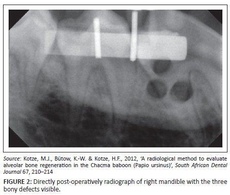
With a trephine burr fitted to a straight hand piece and a surgical unit, three similar bone defects per quadrant (Quadrant 1 [Q1] being the right maxilla, Q2 the left maxilla, Q3 the left mandible and Q4 the right maxilla) were created in the premolar areas of the respective bones (Figures 1 and 2). The alveolar bone defects were 3 mm deep and 2 mm apart from each other and angled at 90 degrees to the surface of the alveolar bone (Kotze et al. 2012).

Radiology
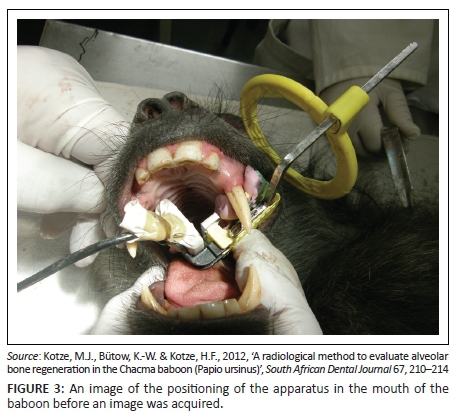
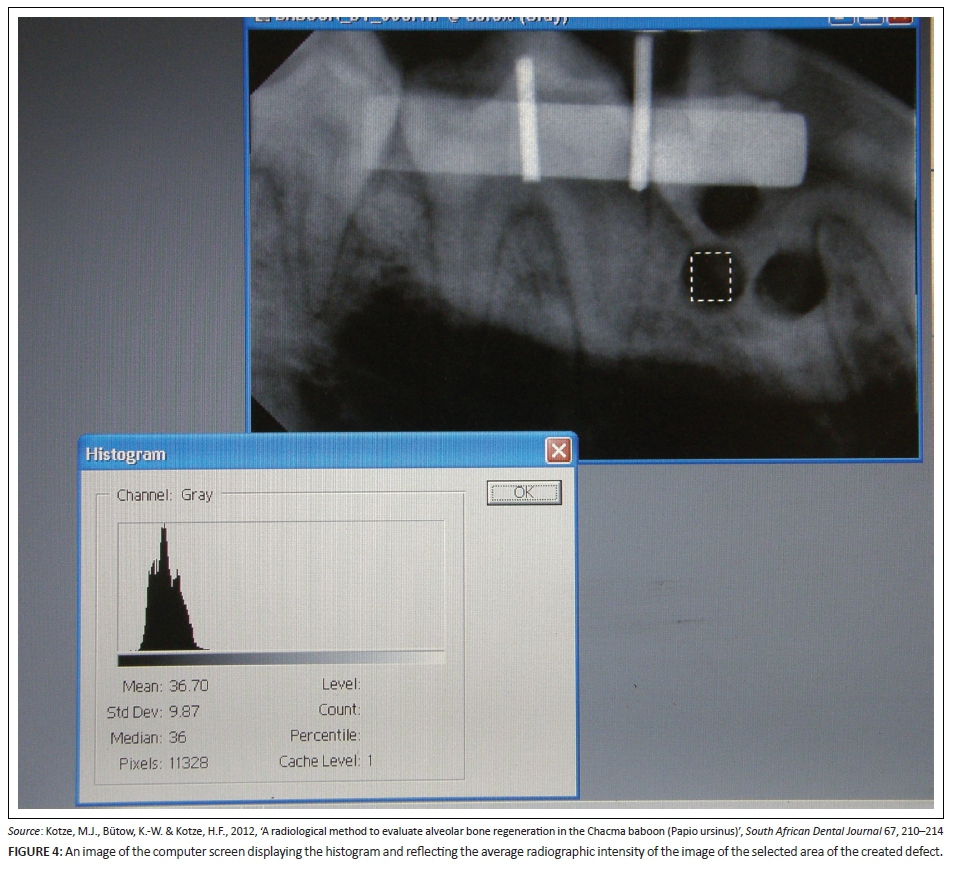
Standardised reproducible radiographs (Figure 2) were taken in the premolar area at each of the four quadrants pre-operatively, directly post-operatively and again after 3 and 6 weeks. To enable reproducible radiographs, a device consisting of a sectional impression tray with impression putty, a bite block (XPC-DS Digital Position System, Gendex, Lake Zurich, Illinois), a sensor (Gendex Visualix EHD Digital intra-Oral x-ray unit - Size 1 with 25.6 line pairs per mm) and a custom made aluminium step wedge (Figure 3) was used. The step wedge was custom made due to the limited space in the mouth of the baboon (Figure 4). Four digital images were acquired per quadrant at the predetermined time intervals. Therefore, records were taken of 16 images per animal at each of the four time periods. The method used for the acquisition of the standardised radiographs was described in detail by Kotze et al. (2012).
Evaluation methods
The images acquired for each quadrant were imported into Adobe Photoshop (V6.0; Adobe Systems Inc., San Jose, CA, USA). Using the histogram, contrast and brightness tools of the Adobe Photoshop software, average grey scale values of the selected regions of interest (ROI) on the image of the aluminium step wedge were standardised across all four images for each quadrant. A transparent sheet was placed over the screen and the ROI was marked on the sheet, so that it was possible to accurately position the radiograph in the exact position with repeated evaluations. An ROI was also selected on each of the defect sites on each of the images taken immediately post-operatively. The average grey scale values for these areas on each of the three defects on each image were determined and recorded. This was done for all images of a quadrant acquired from each of the different time periods (Figure 4). Only one blinded examiner made the measurements and recorded the results. The analysis of the data was standardised and repeatable, which implies that there was no reason to measure inter-observer variation (Kotze et al. 2014). The method used was described by Kotze et al. (2012).
Statistical analyses
Analysis of variance (ANOVA) for repeated measures (analysis of variance considering more than one period) was used to test for possible differences in the data set. Periodic changes (actual and percentage) were analysed using the two-sample t-test within each time point to compare the left and right sides of the mandible and maxilla (Kotze et al. 2014).
Results
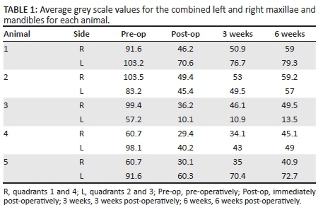
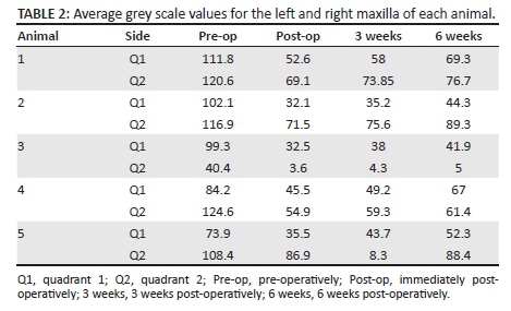
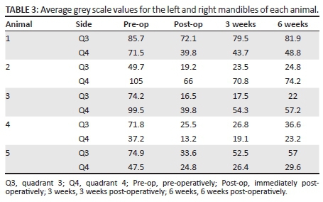
With the use of the ANOVA for repeated measures, the average grey scale values from pre-operatively, immediately post-operatively, 3 weeks post-operatively and 6 weeks post-operatively for the four quadrants from each animal were compared. The results are summarised in Table 1. There was no significant difference when the grey scale averages of the combined first and fourth quadrants (right side) and combined second and third quadrants (left side) were compared for pre-operatively (t = 0.70), immediately post-operatively (t = 0.34), 3 weeks post-operatively (t = 0.40) and 6 weeks post-operatively (t = 0.66). The combined results for comparison of the average grey scale values for all the animals between the first and second quadrant are summarised in Table 2. There was also no significant difference between the values for pre-operatively (t = 0.37), immediately post-operatively (t = 0.30), 3 weeks post-operatively (t = 0.30) and 6 weeks post-operatively (t = 0.38). When the third and fourth quadrants were compared for all the animals (Table 3), there was no significant differences in pre-operatively (t = 0.29), immediately post-operatively (t = 0.69), 3 weeks post-operatively (t= 0.07) and 6 weeks post-operatively (t = 0.06). For animal 3, significant differences were noted between quadrant 3 and quadrant 4 between baseline (pre-operatively) and immediately post-operatively (p = 0.003) on the one hand, and 3 weeks post-operatively (p = 0.005) and 6 weeks post-operatively (p = 0.010) on the other hand.
When the group of animals was compared (Table 4), it was not surprising that the pre-operative density was not significantly different between the left and right sides of the maxilla and mandible (t = 0.70), as similar areas were investigated on both sides. Furthermore, because a similar quantity of bone tissue was removed from all the defect areas, no significant difference was found immediately post-operatively (t = 0.34) between the left and right sides of the mandible and maxilla.
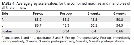
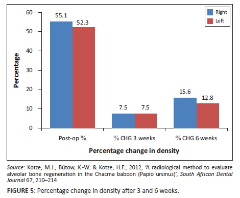
The healing process of the defect areas was similar without any significant differences between the left and right sides after 3 weeks post-operatively (t = 0.040) (p > 0.05) and 6 weeks post-operatively (t = 0.66) (p > 0.05). The rate of healing or the change in density for the first 3 weeks (Figure 5) was similar for the left and right sides. However, the change in density and healing from 3 weeks post-operatively to 6 weeks post-operatively was higher (2.8%) in the right mandible and maxilla, although not significantly so (Figure 5).
Discussion
Bone density and bone regeneration were investigated in the premolar area of the maxilla and mandible in similar areas. Change in density, which is reflected in a change in average grey scale value (AGV), was derived from radiographs in the region of interest. The radiological changes in BD were previously compared with histological and histomorphometrical measurements (Cakıcı et al. 2011; Gulsahi et al. 2010) as well as with digital radiography (Kotze et al. 2012), and a correlation was found between the different approaches. Therefore, the principle of change in radiological density over time can be used as an indicator of bone turnover.
The premolar area in baboons represents the more dense areas in the mandible and maxilla as the density showed a progressive increase from the incisor to the retro molar area (Chugh et al. 2013; Park et al. 2008). Baboon and other non-human primate bones exhibited similar fracture, microstructural and compositional properties to those of humans, compared with other animals such as cats, rodents, rabbits, guinea pigs and mini pigs, sheep, goats and dogs (Turner 2001; Wang, Mabrey & Agrawal 1998).
This increased BD in the premolar area of maxilla and mandible is probably due to more masticatory forces in these areas. Laterality, hand dominance and preferred side to chew may also have an influence on the difference in BD. A positive correlation, with the right hand more dominant, was found between handiness and laterality index in free-ranging bonnet macaque monkeys whilst filling and emptying their cheek pouches (Mangalam, Desai & Singh 2014). No significant difference was found when the left and right sides of the maxilla (p > 0.05) and left and right sides of the mandible (p > 0.05) were compared. However, the laterality, hand dominance and preferred side to chew were not investigated in this study. One animal revealed a difference in BD between the left and right mandibles. A previously published study presented results that alveolar bone remodelling is activated by the flexure produced by mechanical stimulation from mastication through the periodontal membrane (Sato et al. 2005). It is also suggested the cortical bone might increase in density and thicken as masticatory function develops (Sato et al. 2005). The mandible, which is the only moving bone section of the skull, is moved by the muscles of mastication, which are responsible for a variety of three-dimensional movements in the coronal, sagittal and transverse planes. Each movement produces a force or combination of forces transferred through the teeth to the alveolar bone of the mandible and maxilla. It could be possible that this specific animal masticated primarily on the right side, whilst the others were laterality neutral thus, equally left and right, as found in the macaque monkeys (Mangalam et al. 2014). The animals used in the present study were on a natural diet specific for the species. The Chacma baboon is an omnivore with different hardness of food (Hamilton, Buskirk & Buskirk 1978). The molars of the Chacma baboon are positioned relatively far forward to the temporomandibular joint, which results in the baboon being able to exert relatively greater muscle forces during posterior biting in comparison to humans (Mavropoulos et al. 2004). The average weight (19.8 kg ± 4.3 kg) of the animals implied young animals, as the weight of an adult male Chacma baboon can reach up to 40 kg in 5 years (Estes 1992). Therefore, it is presumed that the animals were still in their developing phase, with immature alveolar bone, and possessed an active osteogenic environment that resulted in faster bone healing (Lindaman 2001; Loh, Lee & Yeap 1988).
Conclusion
Radiography made evaluation of BD and healing of iatrogenic bone defects in the premolar areas of both mandibles and maxillae over a 6-week time period possible. Although no significant difference between the left and right sides of the maxilla and mandible was found in alveolar BD and regeneration over 6 weeks, the results showed an increased tendency of the right side to regenerate faster than the left side. Irrespective of the small sample size and utilisation of only the premolar area as the area of interest, the results provide sufficient information for future investigation of the effects of laterality and preferred side of mastication on the rate of healing and alveolar BD in the maxilla and mandible.
Acknowledgements
The personnel of the Animal Facility of North West University who supplied the animals and the company Unique Dental, South Africa, for supplying the radiological equipment and computer software. The Dental Research Education and Development Trust of the South African Dental Association contributed financially. Permission was granted by the Managing Editor of The South African Dental Journal for the use of figures (Kotze et al. 2012).
Competing interests
The authors declare that they have no financial or personal relationships which may have inappropriately influenced them in writing this article.
Authors' contributions
M.J.K., K-W.B. and H.F.K. made substantial contributions to the conception of the present study. S.A.O. performed analysis and interpretation of data besides drafting the manuscript. M.J.K. and H.F.K. led acquisition of data. K-W.B. was involved in revising the manuscript critically and giving final approval of the version to be published. All authors read and approved the final manuscript.
References
Adams, J.E., 2009, 'Quantitative computed tomography', European Journal of Radiology 71, 415-424. http://dx.doi.org/10.1016/j.ejrad.2009.04.074 [ Links ]
Bittar-Cortez, J.A., Passeri, L.A., Bóscolo, F.N. & Haiter-Neto, F., 2006, 'Comparison of hard tissue density changes around implants assessed in digitized conventional radiographs and subtraction images', Clinical Oral Implant Research 17, 560-564. http://dx.doi.org/10.1111/j.1600-0501.2006.01256.x [ Links ]
Blumenthal, N.M., Alves, M.E., Al-Huwais, S., Hofbauer, A.M. & Koperski, R.D., 2003, 'Defect-determined regenerative options for treating periodontal intrabony defects in baboons', Journal of Periodontology 74, 10-24. http://dx.doi.org/10.1902/jop.2003.74.1.10 [ Links ]
Cakıcı, H., Hapa, O., Gideroğlu, K., Ozturan, K., Güven, M., Yüksel, H.Y., et al., 2011 'The effects of leukotriene receptor antagonist montelukast on histological, radiological and densitometric parameters of fracture healing', Eklem Hastalik Cerrahis 22, 43-47. [ Links ]
Carter, D.R., Fyhrie, D.P. & Whalen, R.T., 1987, 'Trabecular bone density and loading history: Regulation of connective tissue biology by mechanical energy', Journal of Biomechanics 20, 785-787. http://dx.doi.org/10.1016/0021-9290(87)90058-3 [ Links ]
Chou, H.-Y., Satpute, D., Müftü, A., Mukundan, S. & Müftü, S., 2015, 'Influence of mastication and edentulism on mandibular bone density', Computer Methods in Biomechanics and Biomedical Engineering 18, 269-281. http://dx.doi.org/10.1080/10255842.2013.792916 [ Links ]
Chugh, T., Ganeshkar, S.V., Revankar, A.V. & Jain, A.K., 2013, 'Quantitative assessment of interradicular bone density in the maxilla and mandible: Implications in clinical orthodontics', Progress in Orthodontics 14, 38. http://dx.doi.org/10.1186/2196-1042-14-38 [ Links ]
Corten, F.G., Van 't Hof, M.A., Buijs, W.C., Hoppenbrouwers, P., Kalk, W. & Corstens, F.H., 1993, 'Measurement of mandibular bone density ex vivo and in vivo by dual-energy X-ray absorptiometry', Archives of Oral Biology 38, 215-219. http://dx.doi.org/10.1016/0003-9969(93)90030-P [ Links ]
Dannan, A. & Alkattan, F., 2007, 'Animal models in periodontal research: A mini-review of the literature', Internet Journal of Veterinary Medicine 5, viewed March 1967 and February 2008, from https://ispub.com/IJVM/5/1/3211 [ Links ]
Estes, R.D., 1992, The behavior guide to African mammals: Including hoofed mammals, carnivores, primates, University of California Press, Berkeley, CA. [ Links ]
Gulsahi, A., Paksoy, C.S., Ozden, S., Kucuk, N.O., Cebeci, A.R.I. & Genc, Y., 2010, 'Assessment of bone mineral density in the jaws and its relationship to radiomorphometric indices', Dentomaxillofacial Radiology 39, 284-289. http://dx.doi.org/10.1259/dmfr/20522657 [ Links ]
Hamilton, I.W.J., Buskirk, R.E. & Buskirk, W.H., 1978, 'Omnivory and utilization of food resources by Chacma baboons, Papio ursinus', The American Naturalist 112, 911-924. http://dx.doi.org/10.1086/283331 [ Links ]
Hylander, W.L., 1979, 'Mandibular function in Galago crassicaudatus and Macaca fascicularis: An in vivo approach to stress analysis of the mandible', Journal of Morphology 159, 253-296. http://dx.doi.org/10.1002/jmor.1051590208 [ Links ]
Kawamura, A., Yamaguchi, M., Kasai, K. & Carter, D., 2005, 'Relationship between masticatory function and internal structure of the mandible based on computed tomography', American Journal of Orthodontics and Dentofacial Orthopedics 128, 766-773. http://dx.doi.org/10.1016/j.ajodo.2005.05.046 [ Links ]
Kinds, M.B., Bartels, L.W., Marijnissen, A.C.A., Vincken, K.L., Viergever, M.A., Lafeber, F.P.J.G., et al., 2011, 'Feasibility of bone density evaluation using plain digital radiography', Osteoarthritis Cartilage 19, 1343-1348. http://dx.doi.org/10.1016/j.joca.2011.08.003 [ Links ]
Kotze, M.J., Bütow, K.-W. & Kotze, H.F., 2012, 'A radiological method to evaluate alveolar bone regeneration in the Chacma baboon (Papio ursinus)', South African Dental Journal 67, 210-214. [ Links ]
Kotze, M.J., Bütow, K.-W., Olorunju, S.A. & Kotze, H.F., 2014, 'A comparison of mandibular and maxillary alveolar osteogenesis over six weeks: A radiological examination', Head & Face Medicine 10, 50. http://dx.doi.org/10.1186/1746-160X-10-50 [ Links ]
Lindaman, L.M., 2001, 'Bone healing in children', Clinics in Podiatric Medicine and Surgery 18, 97-108. [ Links ]
Loh, S.A., Lee, S.T. & Yeap, C.L., 1988, 'Evaluation of the effects of secondary alveolar bone grafting in both adults and children with cleft lip and palate', Annals Academy of Medicine Singapore 17, 400-406. [ Links ]
Mangalam, M., Desai, N. & Singh, M., 2014, 'Do right-handed monkeys use the right cheek pouch before the left?', PLoS One 9, e97971. http://dx.doi.org/10.1371/journal.pone [ Links ]
Mavropoulos, A., Kiliaridis, S., Bresin, A., & Ammann, P., 2004 'Effect of different masticatory functional and mechanical demands on the structural adaptation of the mandibular alveolar bone in young growing rats', Bone 35, 191-197. http://dx.doi.org/10.1016/j.bone.2004.03.020 [ Links ]
Park, H.-S., Lee, Y.-J., Jeong, S.-H. & Kwond, T.-G., 2008, 'Density of the alveolar and basal bones of the maxilla and the mandible', American Journal of Orthodontics and Dentofacial Orthopedics 133, 30-37. http://dx.doi.org/10.1016/j.ajodo.2006.01.044 [ Links ]
Ravosa, M.J., 1996, 'Mandibular form and function in North American and European Adapidae and Omomyidae', Journal of Morphology 229, 171-190. http://dx.doi.org/10.1002/(SICI)1097-4687(199608)229:2%3C171::AID-JMOR3%3E3.0.CO;2-4 [ Links ]
Rosholm, A., Hyldstrup, L., Backsgaard, L., Grunkin, M. & Thodberg, H.H., 2001, 'Estimation of bone mineral density by digital X-ray radiogrammetry: Theoretical background and clinical testing', Osteoporosis International 12, 961-969. http://dx.doi.org/10.1007/s001980170026 [ Links ]
Sato, H., Kawamura, A., Yamaguchi, M. & Kasaic, K., 2005, 'Relationship between masticatory function and internal structure of the mandible based on computed tomography findings', American Journal of Orthodontics and Dentofacial Orthopedics 128, 766-773. http://dx.doi.org/10.1016/j.ajodo.2005.05.046 [ Links ]
Turner, A.S., 2001, 'Animal models of osteoporosis - Necessity and limitations', European Cells & Materials Journal 22, 66-81. [ Links ]
Wang, X., Mabrey, J.D. & Agrawal, C.M., 1998, 'An interspecies comparison of bone fracture properties', Biomedical Materials and Engineering 8, 1-9. [ Links ]
 Correspondence:
Marthinus Kotze
thinus.kotze@up.ac.za
Received: 29 May 2015
Accepted: 16 Mar. 2016
Published: 06 Sept. 2016

