


Serviços Personalizados
Artigo
 Inglês (pdf)
 Artigo em XML
 Referências do artigo
Indicadores

Links relacionados
-
 Citado por Google
 -
 Similares em Google
Compartilhar

 Permalink
Journal of the South African Veterinary Association
versão On-line ISSN 2224-9435
versão impressa ISSN 1019-9128
J. S. Afr. Vet. Assoc. vol.86 no.1 Pretoria 2015
http://dx.doi.org/10.4102/jsava.v86i1.1220
CASE REPORT
Utility of forensic detection of rabies virus in decomposed exhumed dog carcasses
Wanda MarkotterI; Jessica CoertseI; Kevin le RouxII; Joey PeensIII; Jacqueline WeyerIV; Lucille BlumbergIV; Louis H. NelI
IDepartment of Microbiology and Plant Pathology, University of Pretoria, South Africa
IIAllerton Provincial Veterinary Laboratory, Pietermaritzburg, South Africa
IIIState Veterinary Office, Department of Agriculture and Environmental Affairs, South Africa
IVCentre for Emerging and Zoonotic Diseases, National Institute for Communicable Diseases of the National Health Laboratory Services, South Africa
ABSTRACT
This report describes four suspected rabies cases in domestic dogs that were involved in human exposures. In all these cases, the animals were buried for substantial times before rabies testing was performed. Animal rabies is endemic in South Africa and domestic dogs are the main vector for transmission to humans. Diagnosis of rabies in humans is complicated, and diagnosis in the animal vector can provide circumstantial evidence to support clinical diagnosis of rabies in humans. The gold standard diagnostic method, fluorescent antibody test (FAT), only delivers reliable results when performed on fresh brain material and therefore decomposed samples are rarely submitted for diagnostic testing. Severely decomposed brain material was tested for the presence of rabies virus genomic material using a quantitative real-time reverse transcription polymerase chain reaction (q-real-time RT-PCR) assay when conventional molecular methods were unsuccessful. This may be a useful tool in the investigation of cases where the opportunity to sample the suspected animals post mortem was forfeited and which would not be possible with conventional testing methodologies because of the decomposition of the material.
Introduction
Rabies is a fatal and zoonotic disease associated with up to 55 000 human cases per annum worldwide. Many of these cases occur in African countries, where rabies remains uncontrolled in domestic dogs (Knobel et al. 2005). The disease is likely underestimated in African countries because of misdiagnosis and lack of capacity to investigate cases by laboratory testing (Blumberg et al. 2007). Rabies remains endemic in South Africa, where up to 30 human cases are reported annually (Weyer et al. 2011). The disease also continues to re-emerge in localities where it was previously well controlled, including Limpopo, Gauteng and Mpumalanga provinces (Mkhize et al. 2010; Sabeta et al. 2013; Sabeta, Mkhize & Ngoepe 2011).
The majority of animal cases diagnosed occur in the hyperendemic KwaZulu-Natal (KZN) province and neighbouring Mpumalanga and Eastern Cape provinces, where the main vectors are domestic dogs. Rabies control efforts in KZN have been boosted in recent years with support through a Bill and Melinda Gates/World Health Organization (WHO) initiative (Nel 2013).
Subsequently, no human cases were diagnosed from June 2010 to September 2011 - which constituted the first time in more than 20 years that KZN did not report a human rabies case for a period longer than one year. The gold standard for rabies diagnosis remains the fluorescent antibody test (FAT) (Dean, Abelseth & Atansiu 1996). As brain material is required for this test, diagnosis can only be performed post mortem and the FAT yields the most reliable results when performed on fresh brain material. When decomposed brain material is used, the sensitivity of the test is reduced and may even result in false negatives (Robardet et al. 2011). In severely decomposed material there is also no intact brain available and samples are received as a liquid substance that is unsuitable for FAT testing. In some cases, conventional reverse transcription polymerase chain reaction (RT-PCR) and heminested RT-PCR (hnRT-PCR) assays have proven to be useful when other methods such as FAT were not successful (David et al. 2002; Kamolvarin et al. 1993; McElhinney et al. 2014). However, these methods target a comparatively large (over 350 base pair) region of the nucleoprotein gene of the viral genome and in cases of advanced decomposition the viral ribonucleic acid (RNA) may have deteriorated to the extent that these methods are no longer successful. Real-time RT-PCR, in contrast to conventional molecular methods, targets a shorter conserved region of the nucleoprotein gene of the viral genome. Furthermore, quantitative real-time RT-PCR (q-real-time RT-PCR) has previously been shown to be successful for detection of rabies viral RNA in formalin-fixed brain material when other methods have been unsuccessful (Coertse et al. 2011). In 2012, two human cases of rabies from KZN were investigated by the National Institute for Communicable Diseases of the National Health Laboratory Services (NICD-NHLS), South Africa. In both cases, ante-mortem testing for rabies proved problematic because of the rabies vaccination histories of the patients, although incomplete prophylaxis was provided (Mollentze et al. 2013). The dogs associated with the possible exposures died after the exposure events but were buried, and no specimens were submitted for laboratory investigation. To provide circumstantial evidence to support the clinical diagnosis of rabies in these patients, the implicated dogs were exhumed and submitted for laboratory investigation.
In this report, three cases are described in which a q-real-time RT-PCR assay was applied for rabies diagnosis in exhumed dogs when other methods were unsuccessful because of the state of decomposition of the brain material.
Case presentation
Case history
Case 1
On 2 May 2012, a 29-year-old male farmer from Underberg was admitted to a hospital in Pietermaritzburg, KZN. The patient reported contact with a stray puppy some two months before the onset of symptoms. The patient provided shelter for the puppy, but after a few days the animal developed symptoms which in retrospect could have been considered consistent with rabies. The dog subsequently died and was buried on the farm. After consideration of the patient history, rabies was deemed likely. Saliva, skin and cerebrospinal fluid (CSF) were collected over the course of his illness and sent to the NICD-NHLS in Johannesburg but were consistently negative for the presence of rabies virus RNA using conventional as well as real-time RT-PCR methods. To further investigate, the puppy (referred to as Dog GA) was exhumed and the decomposed brain material sent to the University of Pretoria in 50% glycerol-saline solution for molecular testing on 11 May 2012. Rabies-specific IgG was detected in the serum of the patient; however, this was likely as a result of the vaccine he received upon admission. Initial rabies-specific serological tests on CSF were negative. Subsequent CSF samples collected over four weeks indicated the presence of rabies-specific IgG at low titres. The FAT confirmed the presence of rabies virus antigen in a postmortem brain biopsy specimen of the patient. Real-time RT-PCR was also performed on this specimen at the NICD-NHLS and the product sequenced (referred to as SPU 134/12).
Case 2
On 20 May 2012, a 4-year-old child was admitted to a local hospital in KZN. The admitting doctor noticed symptoms suggestive of rabies virus infection, including general weakness, loss of appetite and confusion. Upon retrospective investigation it was found that the child was scratched and bitten by two different dogs on two separate occasions. The first incident took place on 29 March 2012, when a neighbour's dog (referred to as Dog MK) scratched the child on the forehead whilst the child was playing outside. The parents of the child did not seek medical attention. On the same day, Dog MK bit a 5-month-old puppy (referred to as Dog ZC) belonging to another neighbour. In the week after the scratch incident, the owners of Dog MK noticed the dog behaving strangely, including excessive salivation and barking, loose jaw and a weak gait. Dog MK died on 05 April 2012 and was buried by the owner. Dog ZC, the puppy bitten by Dog MK, attacked and bit the 4-year-old child on the right ankle on 23 April 2012 whilst the child was walking. Dog ZC was killed and buried by the owner on the same day of the biting incident. On 28 May 2012, three saliva samples and a nuchal biopsy specimen were collected from the patient and sent to the NICD-NHLS but were negative for the presence of viral RNA. It was decided to exhume both dogs involved in this case for rabies testing. The brain material was subsequently sent to the University of Pretoria in 50% glycerol-saline solution for molecular testing on 20 June 2012.
Case 3
On 28 August 2012, a 21-year-old male was admitted to a local hospital in KZN. Upon investigation, it was found that a dog (referred to as Dog VG) bit the patient on 19 July 2012 whilst he was visiting his girlfriend in the Tshelimnyama area. The owners of Dog VG mentioned that the dog was usually well behaved but suddenly started showing strange behaviour and that the dog was not vaccinated during the recent vaccine campaign in the area. Following these events, the owner chained the dog; however, the dog broke free and went missing. Other people from the neighbourhood reported seeing Dog VG attacking other dogs on several occasions. Dog VG was killed during one of these attacks. The carcass of Dog VG was found slightly submerged in water and in an advanced state of decomposition on 28 August 2012. Brain material was subsequently sent to the University of Pretoria in 50% glycerol-saline solution for molecular testing on 30 August 2012.
Molecular diagnosis
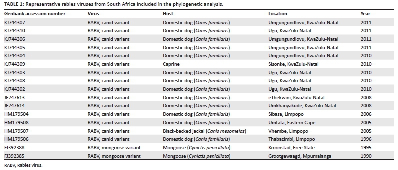
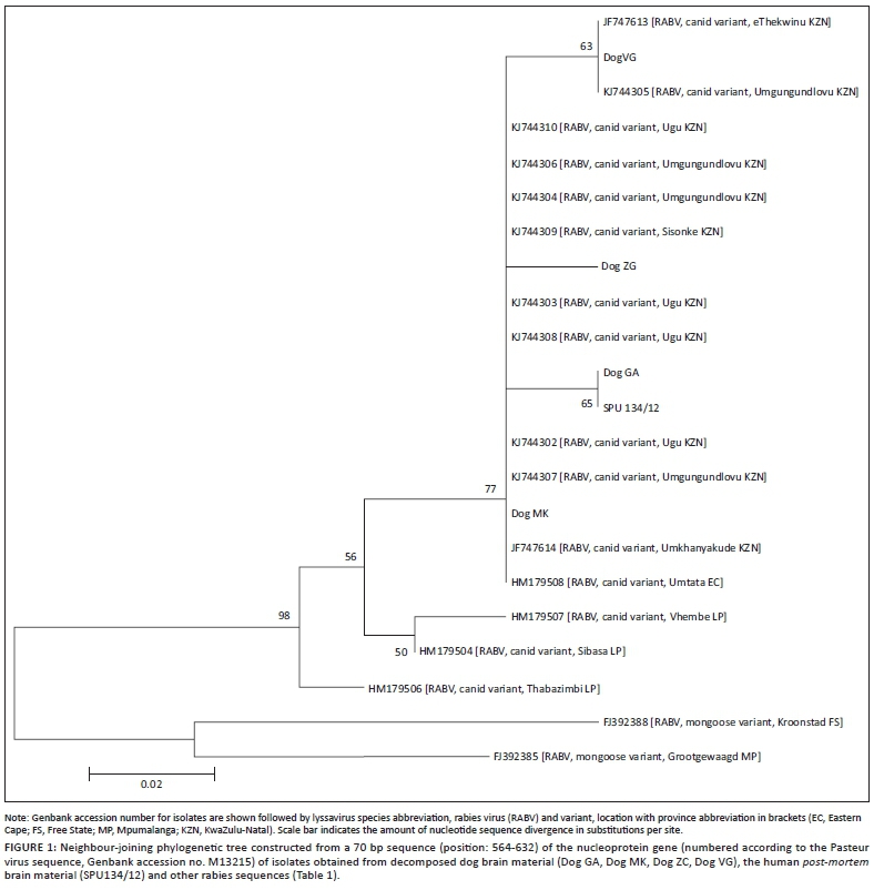
The dogs in these cases were buried for 30-76 days before being exhumed for rabies testing. The FAT was not attempted because of the severe decomposition of the samples, with no intact brain material present in the samples to prepare a smear for FAT. Any results may be unreliable because of the decrease in sensitivity of the FAT when using decomposed material, so it was decided to proceed with molecular testing only. Total RNA was extracted from a decomposed sample from the exhumed dogs by using Trizol reagent (Invitrogen, USA), according to the manufacturer's instructions. Conventional RT-PCR and hnRT-PCR were performed as previously described (Coertse et al. 2010) and were consistently negative. Quantitative real-time RT-PCR (Coertse et al. 2010) was performed on 1 μL RNA and yielded positive results in all four cases. Average viral RNA copy numbers were as follows: Dog GA 50 copies/reaction, Dog MK 213 copies/reaction, Dog ZC 733 copies/reaction and Dog VG 6.5 χ 106 copies/reaction. The amplicons were purified and sequenced and phylogenetic analysis was performed (Markotter et al. 2006) with representative rabies virus sequences available in the public domain (Table 1). Although the resolution after phylogenetic analysis was not ideal as a result of the short region of sequence available from the real-time RT-PCR amplicon, the resulting neighbour-joining (NJ) phylogenetic analysis (Figure 1) confirmed the expected relationship with rabies virus circulating in the KZN domain. The patient bitten by Dog GA died and a viral sequence was generated from samples obtained post mortem. This viral sequence (SPU134/12) was identical to the viral sequence obtained from Dog GA (Figure 1).
Discussion
Three possible human exposures to rabies virus after contact with domestic dogs are described. In all these cases, the dogs had already been buried for a substantial time (up to 76 days) when rabies in the humans was first suspected. In none of the human cases could rabies be confirmed ante mortem, which complicated patient care. The dogs were exhumed and brain material tested using a real-time RT-PCR method. The utility of the real-time RT-PCR protocol in the diagnosis and molecular characterisation of rabies virus RNA from exhumed dog brain material was demonstrated where other methods were unsuccessful. This indicates the importance of using a sensitive real-time PCR assay in suspected rabies cases where other methods are unreliable or will fail because of decomposition of the sample and degradation of the viral RNA. Deoxyribonucleic acid (DNA) sequencing could also be performed on this small amplicon and phylogenetic analysis was used to indicate the epidemiological relationships of these viruses. Conclusive diagnosis in the animal vectors in each of these cases was helpful in eliminating the differential diagnoses in the patients and in providing the appropriate patient care. In addition, the positive diagnosis in dogs was important with regard to re-evaluation of the dog rabies control measures in the affected areas.
Conclusion
A real-time RT-PCR assay was applied in cases where conventional methods were unsuccessful. This method was shown to be very effective for post-mortem rabies diagnosis in severely decomposed carcasses. Subsequent phylogenetic analysis also provided epidemiological information that might otherwise not have been known.
Acknowledgements
This work was funded by the National Research Foundation (NRF) (South Africa) grant number UID 66187 and 78566.
Competing interests
The authors declare that they have no financial or personal relationships which may have inappropriately influenced them in writing this article.
Authors' contributions
W.M. (University of Pretoria) and L.H.N. (University of Pretoria) have made substantial contributions to the conception and design, interpretation of data, drafting the manuscript and revising it. J.C. (University of Pretoria) carried out the molecular diagnostics and phylogenetic analysis and helped draft the manuscript. K.L.R. (Allerton Provincial Veterinary Laboratory) and J.P. (State Veterinary Office) carried out the case investigation and helped draft the animal case reports. J.W. (Centre for Emerging and Zoonotic Diseases, NICD-NHLS) and L.B. (Centre for Emerging and Zoonotic Diseases, NICD-NHLS) carried out the human case investigation and helped draft this report. All authors read and approved the final manuscript.
References
Blumberg, L., Weyer, J., Frean, J. & Ogunbanjo, G.A., 2007, 'Rabies: An evidence-based approach to management', South African Family Practice 49(5), 35-41. http://dx.doi.org/10.1080/20786204.2007.10873599 [ Links ]
Coertse, J., Weyer, J., Nel, L.H. & Markotter, W., 2010, 'Improved PCR methods for detection of African rabies and rabies-related viruses', Journal of Clinical Microbiology 48(11), 3949-3955. http://dx.doi.org/10.1128/JCM.01256-10 [ Links ]
Coertse, J., Nel, L.H., Sabeta, C.T., Weyer, J., Grobler, A., Walters, J. et al., 2011, 'A case study of rabies diagnosis from formalin-fixed brain material', Journal of the South African Veterinary Association 82(4), 250-253. http://dx.doi.org/10.4102/jsava.v82i4.83 [ Links ]
David, D., Yakobson, B., Rotenberg, D., Dveres, N., Davidson, I. & Stram, Y., 2002, 'Rabies virus detection by RT-PCR in decomposed naturally infected brains', Veterinary Microbiology 87, 111-118. http://dx.doi.org/10.1016/S0378-1135(02)00041-X [ Links ]
Dean, D.J., Abelseth, M.K. & Atansiu, P., 1996, 'The fluorescent antibody test', in F.X. Meslin, M.M. Kaplan & H. Koprowski (eds.), Laboratory techniques in rabies, 4th edn., pp. 88-95, World Health Organization, Geneva. [ Links ]
Kamolvarin, N., Tirawatnpong, T., Rattanasiwamoke, R., Tirawatnpong, S., Panpanich, T. & Hemachudh, T., 1993, 'Diagnosis of rabies by polymerase chain reaction with nested primers', Journal of Infectious Diseases 167, 207-210. http://dx.doi.org/10.1093/infdis/167.1.207 [ Links ]
Knobel, D.L., Cleaveland, S., Coleman, P.G., Fèvre, E.M., Meltzer, M.I., Miranda, M.E. et al., 2005, 'Re-evaluating the burden of rabies in Africa and Asia', Bulletin of the World Health Organization 83(5), 360-368. [ Links ]
Markotter, W., Kuzmin, I.V., Rupprecht, C.E., Randles, J., Sabeta, C.T., Wandeler, A.I. et al., 2006, 'Isolation of Lagos bat virus from water mongoose', Emerging Infectious Diseases 12, 1913-1918. http://dx.doi.org/10.3201/eid1212.060514 [ Links ]
McElhinney, L.M., Marston, D.A., Brookes, S.M. & Fooks, A.R., 2014, 'Effects of carcase decomposition on rabies virus infectivity and detection', Journal of Virological Methods 207, 110-113. http://dx.doi.org/10.1016/'.jviromet.2014.06.024 [ Links ]
Mkhize, G.C., Ngoepe, E.C., Du Plessis, B.J., Reininghaus, B. & Sabeta, C.T., 2010, 'Re-emergence of dog rabies in Mpumalanga Province, South Africa', Vector Borne Zoonotic Diseases 10(9), 921-926. http://dx.doi.org/10.1089/vbz.2009.0109 [ Links ]
Mollentze, N., Weyer, J., Markotter, W., Le Roux, K. & Nel, L.H., 2013, 'Dog rabies in southern Africa: Regional surveillance and phylogeographical analyses are an important component of control and elimination strategies', Virus Genes 47(3), 569-573. http://dx.doi.org/10.1007/s11262-013-0974-3 [ Links ]
Nel, L.H., 2013, 'Factors impacting the control of rabies', Microbiology Spectrum 1(2). http://dx.doi.org/10.1128/microbiolspec.OH-0006-2012 [ Links ]
Robardet, E., Picard-Meyer, E., Andrieu, S., Servat, A. & Cliquet, F., 2011, 'International inter laboratory trials on rabies diagnosis: An overview of results and variation in reference diagnosis techniques (fluorescent antibody test, rabies tissue culture infection test, mouse inoculation test) and molecular biology techniques', Journal of Virological Methods 177(1), 15-25. http://dx.doi.org/10.1016/j.jviromet.2011.06.004 [ Links ]
Sabeta, C.T., Mkhize, G.C. & Ngoepe, E.C., 2011, 'An evaluation of dog rabies control in Limpopo Province (South Africa)', Epidemiology & Infection 139(10), 1470-1475. http://dx.doi.org/10.1017/S0950268811001233 [ Links ]
Sabeta, C.T., Weyer, J., Geertsma, P., Mohale, D., Miyen, J., Blumberg, L.H. et al., 2013, 'Emergence of rabies in the Gauteng Province, South Africa: 2010-2011', Journal of the South African Veterinary Association, E1-5. [ Links ]
Weyer, J., Szmyd-Potapczuk, A.V., Blumberg, L.H., Leman, P.A., Markotter, W., Swanepoel, R. et al., 2011, 'Epidemiology of human rabies in South Africa 1983-2007', Virus Research 155, 283-290. http://dx.doi.org/10.1016/jJ.virusres.2010.10.023 [ Links ]
 Correspondence:
Wanda Markotter
Private Bag X20, Hatfield
0028, South Africa
E-mail: wanda.markotter@up.ac.za
Received: 17 June 2014
Accepted: 17 Dec. 2014
Published: 18 May 2015

