


Serviços Personalizados
Artigo
 Inglês (pdf)
 Artigo em XML
 Referências do artigo
Indicadores

Links relacionados
-
 Citado por Google
 -
 Similares em Google
Compartilhar

 Permalink
Journal of the South African Veterinary Association
versão On-line ISSN 2224-9435
versão impressa ISSN 1019-9128
J. S. Afr. Vet. Assoc. vol.85 no.1 Pretoria Jan. 2014
http://dx.doi.org/10.4102/jsava.v85i1.1126
RESEARCH COMMUNICATION
A survey of feline leukaemia virus infection of domestic cats from selected areas in Harare, Zimbabwe
Francis MuchaambaI; Takudzwa H. MutiringindiI; Musavenga T. TivapasiI; Solomon DhliwayoI; Gift MatopeII
IDepartment of Clinical Veterinary Studies, University of Zimbabwe, Zimbabwe
IIDepartment of Paraclinical Veterinary Studies, University of Zimbabwe, Zimbabwe
ABSTRACT
A cross-sectional study was conducted to detect the feline leukaemia virus (FeLV) p27 antigen and to determine risk factors and the haematological changes associated with infection in domestic cats in Zimbabwe. Sera were collected for detection of the p27 antigen, urea, creatinine, alanine aminotransferase and gamma-glutamyl transferase levels, whilst whole blood was collected for haematology. FeLV p27 antigen was detected using a rapid enzyme-linked immunosorbent assay (ELISA) test kit. Data on risk factors were analysed using a logistic regression model. Of the 100 cats tested, 41% (95% CI: 31.19% - 50.81%) (41/100) were positive for the FeLV p27 antigen. Sex and health status of cats were not significantly (p > 0.05) associated with infection. Intact cats (OR = 9.73), those living in multicat housing (OR = 5.23) and cats that had access to outdoor life (OR = 35.5) were found to have higher odds of infection compared with neutered cats, those living in single-cat housing, and without access to outdoor life, respectively. Biochemistry and haematology revealed no specific changes. The results showed that FeLV infection was high in sampled cats, providing evidence of active infection. Thus, it would be prudent to introduce specific control measures for FeLV infection in Zimbabwe.
Introduction
Feline leukaemia virus (FeLV), a retrovirus belonging to the family Retroviridae, subfamily Orthoretrovirinae, genus Gammaretrovirus, is believed to cause widespread infections in cats throughout the world; prevalences of between 1% and 20% have been reported from all the major continents (Bande et al. 2012; Bandecchi et al. 2006; Gabor et al. 2001; Hartmann 2012; Hosie, Robertson & Jarrett 1989; Levy et al. 2006). The virus may be transmitted vertically in utero or horizontally (by secretions and excretions), and young kittens are more susceptible than adults (Aiello & Mays 2011).
Recent studies have indicated that cats exposed to FeLV may progress into any one of four categories: abortive infection (formerly 'regressor cats'), regressive infection (formerly 'transient viraemia' followed by 'latent infection'), progressive infection (formerly 'persistent viraemia') and focal or atypical infection (Hartmann 2012). Abortive infection is likely when some immunocompetent cats are exposed to low doses of the virus, where viral replication may be terminated by an effective humoral and cell-mediated immune response such that neither FeLV antigen nor viral ribonucleic acid (RNA) or proviral deoxyribonucleic acid (DNA) are detected in blood (Major et al. 2010). In regressive infection, transient viraemia (during which the virus is detected in plasma) is terminated within weeks or months of infection, but cannot completely eliminate the virus as the proviral DNA is present in bone marrow (Hartmann 2012). Cats that have progressive infection are persistently viraemic and develop FeLV-associated diseases, with most of them dying within a few years of infection (Hartmann 2012). Progressive FeLV infection is associated with a variety of malignancies that are characterised by development of cytoproliferative and cytosuppressive disorders (Filoni et al. 2003). It clinically manifests as severe immunosuppression, profound anaemia, immune-mediated diseases, reproductive problems and enteritis (Aiello & Mays 2011). Focal or atypical infection is associated with localised viral replication that leads to low-grade viral antigen production that produces a weak positive reaction (Levy et al. 2008). The development of FeLV-associated clinical disease is usually dependent on the age of the cat at the time of infection. Studies have shown that young cats tend to contract progressive infection resulting in severe immunosuppression and death (Hartmann 2012). Mature cats tend to contract abortive or regressive infection or progressive infection with mild and protracted clinical signs (Ettinger & Feldman 2005).
The factors that predispose cats to FeLV infection include young age, intact male cats, having access to outdoor life and living in multicat houses (Bande et al. 2012). Although evidence of FeLV infection in domestic cats (Felis domesticus) has been documented in many regions of the world, there is a lack of information about its prevalence in most parts of Africa, except for reports from Nigeria and South Africa (Bobade, Nash & Rogerson 1988; Schoeman et al. 2001; Schoeman et al. 2005). Similarly, evidence of exposure to FeLV in free-ranging wild felids has not yet been confirmed in Africa but has been documented in wild felids in captivity (Hofmann-Lehmann et al. 1996; Marker et al. 2003; Ramsauer et al. 2007). Anecdotal reports suggest that FeLV is present in domestic cats in Zimbabwe but the prevalence and risk factors for infection have not been studied. Furthermore, conflicting reports about the clinical pathology and haematology results of naturally FeLV-infected cats have been reported in the literature. Therefore, this study was conducted to (1) determine the prevalence of FeLV infection, (2) determine the risk factors associated with infection and (3) investigate the association between infection and clinical pathology in FeLV-infected cats from selected areas in Harare, Zimbabwe.
Materials and methods
Study areas
The study was conducted at eight randomly selected veterinary surgeries and a cat sanctuary (shelter) in Harare between October 2012 and March 2013. The criterion for inclusion in the study was willingness of the management of the veterinary surgeries and the cat sanctuary as well as cat owners to participate in the study.
Sample collection
The study was conducted on randomly selected cats presented to veterinary surgeries for elective treatment and routine examinations (n= 81), cats from a sanctuary (n = 7) and un-owned (stray) cats (n= 12). Because of cost, a randomly selected subset of these cats was evaluated for full blood count (n = 69) and clinical chemistry (n= 74). Blood samples (1 mL - 3 mL) were obtained by venipuncture into ethylenediaminetetraacetic acid (EDTA) and plain vacutainer tubes and transferred under chilled conditions (2 °C - 8 °C) to Diagnopath Laboratory in Harare, where full blood counts were processed within 4 h of collection. Thin blood films were made on microscope slides and stained with a modified Wright's stain for cell morphology and evaluation of blood parasites. Blood in plain tubes was centrifuged at 3000 rpm for 5 min and sera collected into Eppendorf tubes and stored at -20 °C and tested within a week of collection.
Epidemiological data collection
Epidemiological data on factors believed to be associated with FeLV viraemia were collected using patient observation forms immediately after blood sample collection. The information captured included breed (indigenous short and long hair, and exotic), origin (surgeries, sanctuary or un-owned), age, sex, neuter status, lifestyle (access to outdoor life or not), housing type (single-cat or multicat) and health status. The health status was evaluated based on clinical records and general physical examination. For the purpose of this study, cats were broadly classified as 'apparently healthy' if no evidence of illness was noted or the cat had no record of illness and 'sick' if there was clinical evidence of illness, such as high fever, anaemia, lymphadenopathy or loss of body condition.
Laboratory tests
Serology
All the laboratory tests were carried out at Diagnopath Laboratory, Harare. A FeLV antigen enzyme-linked immunosorbent assay (ELISA) rapid test kit (Quicking Biotech, Shanghai, China) was used to detect the p27 antigen in cat sera according to the manufacturer's recommendations. Briefly, all the reagents and test sera were brought to room temperature (approximately 24 °C - 26 °C) before testing. Aliquots of 50 μL test sera were placed into the sample slot on the cassette and immediately mixed with three drops of assay buffer. Results were read within 15 min of adding the buffer by visual appraisal of specified bands as recommended by the manufacturer. The manufacturer reported the test sensitivity and specificity for the p27 antigen to be 99.0% and 98.6%, respectively (Quicking Biotech n.d.).
Haematology and clinical chemistry
Full blood counts were carried out using an Advia-60 haematology analyser (Bayer, Germany), and clinical chemistry was carried out on sera using the Mind-Ray BS-120 analyser (Shenzhen Mindray Biomedical Electronics, Shangai, China), according to the specifications of the manufacturers. For blood counts, cats were evaluated for evidence of anaemia (normal range for haematocrit: 30% - 45%), platelet count (reference range: 3x10¹¹/L - 7x10¹¹/L), eosinophils (reference range: 0% - 0.75%), segmented neutrophils (reference range: 2.5% - 12.5%) and lymphocytes (reference range: 1.5% - 7.0%). To assess liver damage, alanine aminotransferase (ALT) (reference range: 8.3 IU/L - 52.5 IU/L) and gamma-glutamyl transferase (GGT) (reference range: 1.8 IU/L - 12.0 IU/L) levels were determined. For renal insufficiency blood urea nitrogen (BUN) (reference range: 15.4 mg/dL - 31.2 mg/dL) and creatinine levels (reference range: 0.5 mg/dL - 1.9 mg/dL) were measured. All the tests were conducted within 7 days from the date of sample collection.
Statistical analysis
Statistical analyses were performed using STATA/SE version 11.0 (Stata, College Station, Texas, USA). The total number of positive cats was calculated according to breed, origin, lifestyle (access to outdoor life), housing type, age group, sex, and health status of the cat by considering the total number of samples tested and expressed as a percentage. The chi square test was used to assess differences in proportions between generated categories; values of p < 0.05 were considered to be statistically significant.
For univariable analyses, the FeLV infection status of the cat (0 = no; 1 = yes) was used as the dependent variable with sex, age quartile (in years), neuter status, breed, lifestyle, housing type and health status as the independent variables. Independent variables were screened using a two-sided Fisher's exact chi square test. Only variables with p-values < 0.25 and with counts ≥ 5 in each cell as well as no more than 15 missing values were presented to the multivariable logistic regression model.
The multivariable logistic regression model was built using the FeLV infection status of the cat (0 = no; 1 = yes) as the dependent variable and the independent variables identified to have p-values < 0.25 in univariable analyses. The model was manually constructed using a backward selection procedure and statistical contribution of the independent variables to the model were tested using the likelihood ratio test as described by Dohoo, Martin and Stryhn (2003). The logistic regression model was assessed for goodness-of-fit by the Hosmer-Lemeshow test whilst the predictive ability was determined using the receiver operating characteristic (ROC) curve.
Results
Descriptive statistics
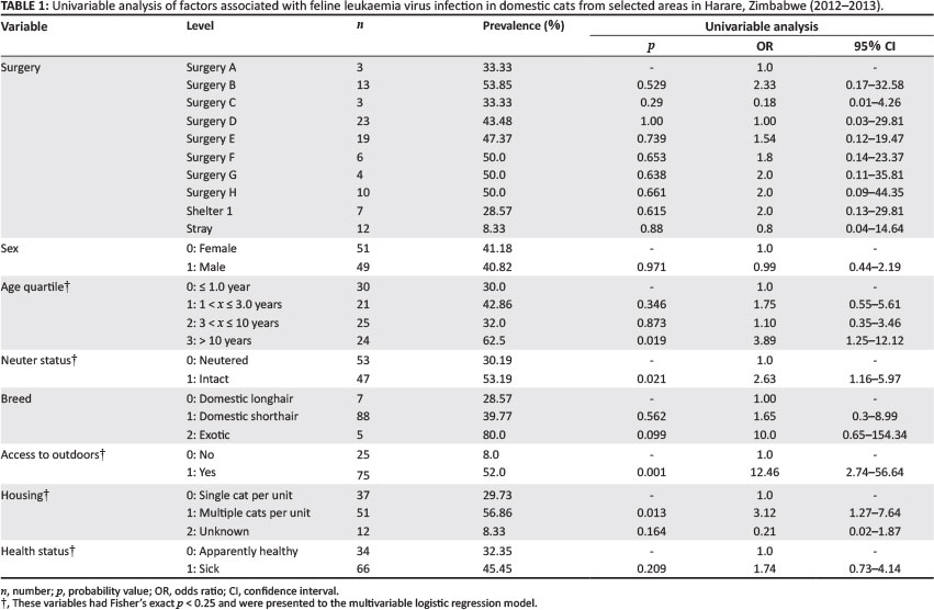
The distribution of cats positive for the p27 antigen relative to those sampled from eight different veterinary clinics (n = 81), one cat sanctuary (n = 7) and un-owned cats (n = 12) are presented in Table 1. Of the 100 cats tested, 41% (95% CI: 31.19%- 50.81%) (41/100) were positive for the FeLV p27 antigen. FeLV infection was significantly (p < 0.05) greater in cats that had access to the outdoors (52%) compared with those that had no access to the outdoors (8%); in multicat (56.86%) compared with single-cat housing (29.73%); and in intact (53.19%) compared with neutered cats (30.19%). Intact cats that were older than 10 years, especially from multicat units, which had access to outdoor life were significantly (p < 0.05) associated with FeLV infection. Sex and health status of cats were not significantly (p > 0.05) associated with infection (Table 1).
Multivariable logistic regression analysis
The final multivariable logistic regression model identified intact cats (not neutered), access to outdoor life and multiple cats per unit to be independently associated with FeLV infection of cats (Table 2). The breed of cat was not included in the model because of insufficient data. Cats that were intact were approximately 10 times (OR = 9.73; 95% CI: 2.63-35.93) more likely to be FeLV positive than neutered cats. Similarly, cats with access to outdoor life (OR = 35.5; 95% CI: 5.52-228.48) and that lived in multicat houses (OR = 5.23; 95% CI: 1.49-18.34) were more likely to be FeLV positive than the controls (Table 2).
Significant interactions between variables and confounders were not detected. The Hosmer-Lemeshow goodness-of-fit test showed that the model fit the data (X2 = 3.02, degrees of freedom 5,p = 0.697) and predictive ability was good (area under the ROC curve = 0.87).
Haematology and clinical chemistry
The following percentages of the cats tested had evidence of anaemia (46.4%; 32/69), thrombocytopenia (46.4%; 32/69), neutrophilia (4.3%; 3/69), eosinophilia (5.8%; 4/69) and lymphopenia (27.5%; 19/69), but there was no association (p > 0.05) with FeLV infection status. Of the cats tested, 13.5% (10/74) had increased liver enzyme activities (ALT and GGT), whilst 6.8% (5/74) had elevated BUN and creatinine levels. Of these, 2.7% (2/74) cats had increased liver enzyme activities, BUN and creatinine levels. No haemoparasites were detected on examination of thin blood films.
Discussion
This study investigated the presence of FeLV p27 antigen and the risk factors associated with FeLV infection in domestic cats mainly from selected veterinary clinics in Harare, Zimbabwe. This information is crucial for control of FeLV infection in domestic cats as there is currently no data on its prevalence in Zimbabwe. The FeLV p27 antigen in blood samples in this study (41%) is higher than those from both developing (Bande et al. 2012; De Almeida et al. 2012) and developed (Bandecchiet al. 2006; Gabor et al. 2001; Lee et al. 2002; Little et al. 2009) countries in other regions where moderate (10% - 20%) to low (< 10%) prevalences have been reported. The observed data may be influenced to an extent by test parameters, a small sample size and sampling bias - as cats presented to veterinary clinics only may not be representative of other apparently healthy cats that did not visit the clinics. However, these results indicated that FeLV infection is circulating in cats from urban Harare areas and highlight the need for implementing ongoing surveillance.
Although the ELISA kit is reported to have a sensitivity and specificity for the p27 antigen of 99.0% and 98.6% respectively and that there is no cross-reactivity with feline immunodeficiency, feline panleukopoenia and feline infectious peritonitis viruses (Quicking Biotech n.d.), data on validation of the test from field studies seems to be lacking. Given the high sensitivity of the test and the inherent low positive predictive value of ELISA kits (Bande et al. 2012), the possibility of false positive cats could not be discounted. However, in large multicat households or in households where cats roam freely outdoors, especially in urban areas, the prevalence of FeLV may be as high as 70% (Little 2006). Considering that 75% of the cats in this study had access to outdoor life and were at high risk of exposure to FeLV, a large proportion of positive cats would be expected if the virus was circulating in the study population. Although the prevalence of FeLV in endemic areas averages 3% in single-cat households, infection rates have been reported to vary according to type of human settlement (urban vs rural setting), geographic region, cat population density, lifestyle and control policies and practices amongst different countries (Bande et al. 2012; De Almeida et al. 2012; Lee et al. 2002; Little 2006). The prevalence and incidence of FeLV has been reported to be low in countries that routinely vaccinate cats and implement other control measures, such as restriction to communal catteries and removal of infected cats (Lutz et al. 2012; Moore et al. 2004). It is also important to determine the feline immunodeficiency virus (FIV) status of cats and cat populations as FeLV vaccines will be less effective in FIV-infected cats as a result of immunosuppression (Bandecchi et al.2006). As noted elsewhere (Bandecchi et al. 2006), the absence of prescribed control and preventive measures against FeLV infection in cats in Zimbabwe may be contributing to high infection rates observed in this study.
In agreement with the findings of Bandecchi et al. (2006), Danner et al. (2007) and Levy et al.(2006), FeLV infection status of cats was independent of sex, but other studies suggested that infection tends to be higher amongst male than female cats (Gleich, Krieger & Hartmann 2009; Levyet al. 2006). These variations might be related to differences in cat subpopulations (Bande et al.2012) and their lifestyles. It has also been reported that younger cats tend to contract progressive infection resulting in severe immunosuppression and death (Hartmann 2012), whilst older cats tend to contract abortive or regressive infection or, less likely, progressive infection with mild and protracted clinical signs (Ettinger & Feldman 2005). Therefore, FeLV infection tended to progressively decrease with age (Arjono et al. 2000). In this study, the observed marginal increase in the odds of infection in cats over 10 years of age could be attributed to other risk factors such as age-related immunosuppression and lifestyle, as 79.17% (19/24) of the cats in that age category had access to outdoor life. This supports the results of Little et al. (2009), who reported higher infection rates in adult cats than in juveniles.
There were increased odds of FeLV infection in intact cats (OR = 9.73), those from multicat housing (OR = 5.23) and cats with access to outdoor life (OR = 35.5) compared with those that were not exposed to these factors, which concur with studies in other regions (Bande et al. 2012; Lee et al.2002). The mode of transmission of FeLV is mainly through saliva, milk, blood and urine, both vertically and horizontally (Lee et al. 2002). Therefore, factors that promote contact with other cats tend to increase the risk of exposure to FeLV infection. It has been reported that intact free-roaming cats, especially male cats, tend to fight for territorial space and also interact intimately during mating, which predisposes them to infection (Little et al. 2009). Thus neutering of these cats may reduce the risk of FeLV infections (Levy et al. 2008; Lutz et al. 2012). Some studies have also established that overcrowding is usually associated with multicat housing and often results in stress, poor hygiene and increased direct contact amongst cats, facilitating FeLV transmission through sharing of food and water containers (Bande et al. 2012).
There was no significant association between FeLV infection and health status, presumably as a result of a small sample size. Little et al. (2009) reported that sick cats were significantly (p < 0.001) more likely to be positive for FeLV p27 antigen compared with healthy ones. As FeLV is immunosuppressive, it is likely that FeLV-infected cats become predisposed to opportunistic or secondary infections and are presented to the hospital as sick (Hartmann 2011). Similarly, the haematology and biochemistry parameters were not significantly associated with FeLV infection. Although anaemia (Hartmann 2012; Markey et al. 1975), leukopenia, neutropenia and eosinopenia (Cotter 1991; Rojko et al. 1979) have been reported frequently in FeLV-infected cats, it appears that FeLV infection is associated with nonspecific clinical pathological changes, as these have not been consistently demonstrated (Hofmann-Lehmann et al. 1997). Despite the fact that microscopic examination of peripheral blood smears did not reveal the presence of haemoparasites such asBabesia spp., haematological and clinical pathological changes as a result of other infectious causes could not be ruled out. The occurrence of co-infections with other pathogens such as haemotropic mycoplasmas, viruses and Haemabartonella felis have been reported frequently in FeLV-infected cats (Cotter 1991; Schoeman et al. 2001).
Conclusion
The study indicated that the prevalence of FeLV p27 was high in sampled cats, providing evidence of active infection circulating in cats from some urban areas in Harare. Whilst infection was independent of sex, intact cats raised in multicat housing that had access to outdoor life were more likely to be FeLV positive. The high FeLV infection rate is of concern in view of the immunosuppressive potential of the pathogen. Thus, the need for introduction of specific control measures such as screening for all cats and vaccination against FeLV in Zimbabwe are recommended. Efforts to increase awareness of FeLV infection in cats amongst veterinarians, animal sanctuaries, rescue organisations and pet owners in Zimbabwe should be considered.
Acknowledgements
The authors gratefully acknowledge the veterinary surgeries and cat owners who participated in this study. Members of staff at Diagnopath (Pvt) Ltd in Harare are thanked for the technical assistance provided in testing the samples.
Competing interests
The authors declare that they have no financial or personal relationship(s) which may have inappropriately influenced them in writing this article.
Authors' contributions
F.M. (University of Zimbabwe) and T.H.M. (University of Zimbabwe) were responsible for the field work, prepared the samples and performed most of the experiments. S.D. (University of Zimbabwe) and M.T.T. (University of Zimbabwe) were responsible for designing the project and supervision of field work and testing of samples. G.M. (University of Zimbabwe) was the project leader and responsible for study design and writing and editing of the manuscript.
References
Aiello, S.E. & Mays, A., 2011, 'Leptospirosis in dogs', in C.M. Kahn (ed.), Merck Veterinary Manual, 10th edn., pp. 555-559, Merck, Philadelphia. [ Links ]
Arjono, A.A., Escolar, E., Soto, I., Barquero, N., Martin, D. & Gomez-Lucia, E., 2000, 'Seroepidemiological survey of infection by feline leukemia virus and immunodeficiency virus in Madrid and correlation with some clinical aspects', Journal of Clinical Microbiology 38, 3448-3449. [ Links ]
Bande, F., Arshad, S.S., Hassan, L., Zakaria, Z., Sapian, N.A., Rahman, N.A. et al., 2012, 'Prevalence and risk factors of feline leukemia virus and feline immunodeficiency virus in peninsular Malaysia', BMC Veterinary Research 8, 33. http://dx.doi.org/10.1186/1746-6148-8-33 [ Links ]
Bandecchi, P., Dell'Omodarme, M., Magi, M., Palamidessi, A. & Prati, M., 2006, 'Feline leukaemia virus (FeLV) and feline immunodeficiency virus infections in cats in the Pisa district of Tuscany, and attempts to control FeLV infection in a colony of domestic cats by vaccination', Veterinary Record158, 555-557. http://dx.doi.org/10.1136/vr.158.16.555 [ Links ]
Bobade, A., Nash, A.S. & Rogerson, P., 1988, 'Feline haemobartonellosis: Clinical, haematological and pathological studies in natural infections and the relationship to infection with feline leukemia virus', Veterinary Record 122, 32-36. http://dx.doi.org/10.1136/vr.122.2.32 [ Links ]
Cotter, S.M., 1991, 'Management of healthy feline leukemia virus-positive cats', Journal of the American Medical Association 199, 1470-1473. [ Links ]
Danner, R.M., Goltz, D.M., Hess, S.C. & Banko, P.C., 2007, 'Evidence of feline immunodeficiency virus, feline leukemia virus, and Toxoplasma gondii in feral cats on Mauna Kea, Hawaii', Journal of Wildlife Diseases 43, 315-318. http://dx.doi.org/10.7589/0090-3558-43.2.315 [ Links ]
De Almeida, N.R., Danelli, M.G.M., Da Silva, L.H.P., Hagiwara, M.K. & Mazur, C., 2012, 'Prevalence of feline leukemia virus infection in domestic cats in Rio de Janeiro', Journal of Feline Medicine and Surgery 14(8), 583-586. http://dx.doi.org/10.1177/1098612X12444693 [ Links ]
Dohoo, I., Martin, W. & Stryhn, H., 2003, Veterinary Epidemiologic Research, pp. 27-407, AVC Inc., Charlottetown. [ Links ]
Ettinger, J.E. & Feldman, E.C., 2005, Textbook of Veterinary Internal Medicine, 6th edn., Elsevier Saunders, St Louis. [ Links ]
Filoni, C., Adania, C.H., Durigon, E.L. & Catao-Dias, J.L., 2003, 'Serosurvey for feline leukemia virus and lentiviruses in captive small neotropic felids in Sao Paulo State, Brazil', Journal of Zoo and Wildlife Medicine 34, 65-68. [ Links ]
Gabor, L.J., Jackson, M.L., Trask, B., Malik, R. & Canfield, P.J., 2001, 'Feline leukemia virus status of Australian cats with lymphosarcoma', Australian Veterinary Journal 79(7), 476-481. http://dx.doi.org/10.1111/j.1751-0813.2001.tb13017.x [ Links ]
Gleich, S.E., Krieger, S. & Hartmann, K., 2009, 'Prevalence of feline immunodeficiency virus and feline leukaemia virus among client-owned cats and risk factors for infection in Germany', Journal of Feline Medicine and Surgery 11, 985-992. http://dx.doi.org/10.1016/j.jfms.2009.05.019 [ Links ]
Hartmann, K., 2011, 'Clinical aspects of feline immunodeficiency and feline leukaemia virus infections', Veterinary Immunology and Immunopathology 143, 190-201. http://dx.doi.org/10.1016/j.vetimm.2011.06.003 [ Links ]
Hartmann, K., 2012, 'Clinical aspects of feline retroviruses: A review', Viruses 4, 2684-2710.http://dx.doi.org/10.3390/v4112684 [ Links ]
Hofmann-Lehmann, R., Fehr, D., Grob, M., Elgizoli, M., Packer, C., Martenson, J.S. et al., 1996, 'Prevalence of antibodies to feline parvovirus, calicivirus, herpesvirus, coronavirus, and immunodeficiency virus and of feline leukemia virus antigen and the interrelationship of these viral infections in free-ranging lions in East Africa', Clinical and Diagnostic Laboratory Immunology 3(5), 554-562. [ Links ]
Hofmann-Lehmann, R., Holznagel, E., Ossent, P. & Lutz, H., 1997, 'Parameters of disease progression in long-term experimental feline retrovirus (feline immunodeficiency virus and feline leukemia virus) infections: Haematology, clinical chemistry and lymphocyte subsets', Clinical Vaccine Immunology 4(1), 33-42. [ Links ]
Hosie, M.J., Robertson, C. & Jarrett, O., 1989, 'Prevalence of feline leukemia virus and antibodies to feline immunodeficiency virus in cats in the United Kingdom', Veterinary Record 125(11), 293-297. http://dx.doi.org/10.1136/vr.125.11.293 [ Links ]
Lee, I.T., Levy, J.K., Gorman, S.P., Crawford, P.C. & Slater, M.R., 2002, 'Prevalence of feline leukemia virus infection and serum antibodies against feline immunodeficiency virus in unowned free-roaming cats', Journal of the American Veterinary Medical Association 220, 620-622. http://dx.doi.org/10.2460/javma.2002.220.620 [ Links ]
Levy, J., Crawford, C., Hartmann, K., Hofmann-Lehmann, R., Little S., Sundahl, E. et al., 2008, 'American Association of Feline Practitioners' feline retrovirus management guidelines', Journal of Feline Medicine and Surgery 10, 300-316. http://dx.doi.org/10.1016/j.jfms.2008.03.002 [ Links ]
Levy, J.K., Scott, H.M., Lachtara, J.L. & Crawford, P.C., 2006, 'Seroprevalence of feline leukemia virus and feline immunodeficiency virus infection among cats in North America and risk factors for seropositivity', Journal of the American Veterinary Medical Association 228(3), 371-376. http://dx.doi.org/10.2460/javma.228.3.371 [ Links ]
Little, S., 2006, Feline leukemia virus , viewed 04 March 2014, fromhttp://www.winnfelinehealth.org/Pages/FeLV_Web.pdf [ Links ]
Little, S., Sears, W., Lachtara, J. & Bienzle, D., 2009, 'Seroprevalence of feline leukemia virus and feline immunodeficiency virus infection among cats in Canada', Canadian Veterinary Journal 50(6), 644-648. [ Links ]
Lutz, H., Addie, D., Belák, S., Boucraut-Baralon, C., Egberink, H., Frymus, T. et al., 2012, 'Feline leukaemia: ABCD guidelines on prevention and management', Journal of Feline Medicine and Surgery11, 565-574. http://dx.doi.org/10.1016/j.jfms.2009.05.005 [ Links ]
Major, A., Cattori, A., Boenzli, E., Rionad, B., Ossent, P., Meli, M.L. et al., 2010, 'Exposure of cats to low doses of FeLV: Seroconversion as the sole parameter of infection', Veterinary Research 41, 17. http://dx.doi.org/10.1051/vetres/2009065 [ Links ]
Marker, L., Munson, L., Basson, P.A. & Quackenbush, S., 2003, 'Multicentric T-cell lymphoma associated with feline leukemia virus infection in a captive Namibian cheetah (Acinonyx jubatus)',Journal of Wildlife Diseases 39(3), 690-695. http://dx.doi.org/10.7589/0090-3558-39.3.690 [ Links ]
Markey, L., Jarrett, W., Jarrett, O. & Laird, H., 1975, 'Anaemia associated with feline leukemia virus infection in cats', Journal of National Cancer Institute 54(1), 209-217. [ Links ]
Moore, G., Ward, M., Dhariwal, J., Wu, C., Glickman, N., Lewis, H. et al., 2004, 'Use of a primary care veterinary medical database for surveillance of syndromes and diseases in dogs and cats', Journal of Veterinary Internal Medicine 18, 386. [ Links ]
Quicking Biotech, n.d., Feline Leukemia Virus antigen test, viewed 06 September 2013, fromhttp://en.quicking.cn/Products/Pet/Feline/Enquicking19.html [ Links ]
Ramsauer, S., Bay, G., Meli, M., Hofmann-Lehmann, R. & Lutz, H., 2007, 'Seroprevhlutz@vetclinics.unizh.chalence of selected infectious agents in a free-ranging, low-density lion population in the central Kalahari Game Reserves in Botswana', Clinical and Vaccine Immunology114(6), 808-810. http://dx.doi.org/10.1128/CVI.00307-06 [ Links ]
Rojko, L.L., Hoover, E.A., Mathes, L.E., Olsen, R.G. & Shaller, J.P., 1979, 'Pathogenesis of experimental feline leukemia virus infection', Journal of the National Cancer Institute 63(3), 759-768. [ Links ]
Schoeman, T., Lobetti, G.R., Jacobson, L.S. & Penzhon, B.L., 2001, 'Feline babesiosis: Signalment, clinical pathology and concurrent infections', Journal of the South African Veterinary Association72(1), 4-11. http://dx.doi.org/10.4102/jsava.v72i1.601 [ Links ]
Schoeman, J.P., Kann, R., Meers, J., Seddon, J., Schoeman, T. & Van Vuuren, M., 2005, 'Seroprevalence of FIV and FeLV infection and determination of FIV subtypes in sick domestic cats in South Africa', 15th Congress of the European College of Veterinary Internal Medicine - Companion Animals, Glasgow, Scotland, September 01-03, 2005, p. 950. [ Links ]
 Correspondence:
Gift Matope
PO Box MP 167
Mount Pleasant
Harare
Zimbabwe
Email: gmatope@vet.uz.ac.zw
Received: 18 Oct. 2013
Accepted: 06 Mar. 2014
Published: 14 Nov. 2014

