


Servicios Personalizados
Articulo
 Inglés (pdf)
 Articulo en XML
 Referencias del artículo
Indicadores

Links relacionados
-
 Citado por Google
 -
 Similares en Google
Compartir

 Permalink
Journal of the South African Veterinary Association
versión On-line ISSN 2224-9435
versión impresa ISSN 1019-9128
J. S. Afr. Vet. Assoc. vol.85 no.1 Pretoria ene. 2014
CASE REPORT
Cutaneous Adenocarcinoma of sebaceous gland in a captive male jaguar (Panthera onca): A case report
Arnab K. Majie; Parswanath Mondal; Swapan K. Ghosh; Dayanarayan Banerjee
Zoological Garden, Alipore, Kolkata, India
ABSTRACT
High incidence of neoplasia in captive jaguar (Panthera onca) has been recorded but there have been no reports of cutaneous adenocarcinoma of the sebaceous gland. A high incidence of neoplasia has been detected in captive jaguars, possibly associated with longevity and husbandry practices in captivity. Neoplasm is a major cause of mortality in jaguar. Tumours of sebaceous gland are common in older domestic felids. A case of cutaneous adenocarcinoma of the sebaceous gland was diagnosed in a male captive jaguar in the Zoological Garden, Alipore, Kolkata, India and was managed successfully. The tumour was observed as a superficial, ulcerated, multilobulated intradermal mass. After preoperative haematological evaluation the tumour was excised through routine surgical procedure under chemical immobilisation. Post-operative management was uneventful. Local tumour recurrence was not noticed till one year after post-operation.
Introduction
Jaguars (Panthera onca) are the largest felid species in the New World (Hoogesteijn & Mondolfi 1992). Neoplasms in captive wild felids, including jaguars, are one of the major causes of mortality (Owston, Ramsay & Rotstein 2008). A high incidence of neoplasia has been detected in captive jaguars, possibly associated with longevity and husbandry practices in captivity (Ramos-Vara, Miller & Preziosi 2000). The neoplasms reported in jaguars include: thyroid adenoma; adenocortical adenoma (Hope & Deem 2006); ovarian cystadenocarcinoma (Maryamma et al. 1974; Munson 1994); haemangio-endothelioma (Ladiges, Foster & Jones 1981); glucagonoma (Ramos-Vara, Miller & Preziosi 2000); squamous cell carcinoma of the tail (Paul et al. 2002); visceral mast cell tumour (Castro et al. 2003); and mammary tumour (Munson & Moresco 2007). To the authors' knowledge there have been no reports of cutaneous adenocarcinoma (skin adenexal tumour) of sebaceous glands in jaguars.
Mammary gland tumours in captive wild felids are mainly associated with extensive use of the progestin (melengesterol acetate) as a contraceptive (Munson & Moresco 2007).Benign tumours of the sebaceous glands have been reported in all domestic animals, but more frequently in old cats and dogs (Strafuss 1976; Vail & Withrow 1996). However, sebaceous carcinomas are rare both in cats and dogs (Gross et al. 2005; McMartin & Gruhn 1977).
This case report is about the diagnosis and management of cutaneous adenocarcinoma of a sebaceous gland in the skin of the right lateral abdominal wall of a 22-year-old captive male jaguar in Alipore Zoological Garden, Kolkata, India (22'53"59° N, 88'33"21° E).
Case history and clinical findings
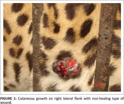
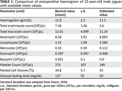
The animal was observed by the keepers of the zoo during the last week of February 2011 to have a round, reddish growth 3 cm in diameter on the skin of the right lateral abdomen with a non-healing type of wound on its surface (Figure 1). Macroscopically, the tumour was a superficial, ulcerated, multilobulated intradermal mass. The animal was brought to the zoo's veterinary hospital from its enclosure for complete surgical excision of the tumour, which was the treatment of choice (Goldschmidt & Hendrick 2003) after preoperative haematological evaluation (Table 1). The estimated values of the different haematological parameters were compared with the available normal (mean) values (Deem 2004).
Treatment
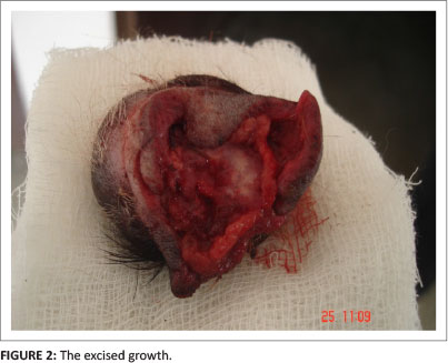
The animal weighed about 60 kg. It was anaesthetised with 0.6 mL injectable xylazine hydrochloride 100 mg/mL (Xyxil 100, Troy laboratory (pty) Ltd, Australia) and 1.8 mL injectable ketamine hydrochloride 100 mg/mL (Ketmin 100, Parnell Australia (pty) Ltd, Australia) administered intramuscularly. Ten minutes after administration of the anaesthetic the animal was restrained in a crush, exposing the area for surgery. The area was prepared for excision and infiltrated with 2% lidocaine (Xylocaine 2%, Astra Zeneca, India). The animal was maintained on intravenous fluid (normal saline, Aravind, India) throughout the process. Routine surgical procedure was followed for complete tumour excision (Morrison 2002). The animal was medicated intravenously with antibiotic Intacef 1 (1 gm Cetriaxone, Intas Pharmaceuticals, India), 2 mL haemostat, adrenochromemonosemicarbazone 1 mg/mL (Chromostat, Life Pharmaceutical, India), 3 mL dexamethasone 4 mg/mL (Decdan, Wockhardt, India), 3 mL meloxicam 5 mg/mL (Melonex, Intas Pharmaceuticals, India,) and injected intramuscularly with 3 mL ranitidine 25 mg/mL (Rantac, J.B. Chemicals & Pharmaceuticals, India). Lastly, 1 mL of injectable yohimbine hydrochloride 10 mg/mL (Riverzine, Bomac (pty) Limited, Australia) was administered intravenously for quick revival from anaesthesia. Observations were noted at half hourly intervals until six hours post surgery. Antibiotic therapy was continued for seven days, and injectable meloxicam and Rantac were given for another three days. The excised tissue (Figure 2) was preserved in 10% formalin and sent for histopathological examination.
The wound was treated regularly with povidone-iodine lotion (Betadine), herbal fly repellent or anti-infective spray (Topicure Spray) and 1 mL injectable pheniramine maleate 22.7 mg/mL (Avil) was administered intramuscularily for three days after the third postoperative day.
Wound healing was uneventful. The skin suture was removed after 12 days and the animal was returned to its original enclosure. Local tumour recurrence was not observed during the year that has elapsed since surgery.
Diagnosis
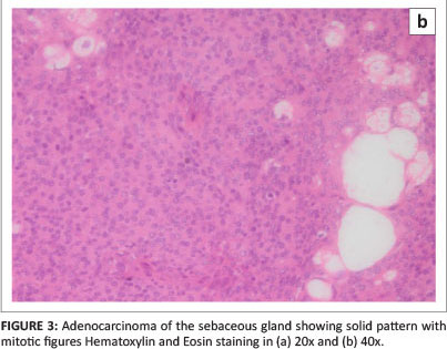
Haematological examination showed no major anomalies (Table 1). Anaesthetic and surgical protocol was satisfactory for this tumour excision. Histopathological examination of the excised tissue showed that the tumour was subdivided by fibrovascular connective tissue trabeculae into lobules of varying size. The section also showed asynchronous atypical growth of cells without planar arrangement. Tumour cells had intracytoplasmic lipid vacuoles with marked solidification of glands (Figure 3a). Nuclei were large and hyperchromatic. Cells were large and pleomorphic with considerable variation in the size and shape of the nuclei. A considerable number of mitotic figures (Figure 3b) were observed in the photomicrograph. Most of the tumour cells did not resemble the basaloid reserve cells of a sebaceous epithelioma.

Discussion
The multilobulated appearance of the section differentiates it from liposarcoma (Goldschmidt & Hendrick 2002). Both the macroscopic and microscopic findings confirmed this case as an adenocarcinoma of the sebaceous gland (Figure 3a and Figure 3b). Sebaceous gland adenocarcinoma recurrence is infrequent; metastasis is rare and occurs only via lymphatics to the regional lymph node (Goldschmidt & Hendrick 2002). In domestic cats, peak incidence of this tumour is between eight to 15 years of age with no breed or sex predilection. Common sites of sebaceous carcinoma in domestic cats are the head, thorax, and perineal region, where it occurs as a multilobulated intradermal mass (Goldschmidt & Hendrick 2002). Sebaceous carcinoma has also been documented in the submandibular salivary gland (Sozmenet al. 2002) and external auditory canal (Terim, Kapakin et al. 2008) of the domestic cat.
Acknowledgements
The authors are thankful to Dr Chandreyee Sen and Dr Pranoti Sharma for their support in histopathological diagnosis of the case.
Competing interests
The authors declare that they have no financial or personal relationship(s) which may have inappropriately influenced them in writing this article.
Author's contribution
All of the authors contributed in the planning of the operation, anesthesia and surgical procedure. A.K.M (Zoological Garden), S.K.G (Zoological Garden) and D.N.B (Zoological Garden), conducted the post-operative procedure. The manuscript was written by A.K.M and P.M (Zoological Garden).
References
Castro, M.B., Werther, K., Godoy, G.S., Borges, V.P. & Alessi, A.C., 2003, 'Visceral mast cell tumor in a captive black jaguar {Panthera onca)', Journal of Zoo and Wildlife Medicine 34, 100-102. PMid:12723809 [ Links ]
Goldschmidt, M.H. & Hendrick, M.J., 2002, 'Tumors of the skin and soft tissues', in D.J. Meuten (ed.), Tumors in domestic animals, 4th edn., pp. 64-67, Iowa State University Press, Ames. http://dx.doi.org/10.1002/9780470376928.ch2 [ Links ]
Gross, T.L., Ihrke, P.J., Walder, E.J. & Affolter, V.R., 2005, Skin diseases of the dog and cat: clinical and histopathologic diagnosis, 2nd edn., Blackwell Publishing, Oxford. http://dx.doi.org/10.1002/9780470752487 [ Links ]
Hoogesteijn, R. & Mondolfi, E., 1992, The jaguar, Armitano Publishers, Caracas. [ Links ]
Hope, K. & Deem, S. L., 2006, 'Retrospective study of morbidity and mortality of captive jaguars (Panthera onca) in North America: 1982-2002', Zoo Biology 25, 501-512. http://dx.doi.org/10.1002/zoo.20112 [ Links ]
International Species Information System, 1999, Medical animal record keeping system, Apple Valley, Minnesota, www.worldzoo.org. [ Links ]
Deem, S.L., 2004, Capture and Immobilization of Free-living Jaguars (Panthera onca), in D. Heard (ed.), Zoological Restraint and Anesthesia, International Veterinary Information Service (www.ivis.org), Ithaca, New York, USA. [ Links ]
Ladiges, W.C., Foster, J.W. & Jones, M.H., 1981, 'Malignant haemangio-endothelioma in a jaguar (Panthera onca), Journal of Zoo Animal Medicine 91, 115-122. [ Links ]
Maryamma, K.I., Sivadas, C.G., Nair, K. & Rajan, A., 1974, 'Cystadenocarcinoma of ovary with leiomyoma of uterus in a jaguar (Panthera onca)', Indian Veterinary Journal 51, 269-270. [ Links ]
McMartin, D.N. & Gruhn, R.F., 1977, 'Sebaceous carcinoma in a horse', Veterinary Pathology 14, 532-534. PMid:919245 [ Links ]
Morrison, W.B., 2002, Cancer in dogs and cats: medical and surgical management, 2nd edn., Teton New Media, Jackson. [ Links ]
Munson L., 1994, 'A high prevalence of ovarian papillary cystadenocarcinomas in jaguars (Panthera onca)', Veterinary Pathology 31, 604. [ Links ]
Munson, L. & Moresco, A., 2007, 'Comparative pathology of mammary gland cancers in domestic and wild animals', Breast Disease 28, 7-21. PMid:18057539 [ Links ]
Owston, M.A., Ramsay, E.C. & Rotstein, D.S., 2008, 'Neoplasia in felids at the Knoxville Zoological Gardens, 1979-2003', Journal of Zoo and Wildlife Medicine 39, 608-13. [ Links ]
Paul, J.C., Ghosh, G.L., Biswas, A., Nandi, S.K. & Biswas, B.K., 2002, 'Surgical management of squamous cell carcinoma of tail in a jaguar', Indian Journal of Animal Health 41, 141-144. [ Links ]
Ramos-Vara, J.A., Miller, M.A. & Preziosi, D., 2000, 'Glucagonoma in a jaguar (Panthera onca)', Journal of Zoo and Wild life Medicine 31, 563-565. PMid:11428406 [ Links ]
Sozmen, M., Brown, P.J. & Eveson, J.W., 2002, 'Sebaceous carcinoma of the salivary gland in a cat', Journal of Veterinary Medicine 49, 425-427. [ Links ]
Strafuss, A.C., 1976, 'Sebaceous gland adenomas in dogs', Journal of the American Veterinary Medical Association 15, 640-642. [ Links ]
Terim Kapakin, K. A., Bozkurt, F. & Haziroglu, R., 2008, 'Sebaceous adenocarcinoma in a cat', Acta Veterinaria, Brno 77, 123-125. http://dx.doi.org/10.2754/avb200877010123 [ Links ]
Vail, M.D. & Withrow, S.J., 1996, 'Tumors of the skin and subcutaneous tissues', in S.J. Withrow & E.G. Macewen (eds.), Small animal clinical oncology, 2nd edn., pp. 167-191, W.B. Saunders Company, Philadelphia. [ Links ]
 Correspondence:
Arnab Majie
Zoological garden, Alipore
Kolkata 700 027, India
Email: majie.arnab@gmail.com
Received: 06 Aug. 2012
Accepted: 11 Apr. 2013
Published: 24 Feb. 2014

