


Servicios Personalizados
Articulo
 Inglés (pdf)
 Articulo en XML
 Referencias del artículo
Indicadores

Links relacionados
-
 Citado por Google
 -
 Similares en Google
Compartir

 Permalink
Journal of the South African Veterinary Association
versión On-line ISSN 2224-9435
versión impresa ISSN 1019-9128
J. S. Afr. Vet. Assoc. vol.84 no.1 Pretoria ene. 2013
CASE REPORT
Osteoarthropathy of unknown aetiology in the long bones of farmed and wild Nile crocodiles (Crocodylus niloticus)
Fritz W. HuchzermeyerI; Herman B. GroenewaldII; Jan G. MyburghI; Johan C.A. SteylI; Martina R. CroleII
IDepartment of Paraclinical Sciences, University of Pretoria, South Africa
IIDepartment of Anatomy and Physiology, University of Pretoria, South Africa
ABSTRACT
Humeri of farmed and wild Nile crocodiles (Crocodylus niloticus) collected during routine post-mortem examinations were boiled, cleaned and examined for lesions. Various degrees of gross bone and articular pathology were found. The lesions were situated predominantly at the proximal and distal epiphyseal and metaphyseal regions of the bone, where growth and bone remodelling occurs. In advanced cases partial collapse of the articular surface could be identified. From the collection of crocodile bones five particular cases are described. Because of the wide distribution of origin of the affected animals, nutritional or toxicological causes seem unlikely. One of the cases presented was associated with mycoplasmosis. These forms of crocodilian bone pathology need further investigation.
Introduction
Crocodiles are nocturnal animals and during the day they either remain submersed or bask and rest on land. Impaired leg mobility is therefore seldom observed, except when accompanied by marked joint swelling as in cases of mycoplasmosis and gout (Huchzermeyer 2003). Nutritional osteomalacia in crocodilian hatchlings is caused by a lack of calcium in the diet and clinically translates into kyphoscoliosis, rubber jaws and glassy teeth (Cowan 1968; Huchzermeyer 1986). Osteodystrophy and osteochondrosis in two adult captive Orinoco crocodiles (Crocodylus intermedius) and one wild American crocodile (Crocodylus acutus) were also believed to have been caused by malnutrition (Blanco 1993). Examination of 342 crocodilian skeletons in museum collections led to the discovery of seven cases of spondyloarthropathy (Rothschild 2010). Similar bone conditions have been found in long bones of older juvenile and adult crocodiles when bones were more closely examined during routine post-mortem examination. Such cases of osteopathy and associated articular pathology of various degrees were found in farmed crocodiles from different farms fed home-made rations, as well as in wild crocodiles. Five selected cases are described.
Materials and methods
During routine post-mortem examination of larger crocodiles, a long bone was excised from each animal, preferably the humerus, as it was shorter than the femur and fitted more easily into an available cooking pan. The bone was boiled in water for about 2 h, after which the soft tissues were scraped off with a knife. It was then left to dry. Other forelegs or cleaned bones were received from post-mortem examinations carried out by pathologists at the Faculty of Veterinary Science, Onderstepoort or from problem crocodiles shot in the wild in Mpumalanga province and treated the same way. The description of the affected areas of the humerus follows the muscle map of the crocodilian humerus (Meers 2003). The cases presented in this article have been selected from a larger number of bones with different degrees of pathology that have been collected since 2003.
Anatomical note
When compared with the humerus of the horse (Zietschmann, Ackerknecht & Grau 1943), the humerus of the crocodile displays some important differences. The caput humeri has three condyles that are not very distinct and the musculus subscapularis inserts on the median condyle. From the lateral condyle, similar to that of the horse, the crista humeri is extended at the point where the musculus supracoracoideus complex and musculus pectoralis insert. However, the crista humeri does not extend as far distally as in the horse. A small tuberosity on the dorsal aspect of the humerus opposite the crista humeri serves as the insertion for the musculus teres major and musculus latissimus dorsi. The distal end of the humerus has two distinct condyles, lateral and medial, but lacks a fossa olecrani.
Case history
Case 1
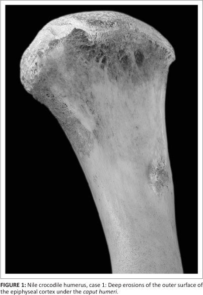
The right humerus of a very large male Nile crocodile (Crocodylus niloticus) was received from a farm in the Northwest Province on 04 September 2003. The post-mortem examination was carried out at the Department of Paraclinical Sciences of the Faculty of Veterinary Science, University of Pretoria, with a diagnosis of generalised mycosis. The senior author had not been able to attend the post-mortem and received only the right foreleg. The humerus showed deep erosion of the outer surface of the epiphyseal cortex on the dorsal aspect just under the c. humeri (Figure 1) and at the distal end a complete collapse of the ventral aspect of the articular surface (Figure 2).

Case 2
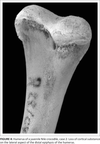
The right humerus of a juvenile crocodile that had died of polyarthritis due to mycoplasmosis was received from a farm in Gauteng on 06 September 2004. The bone showed an almost complete loss of the c. humeri, except for a small piece of articular surface on the median trochanter (Figure 3). There was secondary bone deposition all around the proximal epiphysis, particularly on the dorsal and lateral aspects, and a loss of cortical substance on the lateral (Figure 4) and ventral aspects of the distal epiphysis.

Case 3
The left humerus of a wild adult female crocodile from Flag Boshielo Dam was received on 15 March 2006. The animal was examined at the Department of Paraclinical Sciences, Faculty of Veterinary Science, University of Pretoria, and diagnosed with internal injuries from falling over the dam wall. The dam wall had been raised and the water level had risen sharply owing to very heavy rainfall. Subsequently several crocodiles had appeared to become disoriented and some were seen congregating at the dam wall and trying to climb or swim over it. The bone showed severe loss of cortical substance all around the c. humeri and collapse of about two thirds of the articular surface (Figure 5). There was also some loss of cortical substance around the distal epiphysis.

Case 4
The right humerus of an adult female crocodile (3.20 m long), which had been received from a farm in Gauteng, was examined on 20 August 2007. The necropsy revealed deep bite wounds and severe pleuritis. Upon examination of the bone, severe loss of cortical substance could be identified all around the c. humeri after processing. There was deep erosion on the medial side of the c. humeri at the site of the insertion of the m. pectoralis and also severe loss of cortical substance at its distal end (Figure 6), with partial caving in of the articular surface (Figure 7).


Case 5
The left humerus of an adult female crocodile (2.78 m long), received from a farm in Gauteng, was examined on 08 August 2008. Post-mortem examination revealed consolidated pulmonary haematomas and pleural adhesions, with a large fibrinous mass in the pleural cavity resulting from penetrating injury, presumably bite wounds. The bone showed severe loss of cortical substance all around the c. humeri, a small erosion at the site of the insertion of the m. pectoralis on the medial side of the c. humeri and complete collapse of the distal articular surface (Figure 8).

Discussion
Crocodilians differ from other egg laying archosaurs (dinosaurs and birds) in that they lack medullary bone in the endosteal cavities of the long bones (Schweitzer et al. 2007). The epiphyses of the long bones in crocodiles therefore often appear eroded as more of the remodelling seems to take place on the outside of the bone, as seen in the cases described. Therefore, a certain degree of erosion may have to be considered normal.
In early stages of osteoarthropathy, the periosteal surface of the epiphyses alone appears to be affected and shows deep longitudinal furrows up to and undermining the articular surfaces. In advanced stages, there is collapse of the articular surface, which, in turn, can stimulate an inflammatory reaction in the form of osteoarthritis.
The cases described here originated from routine post-mortem examinations and are therefore random findings without any particular geographical or epidemiological pattern. The wild specimens came from the Limpopo province and the farmed ones from Gauteng and Northwest. The occurrence of the condition both in farmed and in wild crocodiles in three different provinces most likely rules out malnutrition as a major factor, although this was suggested for the South American cases (two captive and one wild) described by Blanco (1993).
Mineral loss due to enteritis as seen in primates has been suggested as a possible cause in the museum cases (Rothschild 2010). However, intestinal infections do not lead to diarrhoea in crocodiles owing to the exudation of fibrin as part of the inflammatory response (Huchzermeyer & Cooper 2000).
A toxic cause of the condition is equally unlikely in view of its occurrence both in farmed and in wild crocodiles from different farms and areas. In the only report of bone abnormality due to pollution with pesticides and their metabolites with endocrine disruptor action in wild American alligators (Alligator mississippiensis), the bones of the affected animals were heavier than those of the controls (Lind et al. 2004). Mycoplasmosis causing polyarthritis in crocodiles (Mohan et al. 1995) could directly provoke articular lesions.
Stress could be a common factor in the cases presented in this article, as has been suggested previously (Huchzermeyer 2002). Glucocorticoid-induced osteoporosis is a common and clinically relevant medical condition (Patschan, Loddenkemper & Butgereit 2001). Socially stressed female cynomolgus monkeys (Macaca fascicularis) were found to have reduced vertebral bone mineral density (Shiveley et al. 1991). In intensively kept egg producing fowls, reduced bone density has been widely reported under the name of cage layer fatigue and recognised as osteoporosis (Riddell et al. 1968; Rowland et al. 1968a). Cage layer fatigue is not related to levels of mineral nutrition (Rowland et al. 1968b) and is apparently stress induced. In farmed crocodiles stress particularly affects growth rate (Elsey et al. 1990). The possibility of a link between stress and the bone pathology in crocodiles described in this article and the related metabolic pathways need further investigation.
Conclusion
Only a few crocodilian cases of osteodystrophy and osteochondrosis have previously been reported (Blanco 1993; Rothschild 2010). The routine examination of a long bone from each post-mortem case over the last few years revealed widespread occurrence of the problem and it is recommended that such an examination be adopted as part of the routine post-mortem procedure for all larger juvenile and adult crocodiles.
Acknowledgements
Sincere thanks are due to all the colleagues who collected and contributed bones for this project. Publication of this article was sponsored by the Faculty of Veterinary Science, University of Pretoria.
Competing interests
The authors declare that they have no financial or personal relationship(s) that may have inappropriately influenced them in writing this article.
Authors' contributions
F.W.H. (University of Pretoria) was the project leader and responsible for identifying the cases, describing the lesions and compiling the manuscript. J.C.A.S. (University of Pretoria) and J.G.M. (University of Pretoria) performed the necropsies, collected bone specimens for further examination and made conceptual contributions. M.R.C. (University of Pretoria) and H.B.G. (University of Pretoria) provided anatomical notes and compiled the photographical collection.
References
Blanco, P.A., 1993, 'Enfermedades degenerativas óseas y articulares en caimán del Orinoco (Crocodylus intermedius) y caimán de la costa (Crocodylus acutus) presentación de tres (3) casos [Degenerative bone and joint diseases in Orinoco crocodiles (Crocodylus intermedius) and American crocodiles (Crocodylus acutus): Presentation of three cases]', Proceedings of the Regional Meeting of the Crocodile Specialist Group of South America and the Caribbean Region, Centro de Innovación Agroindustrial S.C., Villahermosa, Tabasco, Mexico, August 04-07, 1997, pp. 26-37. [ Links ]
Cowan, D.F., 1968, 'Diseases of captive reptiles', Journal of the American Veterinary Medical Association 153, 848-859. PMid:5692920 [ Links ]
Elsey, R.M., Joanen, T., McNease, L. & Lance, V., 1990, 'Growth rate and plasma corticosteroid levels in juvenile alligators maintained at different stocking densities', Journal of Experimental Physiology 25, 30-36. [ Links ]
Huchzermeyer, F.W., 1986, 'Osteomalacia in young captive crocodiles (Crocodylus niloticus)', Journal of the South African Veterinary Association 57, 167-168. PMid:3806562 [ Links ]
Huchzermeyer, F.W., 2002, 'Stress in farmed and captive crocodiles: Stressors and effects', in Crocodiles: Proceedings of the 16th Working Meeting of the Crocodile Specialist Group (unedited), pp. 173-176, IUCN, Gland. [ Links ]
Huchzermeyer, F.W., 2003, Crocodiles: Biology, husbandry and diseases, CABI, Wallingford. http://dx.doi.org/10.1079/9780851996561.0000 [ Links ]
Huchzermeyer, F.W. & Cooper, J.A., 2000, 'Fibriscess, not abscess resulting from a localized inflammatory response to infection in reptiles and birds', Veterinary Record 147, 515-517. http://dx.doi.org/10.1136/vr.147.18.515, PMid:11110493 [ Links ]
Lind, P.M., Milnes, M.R., Lundberg, R., Bermudez, D., Órberg, J. & Guillette, L.J., 2004, 'Abnormal bone composition in female juvenile American alligators from a pesticide-polluted lake (Lake Apopka, Florida)', Environmental Health Perspectives 112, 350-362. [ Links ]
Meers, M.B., 2003, 'Crocodilian forelimb musculature and its relevance to archosauria', Anatomical Record 274A, 891-916. http://dx.doi.org/10.1002/ar.a.10097, PMid:12973714 [ Links ]
Mohan, K., Foggin, C.M., Muvavarirwa, P., Honywill, J. & Pawandiwa, A., 1995, 'Mycoplasma-associated polyarthritis in farmed crocodiles (Crocodylus niloticus) in Zimbabwe', Onderstepoort Journal of Veterinary Research 62, 45-49. PMid:8539034 [ Links ]
Patschan, D., Loddenkemper, K. & Butgereit, F., 2001, 'Molecular mechanisms of glucocorticoid-induced osteoporosis', Bone 29, 498-505. http://dx.doi.org/10.1016/S8756-3282(01)00610-X [ Links ]
Riddell, C., Helmboldt, C.F., Sinsen, E.P. & Matterson, L.D., 1968, 'Bone pathology of birds affected with cage layer fatigue', Avian Diseases 12, 285-297. http://dx.doi.org/10.2307/1588228, PMid:5654587 [ Links ]
Rothschild, B., 2010, 'Macroscopic recognition of nontraumatic osseous pathology in the postcranial skeletons of crocodilians and lizards', Journal of Herpetology 44, 13-20. http://dx.doi.org/10.1670/08-243.1 [ Links ]
Rowland, L.O., Wilson, H.R., Fry, J.L. & Harms, R.H., 1968a, 'A comparison of bone strength of caged and floor layers and roosters', Poultry Science 47, 2013-2015. http://dx.doi.org/10.3382/ps.0472013 [ Links ]
Rowland, L.O., Harms, R.H., Wilson, H.R., Ahmed, E.M., Waldroup, P.W. & Fry, J.L., 1968b, 'Influence of various dietary factors on bone fragility of caged layers', Poultry Science 47, 507-511. http://dx.doi.org/10.3382/ps.0470507 [ Links ]
Schweitzer, M.H., Elsey, R.M., Dacke, C.G., Horner, J.R. & Lamm, E.T., 2007, 'Do egg-laying crocodilian (Alligator mississippiensis) archosaurs form medullary bone?', Bone 40, 1125-1158. http://dx.doi.org/10.1016/j.bone.2006.10.029, PMid:17223615 [ Links ]
Shiveley, C.A., Jayo, N.J., Weaver, D.S. & Kaplan, J.R., 1991, 'Reduced vertebral bone mineral density in socially subordinate female cynomolgus macaques', American Journal of Primatology 25, 250. [ Links ]
Zietschmann, O., Ackerknecht, E. & Grau, O., 1943, Ellenberger-Baum, Handbuch der Anatomie der Haustiere [Ellenberger-Baum, Handbook of the Anatomy of Domestic Animals], Springer Verlag, Berlin. [ Links ]
 Correspondence:
Fritz Huchzermeyer
Private Bag X04
Onderstepoort 0110, South Africa
Email: crocvet@vodamail.co.za
Received: 06 Jan. 2013
Accepted: 25 June 2013
Published: 13 Nov. 2013

