


Servicios Personalizados
Articulo
 Inglés (pdf)
 Articulo en XML
 Referencias del artículo
Indicadores

Links relacionados
-
 Citado por Google
 -
 Similares en Google
Compartir

 Permalink
Journal of the South African Veterinary Association
versión On-line ISSN 2224-9435
versión impresa ISSN 1019-9128
J. S. Afr. Vet. Assoc. vol.84 no.1 Pretoria ene. 2013
CASE REPORT
Multiple myeloma in a captive lion (Panthera leo)
Adrian S.W. TordiffeI, II; Nicky CasselII; Emily P. LaneI; Fred ReyersIII
IDepartment of Research and Scientific Services, National Zoological Gardens of South Africa, South Africa
IIDepartment of Companion Animal Clinical Studies, University of Pretoria, South Africa
IIISchool of Life Sciences, University of Lincoln, United Kingdom
ABSTRACT
Multiple myeloma is a rare, systemic proliferation of neoplastic plasma cells. A case was reported in an 11-year-old male captive lion (Panthera leo) at the National Zoological Gardens of South Africa, Pretoria. The classic features of symptomatic multiple myeloma were all evident in this case; namely osteolytic lesions, monoclonal gammopathy in the serum with excretion of monoclonal proteins in the urine, neoplastic plasma cells in the bone marrow and associated renal failure and anaemia. In addition, similar to the common pattern of this disease in domestic felids, at least three extramedullary tumours were found and several organs were infiltrated by neoplastic plasma cells. The cytoplasm of approximately 50% of the neoplastic round cells, including a few giant myeloma cells, stained weakly to strongly using immunohistochemical stains for B-lymphocytes (CD79a). The normal haematological parameters and lack of any osteolytic lesions in the lion at the time of the first evaluation suggest that the primary neoplastic cells could have originated from one of the extramedullary tumour sites. Only two cases of multiple myeloma have previously been reported in captive wild felids. To the authors' knowledge, there are no case reports of multiple myeloma in lions.
Introduction
Multiple myeloma is the most common of several rare myeloma-related disorders involving the malignant transformation of terminally differentiated B-lymphocytes. It is classically characterised in dogs and humans by: (1) bone marrow plasmacytosis, (2) monoclonal gammopathy on serum protein electrophoresis, (3) osteolysis and (4) light-chain Bence-Jones proteinuria.
Myeloma-related disorders are rare in domestic cat species, making up between 0.0012% and 0.0025% of malignant neoplasms found (Carpenter, Andrews & Holzworth 1987; Engel & Brodey 1969). The incidence of multiple myeloma is more frequent in older cats, with a mean age of 12 years and an equal sex distribution recorded in one study (Mellor et al. 2006). The presenting clinical signs are normally vague, but commonly include weight loss, anorexia and lethargy. In contrast to humans and dogs with multiple myeloma, osteolytic lesions are rarely detected in domestic cats and additional extramedullary plasma cell neoplasias are more frequently a feature (Mellor et al. 2006; Mellor et al. 2008).
Only two other cases of multiple myeloma have been recorded in other captive non-domestic felids: a 21-year-old jaguar (Panthera onca) (Port et al. 1981) and a 6-year-old snow leopard (Uncia uncia) (Kappeli et al. 2009). To the authors' knowledge, there are no reports of multiple myeloma in lions (Panthera leo).
Case History
A captive-born male lion, which was housed at the National Zoological Gardens of South Africa (NZG) in Pretoria, developed lethargy and a reduced appetite. He weighed 180 kg, was 11-year-old and was vasectomised. Initial observations revealed no obvious gait or other abnormalities. The lion was immobilised (day 0) with a combination of 0.56 mg/kg tiletamine and zolazepam (Zoletil, Virbac RSA [Pty] Ltd, Halfway House, 100 mg/mL) and 17 µg/kg medetomidine (Medetomidine, Kyron Laboritories [Pty] Ltd, Johannesburg, 20 mg/mL). Urine was collected by catheterisation and was found to have a specific gravity of 1.060 and, apart from a moderate (2+) proteinuria, all the dipstick parameters were within normal limits. The urine sediment was evaluated using light microscopy, but no abnormalities were found. Haematology and selected serum biochemistry values were considered; except for minor elevations in amylase and total bilirubin, all other blood parameters were unremarkable (Table 1). In order to evaluate any potential spinal or musculoskeletal pain, the lion was medicated with 0.17 mg/kg of meloxicam (Coxflam, CiplaMedpro, Durbanville) once daily for five days, but his general habitus failed to improve significantly.
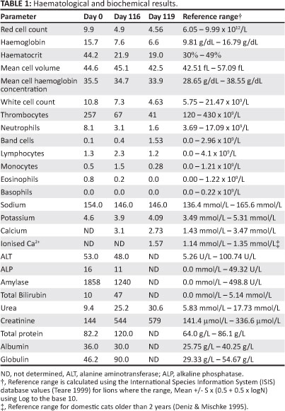
Over the next few months, he intermittently walked with a stiff gait and was slow in rising and lying down. Two months later (day 62), he was immobilised again with the same doses of zolazepam/ tiletamine and medetomidine and transported to the Onderstepoort Veterinary Academic Hospital (OVAH) for survey spinal and pelvic radiographs. No radiographic abnormalities were detected. However, his appetite further deteriorated and progressed to complete anorexia. On day 116 he was immobilised with the same drug combination and was moved to an enclosure within the NZG veterinary hospital. He had lost approximately 10 kg in body weight. His mucous membranes were pale-white and mild icterus was noted. In contrast to the day 0 tests, day 116 serum biochemistry results (Table 1) showed severe azotaemia with marked hyperglobulinaemia, and a moderate non-regenerative anaemia and thrombocytopenia were noted. Piroplasms, with morphology compatible with that of Babesia leo, were visible at low density (< 0.015%) on both peripheral and central blood smears. There was limited evidence of erythrophagocytosis and toxic granulation of the neutrophils (as well as Dohle bodies). However, the most striking feature on the smears was the protein precipitate in the background and the substantial rouleaux formation.
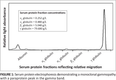
Additional laboratory tests included serum coronavirus antibody titre, which was low (1:320), and serum protein electrophoresis (SPE), which demonstrated a clear monoclonal gammopathy with a paraprotein peak in the gamma (γ) band (Figure 1), suggestive of multiple myeloma.
On day 119, the lion was again immobilised and transported to the OVAH for further diagnostic procedures. Blood samples were again collected to repeat some of the serum chemistry and haematology tests (Table 1). The results indicated a further deterioration in both the erythrocyte, leukocyte and platelet parameters. Serum calcium was within the normal reference range, but ionised calcium was mildly elevated when compared with the reference range established for domestic cats (Deniz & Mischke 1995). Feline leukaemia virus (FeLV) and feline immunodeficiency virus (FIV) tests (SNAPCombo Plus Feline Leukemia Virus Antigen/Feline ImmunodeficiencyVirus AntibodyTest Kit, IDEXX Corp.) performed on the serum were both negative.
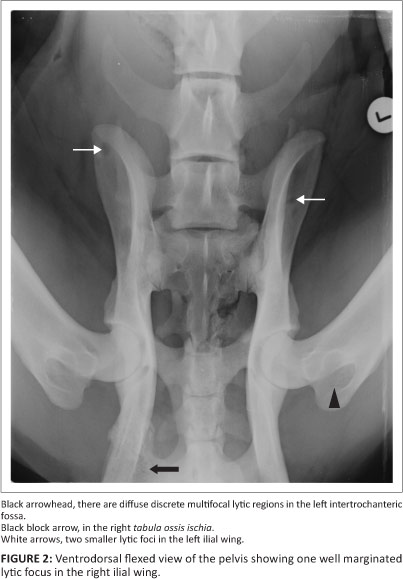
Survey radiographs showed a 12.5 cm x 6.0 cm sub-lumbar soft tissue opacity compressing the dorsal margin of the terminal descending colon and multiple circular to oval (up to 20 cm x 13 cm) lytic foci in the left and right ilial wings (Figure 2), with similar suspect lytic foci in the spinous process of the third thoracic vertebra and several mid-thoracic vertebrae. Multifocal to coalescing lytic foci were seen in the left inter-trochanteric fossa and the right tabula ossis ischii (Figure 2). Abdominal ultrasound was performed with the patient in left lateral recumbency. This confirmed the presence of a well-marginated, oval, 8.0 cm x 4.3 cm homogenously hypoechoic sub-lumbar mass, which was believed to be an enlarged medial iliac lymph node. Ultrasound-guided fine needle aspirates were obtained from the spleen, but due to patient positioning and depth of the medial iliac lymph node, this lymph node could not be aspirated.
Urine was collected via cystocentesis and submitted for the detection of Bence-Jones proteins. The results were positive for both bound and free light chain paraproteins, with a total urine protein content of 4.36 g/L. Bone marrow biopsies collected from the proximal left tibia were almost acellular, with only small numbers of megakaryocytes and few leukocytes and erythrocyte precursors visible. Fine needle aspirate of the spleen yielded a large number of nucleated cells mainly consisting of anaplastic plasma cells showing the following criteria of malignancy: anisocytosis, anisokaryosis, coarse chromatin, and prominent and multiple nucleoli. Some plasma cells were bi-nucleated and one plasma cell was noticed with three nuclei of unequal size.
Pathological findings
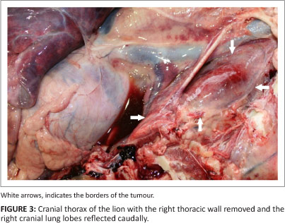
The lion was euthanased at the NZG veterinary hospital and a post mortem examination was performed. A large mass in the cranioventral thorax was present in the area of the thymus (180 mm x 50 mm x 50 mm) (Figure 3). A second mass was present in the caudal abdomen, immediately ventral to the sub-lumbar muscles (50 mm x 35 mm x 45 mm). Both had a gelatinous outer surface and a more fibrous inner core. A smaller third mass (2 cm in diameter) was found on the pleural surface of the costochondral junction of the fourth right rib. Several small nodules (3 mm - 5 mm in diameter) were scattered in various liver lobes. Bone marrow taken from the right femur appeared yellowish-white in colour. The gastric mucosa had numerous small ulcers, possibly due to the chronic uraemia.
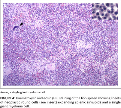
Formalin-fixed tissues were processed routinely and stained with haematoxylin and eosin. Sections of all three masses, and similar masses in the liver and bone marrow (5 mm diameter), consisted of sheets of round cells with clear cytoplasmic margins, medium-sized often polar nuclei with clumped chromatin and abundant lightly basophilic cytoplasm with occasional pale peri-nuclear foci (Figure 4). The mitotic index was low (> 1/HPF). Small numbers of large cells with distinct cell borders, abundant clear to faintly basophilic cytoplasm and large, often lobulated hyperchromatic nuclei (interpreted as giant myeloma cells) occurred throughout all four masses. Similar cells expanded splenic sinusoids and formed rare aggregates in bone marrow, pulmonary alveolar and adrenocortical capillaries. The cytoplasm of approximately 50% of the neoplastic round cells, including a few giant myeloma cells, stained weakly to strongly using immunohistochemical stains for B-lymphocytes (CD79a); strongly stained follicular cells provided a good internal positive control since this stain has not been validated for lions. Small numbers of cells that stained strongly with CD3 immunohistochemical stain (a T-cell marker) lining splenic trabeculae were interpreted as remnant T-cell populations marginalised by the neoplastic cells.
In addition, renal tubules contained variably eosinophilic (proteinaceous) material and were lined by epithelial cells showing mild hydropic degeneration (consistent with protein-losing nephropathy). Findings that may be associated with the myeloma included mild diffuse global membranous glomerulonephritis and moderate multifocal midzonal gastric metastatic mineralisation (uraemic gastropathy).
Ethical considerations
This case report was written as a retrospective clinical case report of an NZG owned animal. All diagnostic tests and treatments were performed as part of the routine work up and treatment of the patient. None of the diagnostic procedures and treatments were performed for research purposes.
Discussion
The classic features of symptomatic multiple myeloma were all demonstrated in this case, namely: osteolytic lesions; monoclonal gammopathy in the serum, with excretion of light chain proteins in the urine; neoplastic plasma cells in the bone marrow; and associated renal failure and anaemia. In addition, similar to the common pattern of this disease in domestic felids, at least three extramedullary tumours were found and several organs were infiltrated by neoplastic plasma cells.
Only two cases of multiple myeloma have been previously reported in captive wild felids. A 21-year-old male jaguar (Panthera onca) died in a North American Zoo 13 days after presenting with progressive hind limb lameness, anorexia, ataxia and depression. Elevated serum total proteins and a gamma globulin peak, demonstrated on serum protein electrophoresis, suggested a diagnosis of multiple myeloma. This was confirmed on evaluation of bone marrow aspirates at the time of the post mortem examination (Port et al. 1981). A 6-year-old male captive snow leopard (Uncia uncia) in Switzerland developed progressive hind limb weakness, ataxia and loss of proprioception. Radiographs of the spine showed a poorly defined radiolucent area within the 12th thoracic vertebral body, with compression of the spinal cord evident on the myelogram. Total serum proteins were, however, normal in this patient and serum protein electrophoresis did not reveal a monoclonal gammopathy. A diagnosis of multiple myeloma was confirmed by histopathology and immunology of the mass found in the 12th vertebra (Kappeli et al. 2009). In both cases the neoplastic cells were restricted to the skeletal system.
In a retrospective study of 24 cases of myeloma-related disorders in domestic cats, extramedullary plasma cell neoplasia was found to be common, occurring in 16 (67%) of the cats. Radiography was performed in 13 of these cats (4 complete and 9 partial skeletal surveys) and osteolytic lesions were only observed in a single case (Mellor et al. 2006). Therefore, the presentation and development of myeloma-related disorders in both domestic felids and in the small number of wild felids reported so far appears to be rather diverse, with a wide spectrum of dissemination.
There is substantial immunological cross-reactivity between antibodies used in the identification of human immunoglobulins and immunoglobulin components, those same immunoglobulins and immunoglobulin components in dogs and cats (Darbès, Majzoub & Hermanns 1997; Sinkora et al. 2007). It was therefore reasonable to assume that this might be the case in the lion as well, and so the immunofixation tests were run. The fact that the procedure revealed the presence of light chains by this method, using human reagents, vindicates the assumption, because if there had been no cross-reactivity, no immunofixation band would have been revealed. A positive result cannot be false positive (in terms of incorrectly identifying the presence of light-chain proteins) but can significantly underestimate the quantity present if the cross-reactivity is poor. The test, however, is essentially binary (positive or negative) but is interpreted semi-quantitatively when used with samples from humans. In this case report, the binary interpretation was applied.
The normal haematological parameters and lack of any osteolytic lesions in the lion at the time of the first evaluation suggest that the primary neoplastic cells could have originated at one of the extramedullary tumour sites. The largest of the three extramedullary tumours, found in the area of the thymus, would therefore be the most probable primary site of neoplastic plasmacyte proliferation. Renal failure is common in human multiple myeloma patients, primarily due to the toxic effects of the monoclonal light chain proteins on the renal tubules and to a lesser extent on the glomeruli (Dimopoulos et al. 2008). The renal disease in this animal may have been caused by a combination of Bence-Jones proteinuria and diminished renal perfusion due to hyperviscosity syndrome and dehydration. Other causes of renal failure in multiple myeloma cases, such as hypercalcaemia and amyloidosis, were not noted in this lion. The normal serum urea and creatinine values and the high urine specific gravity recorded in the patient on day 0 suggest that renal function was relatively normal prior to the development of the multiple myeloma.
The rapid deterioration, severe haematopoetic abnormalities, renal failure and poor appetite of this lion were all considered to be poor prognostic indicators and resulted in the decision to euthanase the animal rather than attempt an form of treatment. The variable CD79a expression by neoplastic plasma cells is consistent with the poor prognosis in this case. Although the mechanism is not yet understood, loss of CD79a expression in human myelomas is associated with thrombocytopenia, an indicator of prognosis (Tanaka et al. 2009).
The risk factors associated with the development of multiple myeloma in companion animals are unknown. There is currently no evidence to suggest that FeLV, FIV, or feline infectious peritonitis virus (FIP) are involved in the pathogenesis of this disease in domestic cats (Mellor et al. 2006). In a study on the occurrence of multiple myeloma in humans, exposure to petroleum products and industrial emissions appear to be risk factors (Speer et al. 2002). Dietary factors, however, also seem to play a role (Wang et al. 2012). A review of neoplasia in felids at the Knoxville Zoological Gardens in the United States reported a high incidence of malignant neoplasia (25% of felids affected over a 24 year period) (Owston, Ramsay & Rotstein 2008). They speculated that the location of the zoo, in the middle of the city and adjacent to a busy interstate highway, might have exposed the felids to high levels of potentially carcinogenic vehicle emissions. However, no cases of myeloma-related disorder were found in that study (40 neoplasms in 26 zoo felids). Over the last 22 years, neoplasia was diagnosed on post mortem examination in two out of three adult lions (66%) and two out of three (66%) adult tigers that died or were euthanased in that period at the NZG. The incidence of neoplasia in large felids at the NZG is therefore relatively high and although many factors may be involved, it is possible that environmental pollutants, associated with the inner city location of this facility, may play a role.
Acknowledgements
We, the authors would like to thank Dr Angela Brüns from the Veterinary Department at the NZG, Prof. Andrew Leisewitz and Ms Carien Muller from the Department of Companion Animal Clinical Studies, Faculty of Veterinary Science, University of Pretoria for their valuable assistance in this case. We are also indebted to the histopathology lab of the Pathology Section, Department of Paraclinical Sciences, Faculty of Veterinary Science, University of Pretoria for excellent technical assistance. Publication of this article was sponsored by the Wildlife Group (http://www.vets4wildlife.co.za) of the South African Veterinary Association.
Competing interests
The authors declare that they have no financial or personal relationship(s) that may have inappropriately influenced them in writing this article.
Authors' contribution
A.S.W.T. (National Zoological Gardens of South Africa & University of Pretoria) was the clinician responsible for the case, collected and submitted most of the samples and wrote most of the manuscript. N.C. (University of Pretoria) performed the ultrasound and radiographic examinations and contributed toward the relevant sections of the manuscript. E.P.L. (National Zoological Gardens of South Africa) performed the histopathology and contributed toward the relevant sections of the manuscript. F.R. (University of Lincoln) evaluated the haematology and serum biochemistry results, provided valuable advice on the case and reviewed the manuscript.
References
Carpenter, J.L., Andrews, L.K. & Holzworth, J., 1987, 'Tumors and tumor-like lesions', in J. Holzworth (ed.), Diseases of the cat: Medicine and surgery, p. 406, WB Saunders, Philidelphia. [ Links ]
Darbès, J., Majzoub, M. & Hermanns, W., 1997, 'Evaluation of the cross-reactivity between human and feline or canine leucocyte antigens using commercially available antibodies', Journal of Veterinary Diagnostic Investigation 9, 94-97. http://dx.doi.org/10.1177/104063879700900120, PMid:9087936 [ Links ]
Deniz, A. & Miscke, R., 1995, 'Ionized calcium and total calcium in the cat', Berliner und Múnchener Tierártzliche Wochenschrift 108, 105-108. PMid:7646424 [ Links ]
Dimopoulos, M.A., Kastritis, E., Rosinol, L., Blade, J. & Ludwig, H., 2008, 'Pathogenesis and treatment of renal failure in multiple myeloma', Leukemia 22, 1485-1493. http://dx.doi.org/10.1038/leu.2008.131, PMid:18528426 [ Links ]
Engel, G.C. & Brodey, R.S., 1969, 'A retrospective study of 395 feline neoplasms', Journal of the American Animal Hospital Association 5, 21-31. [ Links ]
Kappeli, U., Eulenberger, U., Nitzl, D., Bley, C.R., Steffen, F., Sydler, T. et al., 2009, 'Clinical challenge. Multiple myeloma', Journal of Zoo and Wildlife Medicine 40, 398-401. http://dx.doi.org/10.1638/2008-0120.1, PMid:19569496 [ Links ]
Mellor, P.J., Haugland, S., Murphy, S., Smith, K.C., Holloway, A., Archer, J. et al., 2006, 'Myeloma-related disorders in cats commonly present as extramedullary neoplasms in contrast to myeloma in human patients: 24 cases with clinical follow-up', Journal of Veterinary Internal Medicine 20, 1376-1383. http://dx.doi.org/10.1111/j.1939-1676.2006.tb00754.x, PMid:17186853 [ Links ]
Mellor, P.J., Haugland, S., Smith, K.C., Powell, R.M., Archer, J., Scase, T.J. et al., 2008, 'Histopathologic, immunohistochemical, and cytologic analysis of feline myeloma-related disorders: Further evidence for primary extramedullary development in the cat', Veterinary Pathology 45, 159-173. http://dx.doi.org/10.1354/vp.45-2-159, PMid:18424828 [ Links ]
Owston, M.A., Ramsay, E.C. & Rotstein, D.S., 2008, 'Neoplasia in felids at the Knoxville Zoological Gardens, 1979-2003', Journal of Zoo and Wildlife Medicine 39, 608-613. http://dx.doi.org/10.1638/2008-068.1, PMid:19110704 [ Links ]
Port, C.D., Maschgan, E.R., Pond, J. & Scarpelli, D.G., 1981, 'Multiple neoplasia in a jaguar (Panthera onca)', Journal of Comparative Pathology 91, 115-122. http://dx.doi.org/10.1016/0021-9975(81)90051-7 [ Links ]
Sinkora, J., Samankova, P., Kummer, V., Leva, L., Maskova, J., Rehakova, Z. et al., 2007,' Commercially available rabbit anti-human polyclonal antisera as a useful tool for immune system studies in veterinary species', Veterinary Immunology and Immunopathology 119, 156-162. http://dx.doi.org/10.1016/j.vetimm.2007.06.023, PMid:17659784 [ Links ]
Speer, S.A., Semenza, J.C., Kurosaki, T. & Anton-Culver, H., 2002, 'Risk factors for acute myeloid leukemia and multiple myeloma: A combination of GIS and case-control studies', Journal of Environmental Health 64, 9-16. PMid:11901667 [ Links ]
Tanaka, T., Ichimura, K., Sato, Y., Takata, K., Morito, T., Tamura, M. et al., 2009, 'Frequent down-regulation or loss of CD79a expression in plasma cell myelomas: Potential clue for diagnosis', Pathology International 59, 804-808. http://dx.doi.org/10.1111/j.1440-1827.2009.02448.x, PMid:19883431 [ Links ]
Teare, A.J. (ed.), 1999, International Species Information System: Physiological data reference values, Apple Valley, USA. [ Links ]
Wang, Q., Wang, Y., Zhaohua, J., Xiequn, C., Yaozhu, P., Guangxun, G. et al., 2012, 'Risk factors for multiple myeloma: A hospital-based case-controlled study in Northwest China', Cancer Epidemiology 36, 339-344. http://dx.doi.org/10.1016/j.canep.2012.05.002, PMid:22673750 [ Links ]
 Correspondence:
Adrian Tordiffe
PO Box 754
Pretoria 0001, South Africa
Email: adrian@nzg.ac.za
Received: 23 Oct. 2012
Accepted: 12 Apr. 2013
Published: 26 Sept. 2013

