


Serviços Personalizados
Artigo
 Inglês (pdf)
 Artigo em XML
 Referências do artigo
Indicadores

Links relacionados
-
 Citado por Google
 -
 Similares em Google
Compartilhar

 Permalink
Journal of the South African Veterinary Association
versão On-line ISSN 2224-9435
versão impressa ISSN 1019-9128
J. S. Afr. Vet. Assoc. vol.84 no.1 Pretoria Jan. 2013
SHORT COMMUNICATION
Intestinal dermoid cyst in a German shepherd dog
Mehdi SaberiI; Omid AzariI; Reza KheirandishII; Rokhsana RasouliIII; Maryam AghazamaniIV; Elham MohebbiV
IDepartment of Clinical Sciences, Shahid Bahonar University of Kerman, Iran
IIDepartment of Pathobiology, Shahid Bahonar University of Kerman, Iran
IIILeishmaniasis Research Centre, Kerman University of Medical Sciences, Iran
IVResearch Centre for Modelling in Health, Institute for Futures Studies in Health, Kerman University of Medical Sciences, Iran
VResearch Center for Modling Health, Institute for Futures Studies in Health, Kerman University of Medical Sciences, Iran
ABSTRACT
A two-year-old male German shepherd dog was admitted to Shahid Bahonar Veterinary Hospital with clinical signs that included lethargy, anorexia, vomiting, abdominal pain and dehydration. Physical examination revealed nothing significant. Routine paraclinical tests only revealed a stress leukogram. Radiography revealed a mass in the stomach. Whilst performing a laparotomy, the surgeon observed an unusual mass in the subserosal layer of the proximal part of the jejunum. The histopathology of the mass revealed some scattered sebaceous and sweat glands associated with the cyst wall that confirmed the diagnosis of a dermoid cyst. Intestinal dermoid cysts are very rare and to our knowledge this is the first report of an intestinal dermoid cyst in a dog.
Introduction
The dermoid cyst is an uncommon developmental anomaly that has been reported in dogs, cats, horses, and cattle (Akhtardanesh, Kheirandish & Azari 2012; Maxie, Jubb & Kennedy 2007). A dermoid cyst is usually a congenital or hereditary lesion (Bailey, Holmberg &Yager 2001; Chénier, Quesnel & Girard 1998). In dogs, cutaneous dermoid cysts are common in some breeds and are usually diagnosed in young animals (Burrow 2004; Cornegliani, Jommi & Vercelli 2001; Sturgeon 2008; Tshamala& Moens 2000). Intestinal dermoid cysts have not, to our knowledge, been reported in dogs. This report describes the macroscopic and microscopic characteristics of what is believed to be the first known case of an intestinal dermoid cyst in a dog.
Case report
A two-year-old male German shepherd dog was referred to the Shahid Bahonar academic veterinary hospital with lethargy, anorexia, vomiting, abdominal pain and dehydration. The owner reported that the dog had suffered mild anorexia and vomiting over the past ten days. Metoclopramide and ranitidine were prescribed for acute gastritis, but the dog did not improve and was referred to the academic veterinary hospital. Physical examination revealed nothing significant. Haematology revealed a stress leukogram. Plain and barium radiographs showed a large round foreign body in the stomach.
The dog was premedicated with 0.05 mg/kg acepromazine (Kempisch Laboratorium [KELA Laboratoria]) intramuscularly and anaesthesia was induced intravenously with 10 mg/kg thiopental sodium (Sandoz) and maintained with 2% halothane (Halothane BP, Nicholas Piramal). After midline laparatomy and removal of the foreign object by gastrotomy, routine abdominal exploration was performed. On gross examination of the intestines, an unusual mass was observed in the subserosal layer of the proximal part of the jejunum (Figure 1).
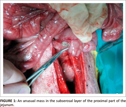
The mass was dissected from the jejunal wall and excised. Grossly, the solitary mass (3 mm in diameter) was well-defined, nonpedunculated, firm, and grey, with a smooth and shiny surface. Upon incision, cystic structures filled with flaky material were observed.
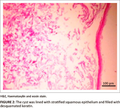
For routine histopathological examination, the mass was fixed in 10% buffered formalin and embedded in paraffin, sectioned at 4 µm, and stained with haematoxylin and eosin (H&E).
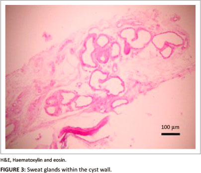
Histologically, the cyst was lined by flattened epithelium consisting of 3-5 layers of squamous cells and its lumen was filled with concentric layers of desquamated keratinous material (Figure 2). Some scattered sebaceous and sweat glands were associated with the cyst wall. No evidence of an inflammatory reaction was observed around the cyst. All these histological features were consistent with a dermoid cyst (Figure 3). The dog healed without complication and the cyst has not recurred 12 months postoperatively.
The majority of dermoid cysts occur on the dorsal midline because of incomplete separation of skin and neural tube during embryonic development, but they may also occur in other locations. Dermoid cysts are sometimes present in the flank instead of along the dorsal midline, possibly as a result of embryogenic defects of adjacent dermatome fusion. In this case, the dog was two years old at the time of presentation. It was not known how long the cyst had been present in the dog but, considering its young age, it was probably a congenital disorder; it had possibly grown in size recently prior to presentation.
Histologically, dermoid cysts are lined with stratified epithelium resembling normal skin with adnexa and filled with keratinous material (Muller, Kirk, && Scott 1983). The cyst usually contains hair, keratin and sebum, and these materials may produce progressive enlargement of the structure so that it becomes clinically apparent (Maxie, Jubb & Kennedy 2007; Schaer 1983).
Dermoid cysts have been classified according to depth of penetration of the sinus. Class I cysts extend from the skin to the supraspinous ligament, class II cysts do not extend as deeply but are connected to the supraspinous ligament by a fibrous band, and class III cysts are similar to class II cysts but have no connecting band to the ligament (Angarano& Swaim 1993; Baker & Thomsett 1990). The present dermoidcyst belonged to class III.
The differential diagnoses of cysts in the abdominal area, such as polyps, foreign bodies and neoplasms, should be considered. It is difficult to diagnose dermoid cysts by imaging only, and histological analysis is essential.
The surgical treatment of a dermoid cyst is straightforward and the only way to cure it involves careful excision of the cyst. The recurrence of these anomalies is due to incomplete surgical excision and is usually evident within one month of surgery. In this case, the cyst has not recurred 12 months postoperatively.
Acknowledgements
The authors would like to acknowledge the reviewers of our drafts and the teaching veterinary hospital of Shahid Bahonar University of Kerman, Iran.
Competing interests
The authors confirmed that they have no financial or personal relationship(s) which may have inappropriately influenced them in writing this article.
Authors' contributions
M.S. (University of Kerman) was the project leader, M.S., R.K.H. (University of Kerman) and O.A. (University of Kerman) were responsible for all clinical procedures. M.A. (University of Kerman) and R.R. (University of Kerman) provided slides. R.KH. performed histopathological assessments and confirmed the intestinal dermoid cyst and E.M. (Kerman University of Medical Sciences) wrote the manuscript.
References
Akhtardanesh, B., Kheirandish, R. & Azari, O., 2012, 'Dermoid cyst in a domestic shorthair cat', Asian Pacific Journal of Tropical Biomedicine 2, 247-249. http://dx.doi.org/10.1016/S2221-1691(12)60051-3 [ Links ]
Angarano, D.W. & Swaim, S.F., 1993, Congenital skin diseases, Academic Press, Philadelphia. [ Links ]
Bailey, T.R., Holmberg, D.L. & Yager, J.A., 2001, 'Nasal dermoid sinus in an American cocker spaniel', Canadian Veterinary Journal 42, 213-215. PMid:11265192, PMCid:1476468 [ Links ]
Baker, K.P. & Thomsett, L.R., 1990, Canine and feline dermatology (1st edn.), Blackwell, Boston. [ Links ]
Burrow, R.D., 2004, 'A nasal dermoid sinus in an English bull terrier', Journal of Small Animal Practice 45, 572-574. http://dx.doi.org/10.1111/j.1748-5827.2004.tb00207.x, PMid:15553197 [ Links ]
Chénier, S., Quesnel, A., Girard, C., 1998, 'Intracranial teratoma and dermoid cyst in a kitten', Journal of Veterinary Diagnostic Investigation 10, 381-384. http://dx.doi.org/10.1177/104063879801000417, PMid:9786533 [ Links ]
Cornegliani, L., Jommi, E. & Vercelli, A.,2001, 'Dermoid sinus in a golden retriever', Journal of Small Animal Practice 42, 514-516. [ Links ]
Maxie, M.G., Jubb, K.V.F., Kennedy, P.C. & Palmer, N., 2007, Jubb, Kennedy and Palmer's pathology of domestic animals, Elsevier Saunders, Edinburgh. PMid:17459839 [ Links ]
Muller, G.H., Kirk, R.W. & Scott, D.W., 1983, Small animal dermatology (3rd edn.), Saunders, Philadelphia. [ Links ]
Schaer, M., 1983, Clinical signs in small animal medicine, Manson, London. PMCid:1066306 [ Links ]
Sturgeon, C., 2008, 'Nasal dermoid sinus cyst in a shih tzu', Veterinary Record 163, 219-20. http://dx.doi.org/10.1136/vr.163.7.219, PMid:18708657 [ Links ]
Tshamala, M. &Moens, Y., 2000, 'True dermoid cyst in a Rhodesian ridgeback', Journal of Small Animal Practice 41,352-353. http://dx.doi.org/10.1111/j.1748-5827.2000.tb03217.x, PMid:11002937 [ Links ]
 Correspondence:
Mehdi Saberi
PO Box 7619813159, Kerman, Iran
Email: mehdi.saberi75@gmail.com
Received: 25 July 2012
Accepted: 26 Mar. 2013
Published: 25 June 2013

