


Services on Demand
Article
 English (pdf)
 Article in xml format
 Article references
Indicators

Related links
-
 Cited by Google
 -
 Similars in Google
Share

 Permalink
Journal of the South African Veterinary Association
On-line version ISSN 2224-9435
Print version ISSN 1019-9128
J. S. Afr. Vet. Assoc. vol.84 n.1 Pretoria Jan. 2013
CLINICAL COMMUNICATION
Corynebacterium pseudotuberculosis associated with otitis media-interna in goats
Rhoda Leask; David J.C. Blignaut; Maria J. Grobler
Department of Production Animal Studies, University of Pretoria, South Africa
ABSTRACT
Corynebacterium pseudotuberculosis or caseous lymphadenitis is a common condition in sheep and goats. Two cases are described involving otitis media-interna and, in one case, cerebellar abscessation. The first case began with otitis externa and progressed to cerebellar abscessation, presumably as a result of C. pseudotuberculosis infection based on the macroscopic appearance of the abscess. The second case of otitis media-interna involved C. pseudotuberculosis with parasitic encephalitis or secondary meningo-encephalitis. Caseous lymphadenitis is a worldwide problem in livestock and also has zoonotic implications. Antimicrobial therapy of abscesses is often unrewarding due to the thick encapsulation of the abscesses and the extremely contagious nature of the organism. Alternative measures of treating this condition must be sought. In flocks or herds where caseous lymphadenitis has been diagnosed, it should be considered as a differential diagnosis for neurological conditions. The potential for spread must be kept in mind when it is suspected to be the cause of otitis in livestock.
Introduction
Otitis media is relatively common, albeit under-reported, in food-producing animals (Bath & De Wet 2000; Coetzer, Thomson & Tustin 1994; Kusiluka & Kambarage 1996; Oliver, Lorenz & Kornegay 1997). This includes lambs (Jensen et al. 1982; Macleod, Wiener & Barlow 1972), especially those that are bottle-fed (Macleod, Wiener & Barlow 1972). Several organisms have been implicated as a cause; however, Corynebacterium pseudotuberculosis seems rarely to be diagnosed as a cause. Otitis media-interna and secondary meningitis associated with C. pseudotuberculosis have been reported in horses (Rand et al. 2012) and cattle (Jensen et al. 1983). Trueperella (Arcanobacterium) pyogenes has been identified as a cause of otitis externa in cats (Billington, Post & Jost 2002).
Cerebral abscesses or pyogranulomas due to a number of bacterial organisms have previously been reported in goats and other species (Altman & Bogokovsky 1973; Amand, Ansley & Johnson 1973; Glass, De Lahunta & Jackson 1993; Moriwaki et al. 1972; Morris, Uzal & Cipolla 2005; Smith & Sherman 1994). Although C. pseudotuberculosis, also known as caseous lymphadenitis (CLA), infection is common in goats (Bath, Van Wyk & Pettey 2005; Luvizotto et al. 2009; Williamson 2001), and has been implicated in causing brain abscesses (Glass, De Lahunta & Jackson 1993), to the knowledge of the authors it has not previously been described as the causative agent of otitis media or interna. Two cases involving otitis are described.
In the first case it is presumed that the otitis externa led to cerebellar abscessation and in the second case it possibly resulted in a secondary meningo-encephalitis (complicated by parasitic involvement).
Clinical cases
Case 1
An eight tooth (approximately four years old) female dwarf Cameroon crossbred goat was presented at the Onderstepoort Veterinary Academic Hospital (OVAH) for pregnancy diagnosis. No behavioural or postural abnormalities were detected at the time of examination. One month later, the goat returned, as she was unsure of her footing. Neurological examination revealed a wide-based stance, but no other abnormalities. Excessive wax build-up was observed on otoscopic examination and the tympanic membrane could not be visualised in the left ear. Swabs were taken and smears made from both ears. These smears were stained with Cams-Quick stain (Kyro-Quick Solution, Kyron laboratories [Pty] Ltd. South Africa). Malassezia was identified on both smears. Due to financial constraints the Malassezia was not identified to a specific species level. The owner was given Epi-otic (Virbac RSA [Pty] Ltd) and instructed to clean the ear in order to visualise the eardrums better and to return with the goat after a few days.
Twoweeks later, the owner returned with the goat, which now showed a slight head tilt to the left, mild ataxia, hypermetria and an intention tremor. A diagnosis of otitis media-interna was made (left ear) and the goat was treated with Clamoxyl® RTU (Pfizer Laboratories [Pty] Ltd. South Africa) 20 mg/kg once daily, Prednisolone 1% Kela (Bayer [Pty] Ltd, South Africa) 1 mg/kg daily and Epi-otic for five days. There was no improvement in the clinical signs after five days of treatment. In order to rule out brain lesions, a computed tomography (CT) scan was performed for academic interest at no additional cost to the owner. Numerous multiplanar reformatting (MPR) and reconstructions were performed and viewed in a soft tissue and bone window with smooth and sharp kernels. A spherical mass 24 mm x 31 mm x 26 mm was seen, beginning just caudal to the external acoustic meatus and extending to the occipital bone in the middle of the cerebellum (Figure 1).
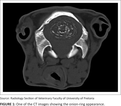
The lesion had a layered appearance with a hyperdense centre and irregular layers of hyperdense tissue interspersed with areas of hypodensity. The remaining tissue had an isodense appearance in keeping with the rest of the brain.
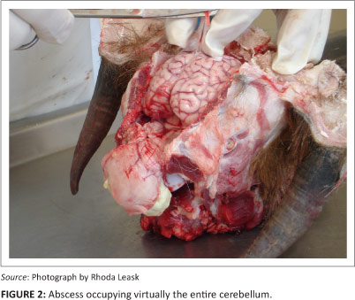
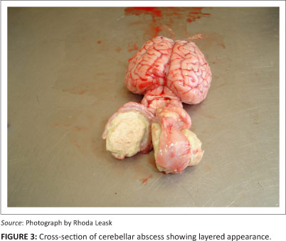
Chronic parasitic migration, C. pseudotuberculosis, calcinosis and mineralised benign neoplasia were considered as differential diagnoses for the lesion seen. Three weeks later, the neurological signs had progressed to falling to the left, a pronounced head tilt and eventual inability to stand. The goat was humanely euthanased by overdose of Euthapent (Kyron laboratories [Pty] Ltd, South Africa). A macroscopic post mortem examination was performed and the lesion was found to be an abscess (Figure 2) that extended towards the left ear canal, with the typical onion-ring appearance of CLA (Figure 3). However, cultures were not performed due to financial constraints.
Case 2
An adult male Boer goat was presented at the OVAH with a history of circling. It had been treated by the owner with 60 ml praziquantel (Brutel, Bayer Animal Health Division, South Africa) the previous day. The goat died and the owner requested a post mortem examination. Abscesses were found in the right ear canal and sub-cutis at the level of the tempero-mandibular joint. Severe meningeal congestion was also present. A single immature cyst of Taenia multiceps was found on histopathological examination. A pure culture of C. pseudotuberculosis was isolated from pus taken from the inner ear. A final diagnosis of otitis media and parasitic encephalitis was made. The possibility that the condition seen in this animal was a result of inner ear infection with secondary meningo-encephalitis could not be ruled out with standard post mortem procedures.
Discussion
Although Malassezia was identified on the smears of the external auditory meatus in the first case, it is unlikely that this was the cause of the otitis. Malassezia is usually an opportunistic pathogen and some initial trauma to the ear or infection by other organisms is usually observed (Pier et al. 2000; Shiota et al. 2009). Jensen et al. (1982) mentioned three possible routes of infection into the middle ear, namely: the external acoustic meatus, the auditive tubes and the haematogenous route. The most common cause of otitis media is otitis externa due to bacterial infections (Oliver, Lorenz & Kornegay 1997); however, otitis media-interna may develop in large animals without a history of otitis externa (Jensen et al. 1982; Oliver, Lorenz & Kornegay 1997). In the first case it is suspected that trauma to the ear canal (perhaps tick bites) may have been the initial port of entry for the organism (presumably C. pseudotuberculosis), although culture is recommended in such cases as other organisms may have a similar appearance.
Computed tomography scanning is not practical in food-producing animals mainly due to the costs involved and specialised equipment required. It is not recommended that all animals that present with neurological signs suggesting vestibular or cerebellar involvement be screened in this manner. However, this case shows that brain involvement should be considered in cases of otitis. Although samples from the cerebellar abscess were not cultured, it was presumed from the typical macroscopic appearance of the cerebellar abscess and the occurrence of the second case that otitis externa involving C. pseudotuberculosis may progress to otitis media-interna and even cerebellar abscessation, and should be considered as a possible cause in neurological cases where CLA has been diagnosed as a flock or herd problem.
Conclusion
It is difficult to treat C. pseudotuberculosis, this is probably due to the nature of the abscesses that are formed (layered and encapsulated) and consequently the penetration required for any antimicrobial agent to be effective. Difficulty in identifying subclinical cases and the ability of the organism to persist in the environment also play a role in its persistence (Williamson 2001). Vaccines are available for infected flocks and it has been suggested that such vaccines may assist not only in prevention of CLA, but also as a treatment and possibly a cure (Prof Ken Pettey, pers. comm., 10/12/2012). More research is required in this field.
Acknowledgement
The authors would like to thank the Pathology Section and the Radiology Section of the Faculty of Veterinary Science, University of Pretoria, for the information regarding post mortem findings and CT scans respectively.
Competing interests
The authors declare that they have no financial or personal relationship(s) that may have inappropriately influenced them in writing this article.
Authors' contributions
R.L. (University of Pretoria) wrote the manuscript and collected the data on the clinical cases, contributed towards the introduction and discussion and references; D.J.C.B. (University of Pretoria) contributed towards the information in the introduction and discussion as well as references; M.J.G. (University of Pretoria) contributed towards the case studies.
References
Altmann, G. & Bogokovsky, B., 1973, 'Brain abscess due to Corynebacterium haemolyticum' Lancet 1, 378-379. http://dx.doi.org/10.1016/S0140-6736(73)90177-3 [ Links ]
Amand, W., Ansley, J. & Johnson, J., 1973, 'Brain abscess in a Barbados sheep', Journal of the American Veterinary Medical Association 163, 562-564. PMid:4742077 [ Links ]
Bath, G.F. & De Wet, J., 2000, Sheep and goat diseases, Tafelberg Publishers Limited, Cape Town. [ Links ]
Bath, G.F., Van Wyk, J.A. & Pettey, K.P., 2005, 'Control measures for some important and unusual goat diseases in southern Africa', Small Ruminant Research 60, 127140. http://dx.doi.org/10.1016/j.smallrumres.2005.06.007 [ Links ]
Billington, S.J., Post, K.W. & Jost, B.H., 2002, 'Isolation of Arcanobacterium (Actinomyces) pyogenes from cases of feline otitis externa and canine cystitis', Journal of Veterinary Diagnostic Investigation 14, 159-162. http://dx.doi.org/10.1177/104063870201400212, PMid:11939339 [ Links ]
Coetzer, J.A.W., Thomson, G.R. & Tustin, R.C. (eds.), Infectious diseases of livestock with special reference to Southern Africa, Oxford University Press, Cape Town. [ Links ]
Glass, E.N., De Lahunta, A. & Jackson, C., 1993, 'Brain abscess in a goat', Cornell Veterinarian 83, 275-282. PMid:8306650 [ Links ]
Jensen, R., Pierson, R.E., Weibel, J.L., Tucker, J.O. & Swift, B.L., 1982, 'Middle ear infection in feedlot lambs', Journal of the American Veterinary Medical Association 181, 805-807. PMid:7141977 [ Links ]
Jensen, R., Maki, L.R., Lauerman, L.H., Raths, W.R., Swift, B.L., Flack, D.E. et al., 1983, 'Cause and pathogenesis of middle ear infection in young feedlot cattle', Journal of the American Veterinary Medical Assocation 182, 967-972. PMid:6853319 [ Links ]
Kusiluka, L. & Kambarage, D., 1996, Diseases of small ruminants: A handbook. Common diseases of sheep and goats in Sub-Saharan Africa, VETAID, Midlothian, viewed 30 October 2012, from www.fao.org/docs/eims/upload/agrotech/1906/DiseasesofSmallRuminants.pdf. [ Links ]
Luvizotto, M.C.R., Carreira, V.S., Ferrari, H.F., Ribeiro, D., Vallim, M.A., Azevedo, V. et al., 2009, 'Brain and lung cryptococcoma and concurrent Corynebacterium pseudotuberculosis infection in a goat: a case report', Journal of Venomous Animals and Toxins including Tropical Diseases 15, On line version, viewed 30 October 2012, from www.scielo.br/scielo.php?script=sci_arttext&pid=S1678-91992009000300015. [ Links ]
Macleod, N.S.M., Wiener, G. & Barlow, R.M., 1972, 'Factors involved in middle ear infection (otitis media) in lambs', Veterinary Record 91, 360-362. http://dx.doi.org/10.1136/vr.91.15.360, PMid:5079245 [ Links ]
Moriwaki, M., Watase, H., Fukumoto, M. & Hyashi S., 1972, 'Exophthalmos due to rete mirabile abscess caused by infection with Corynebacterium pyogenes in cattle', National Institute of Animal Health Quarterly 13, 14-22. [ Links ]
Morris, W.E., Uzal, F.A. & Cipolla, A.L., 2005, 'Pyogranulomatous meningoencephalitis in a goat due to Corynebacterium ulcerans', Veterinary Record 156, 317-318. PMid:15786922 [ Links ]
Oliver, J.E., Lorenz, M.D. & Kornegay, J.N., 1997, Handbook of veterinary neurology, (3rd edn.), W.B. Saunders Company, Philadelphia. [ Links ]
Pier, A.C., Cabañes, F.J., Chermette, R., Ferreiro, L., Guillot, J., Jensen, H.E. et al., 2000, 'Prominent animal mycoses from various regions of the world', Medical Mycology 38, 47-58. PMid:11204164 [ Links ]
Rand, C.L., Hall, T.L., Aleman, M. & Spier, S.J., 2012, 'Otitis media-interna and secondary meningitis associated with Corynebacterium pseudotuberculosis infection in a horse', Equine Veterinary Education 24, 271-275. http://dx.doi.org/10.1111/j.2042-3292.2011.00273.x [ Links ]
Shiota, R., Keneko, T., Yano, H., Takeshita, K., Nishioka, K. & Makimura, K., 2009,'Malassezia ‘®^‹Û‚ÌŠÖ—^‚ª‹^‚í‚ꂽŠOŽ¨“¹‰Š‚ÌŒŸ“¢f' [A study of otitis externa associated with Malassezia]. Nihon Ishinkin Gakkai Zasshi 50, 109-116. http://dx.doi.org/10.3314/jjmm.50.109 [ Links ]
Smith, M.C. & Sherman, D.M., 1994, Goat medicine, Lea & Febiger, Philadelphia. [ Links ]
Williamson, L.H., 2001, 'Caseous lymphadenitis in small ruminants', Veterinary Clinics of North America Food Animal Practice 17, 359-371. PMid:11515406 [ Links ]
 Correspondence:
Rhoda Leask
Private Bag X04
Onderstepoort 0110, South Africa
Email: rhoda.leask@up.ac.za
Received: 12 Dec. 2012
Accepted: 11 April 2013
Published: 07 Aug. 2013

