


Serviços Personalizados
Artigo
 Inglês (pdf)
 Artigo em XML
 Referências do artigo
Indicadores

Links relacionados
-
 Citado por Google
 -
 Similares em Google
Compartilhar

 Permalink
Journal of the South African Veterinary Association
versão On-line ISSN 2224-9435
versão impressa ISSN 1019-9128
J. S. Afr. Vet. Assoc. vol.84 no.1 Pretoria Jan. 2013
CLINICAL COMMUNICATIONS
Clinical, ultrasonography and haematology of aglepristone-induced mid-gestation pregnancy terminations in rabbits
Gözde R. ÖzalpI; Ethem M. TemizelII; Elçin Özocak-BatmazIII
IDepartment of Obstetrics and Gynaecology, Uludağ University, Turkey
IIDepartment of Internal Medicine, Uludağ University, Turkey
IIIDepartment of Surgery, Uludağ University, Turkey
ABSTRACT
Aglepristone is a safe abortifacient in cats, dogs and rabbits. Although no serious side effects have been reported, there is no information available about the effects of the medicine on haematological parameters. For the first time clinical and ultrasonographic features and haematological profiles were evaluated in rabbits treated with aglepristone 15 and 16 days after mating. Ten healthy 10-14 month-old New Zealand White female rabbits were mated with fertile bucks and pregnancies were confirmed by ultrasound 15 days later. Of these, 5 does were treated with aglepristone (test group, n = 5) whilst the remaining five (control group, n = 5) were treated with a saline solution (0.9% NaCl). The treatment dose was 10 mgkg body weight, administered subcutaneously once daily on two consecutive days (day 15 and 16 post mating). Ultrasonographic, clinical and haematological assessments were performed daily. Aglepristone treatment induced embryonic fluid resorptions without foetal death in mid-gestation terminations. Following ultrasonographic and haematological examinations, it was established that aglepristone is a safe abortifacient in rabbits.
Introduction
The efficacy of aglepristone, a progesterone receptor blocker, has previously been investigated in mid-term termination of pregnancy and in the prevention of implantation in rabbits (Özalp et al. 2008; Özalp et al. 2010). Aglepristone is a synthetic steroid with anti-progesterone activity and it is widely used as the first licensed antiprogestin in small animal practice in most European, Latin American and Pacific countries.
Although aglepristone is administered in cats and dogs, there are well-documented side effects. Anorexia, necrosis, itching at injection sites and a decrease in body temperature have been reported (Corrada et al. 2005; Georgiev & Wehrend 2006; Hubler & Arnold 2000). Transient and reversible haematological changes have been reported in 4.5% of dogs after the administration of aglepristone, including neutrophilia, neutropaenia, thrombocytosis, haematocrit variation, lymphocytosis and lymphopaenia (Fieni et al. 1996). However, in rabbits, aglepristone's abortifacient effects and prevention of implantation, without any clinical side effects, even on future mating behaviour, pregnancy and parturition rates, are well documented (Özalp et al. 2008, 2010).
Embryonic or foetal development and placental growth can be assessed by ultrasonographic examinations (Chavatte-Palmer et al. 2008). Pregnancy can be confirmed by means of ultrasonography based on the detection of embryonic vesicles on day 6 of pregnancy and heartbeat of an embryo on day 14 post mating in rabbits (Gutierrez & Zamora 2004; Özalp et al. 2010). Ultrasonography is also an important method for monitoring embryonic or foetal viability and is therefore useful in determining foetal mortality in induced abortions in rabbits.
Haematological examinations, in turn, are used to assess general health and physiological changes in animals. During treatment, routine haematological monitoring is recommended to enable identification and control of any toxic side effects of the medication. Although no serious side effects of aglepristone have been observed clinically, there is no information on haematological changes during aglepristone application in rabbits. This study therefore aimed to describe the clinical features and haematological profiles after aglepristone-induced abortions by monitoring the foetus, foetal membranes and embryonic vesicles ultrasonographically during the abortion process.
Materials and methods
Ten healthy 10-14 month-old New Zealand White female rabbits were used in this study. They were housed individually under daylight conditions at 25 °C - 28 °C. The does were fed a standard commercial dry food daily (5 g/100 g body weight) and given water ad libitum.
Each doe was placed in a cage with a fertile buck for 24 h. In all cases, the first mating was observed within an hour and this day was recorded as day 0 of pregnancy. Pregnancy was confirmed by ultrasonographic detection of gestational sacs and embryonic structures 15 days after mating (5 MHz - 7.5 MHz linear array transducer, Dynamic Imaging MCV-Concept, United Kingdom). Hair on the ventral abdomen was clipped and the does were placed in a standing position for ultrasonographic examination.
Pregnant does were divided randomly into two equal groups. The animals were treated either with aglepristone (Alizin, Virbac, Germany) (test group: n = 5) or with a saline solution (0.9% NaCl) (Baxter, Turkey) (control group: n = 5). Aglepristone and saline solution injections were administered at 10 mgkg body weight subcutaneously once daily on two consecutive days (day 15 and 16 post-mating).
Ultrasonographic examinations were performed from day 15 post-mating until foetal death or abortion occurred. On the first three days after administration, the does were examined ultrasonographically twice daily. The clinical status of the does was checked daily and recorded. In addition to clinical and ultrasonographic evaluation, routine haematology was run on blood samples collected from the ear vein on days 15, 17, 19, 21 and 23 post-mating. The ear vein area was wiped with an alcohol swab and blood samples (2 mL) were drawn into EDTA vacuum tubes using 30 g needles. Using an autoanalyser (Abbot Cell Dyn 3500 Hematology Analyzer, Santa Clara, CA), the samples were analysed for red blood cell (RBC) counts, haemoglobin (Hb) concentration, packed cell volume (PCV), total leukocyte count (TLC) and differential leukocyte count. Erythrocyte indices including mean corpuscular volume (MCV), mean corpuscular haemoglobin concentration (MCHC) and mean corpuscular haemoglobin (MCH) were also reported.
Statistical analysis
Data were expressed as mean ± s.d and analysed using one-way repeated measures analysis of variance, followed by post hoc. Tukey tests. Student's t-tests were used to test the significance of differences between the study and control groups. A p-value < 0.05 was considered significant. All statistical analyses were performed using the Sigma Stat 3.1 for Windows statistical package (Systat Software, Point Richmond, CA). Statistical analysis for food consumption and body temperature was performed with Student's t-test (Minitab). The level of significance was set at p < 0.001.
Ethical considerations
Approval for using the animals was obtained from the ethical committee of the Uludağ University (19.09.2005/3).
Results
Pregnancy was confirmed by ultrasonograpic detection of clear gestational sacs and embryos. on day 15 post-mating. In the test group, the diameter of embryonic sacs was measured as 18.3 mm, 15.7 mm, 20.6 mm, 19.2 mm and 18.7 mm in the respective does (mean = 18.5 mm ± 1.79 mm) before aglepristone was administered. In the control group, the diameter of the embryonic sacs was measured as 19.3 mm, 15.2 mm, 20.4 mm, 17.3 mm and 18.2 mm (mean = 1.1 mm ± 0.77 mm) in the respective does before the saline solution was administered.
Vaginal bleeding and discharge started within 22 h - 51 h (mean = 31.6 h ± 11.3 h) after the initial injection of aglepristone. Once vaginal bleeding had been observed, the first ultrasound examination was performed 37.5 h after the initial application in the rabbits. The diameter of the embryonic sacs were measured as 15.4 mm, 15.6 mm, 15.1 mm, 18.4 mm and 18.1 mm, respectively (mean = 16.5 mm ± 1.59 mm). Ultrasound examination showed the foetal membranes and foetuses with a decreasing amount of foetal fluid on consecutive examinations. Foetal heartbeats were clearly detected although abortion had started. No dead foetuses were observed during the examinations until the abortions were complete. The foetuses were totally expelled within two to four days (mean = 3 days ± 0.7 days). In one doe, the foetuses and foetal membranes and placentas were expelled separately, whilst in the other four the foetuses were expelled with the foetal membranes. There was no gross abnormality in the aborted foetuses observed during necropsy. The mean (± s.d.) crown rump length of the foetuses was 1.77 cm (± 0.16 cm; range = 1.52 cm - 1.96 cm). The mean number of aborted foetuses per doe was 4.2 cm ± 1.09 mm (range: 3-6). Control does remained pregnant and whelped normal litters after a mean pregnancy length of 31.2 ± 0.37 days (range = 30-32 days). Mean litter size was 4.8 ± 1.30 kittens.
Measurements of daily body temperatures showed no statistically significant differences between the two groups. Interestingly, diarrhoea was observed in three does that received aglepristone although there was no change in their diets. Three does exhibited maternal behaviour, including pulling hair, during the abortions, but they did not cannibalise the foetuses'.
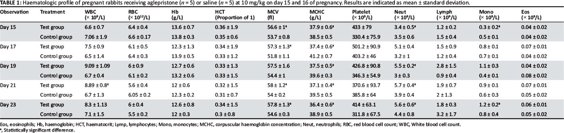
Haematology parameters as measured in the test group and the control group are shown in Table 1. White blood cell counts tended to increase after aglepristone administrations. However, there were significant differences in repeated measurements between day 15 and 21 only in the aglepristone group. In addition, there were significant differences between the test group and the control group for neutrophil counts (p < 0.05) as measured on day 19, day 21 and day 23. There were no statistical differences for RBC count, PCV, Hb or lymphocyte, platelet and eosinophil counts. These parameters were found to be within the respective reference limits. MCV levels were detected on the basal limit in both the study and the control group; significant differences were observed in measurements on days 17, 21 and 23 (p < 0.05). The MCHC levels detected were within the reference limit, although statistical differences were observed on day 17, 19, 21 and 23.
Discussion
Aglepristone is as effective as an abortifacient in rabbits as it is in dogs and cats (Galac et al. 2000; Özalp et al. 2008). All pregnancies terminated within 1-4 days after the first treatment. This observation is in agreement with previous observations in rabbits (Özalp et al. 2008). It was interesting that diarrhoea was observed in some aglepristone-treated rabbits, although neither the diet was changed nor a decrease in food consumption was observed. Özalp et al. (2008) noted a slight decrease in food consumption after treatment, but no information was given about diarrhoea being observed. This clinical problem was the only symptom observed in the test group. There are no reports about the occurrence of diarrhoea in treated dogs and cats. The reason for this pathology remains unclear.
A decrease in body temperature was detected after aglepristone treatment in dogs (Corrada et al. 2005). In our study no differences were recorded in the body temperatures of rabbits. This finding agrees with another study that reported stable body temperature in dogs (Akbas & Kirsan 2007). An influence of aglepristone on the central nervous system was postulated in dogs, but this cannot be speculated on without further studies in rabbits.
An increase in nonspecific immunity after aglepristone treatment (Sugiura et al. 2004) is one of the factors that may accelerate recovery. Blocking the progesterone receptors by aglepristone can affect the progesterone activity in immunological cell function, but its direct influence on these cells remains unknown. In a study reported by Jurka, Faundez and Winnicka (2010), application of aglepristone over seven days caused significant differences in total leukocyte, monocyte, and granulocyte counts.
In the present study, white blood cell counts and neutrophil values tended to increase. This finding could be consistent with infection and it agrees with previous reports of haematological values after aglepristone applications (Jurka et al. 2010). Although the abortion process was completed in four days in all does, the haematological parameters were monitored for 10 more days (until day 23 post-mating), because progesterone concentrations decreased until day 21 (Özalp et al. 2008). The clinical picture returned to normal upon completion of the process.
The ultrasonographic examinations showed that the foetuses were not dead during the abortion; heartbeats were observed at each examination. The effect of aglepristone is possibly due to the resorption of embryonic fluids. The daily ultrasonographic examinations and measurements of embryonic vesicles supported this finding, which corresponds to the plasma progesterone concentrations that were measured in previous studies. The results of these studies showed that aglepristone acted as a progesterone antagonist in the uterus, thereby modifying plasma progesterone concentrations and inducing pregnancy termination without any immediate effects on luteal function. Owing to its high affinity for progesterone receptors, aglepristone might act as an antagonist on uterine receptors at this time (Özalp et al. 2010). The inhibition of progesterone production in the second trimester of human or the third trimester of primate pregnancies has been observed to be followed by a decrease in maternal foetal and amniotic fluid progesterone and preterm labour and delivery (Haluska, Cooka & Novy 1997). The excellent agreement between the decrease of progesterone and the increase of foetal resorption has also been reported in sheep (Nishi et al. 1976). In a study on cats, ultrasonography was performed during abortion in queens after aglepristone applications. It was reported that aborted foetuses were still alive inside the foetal membranes during the examinations (Georgiev & Wehrend 2006). In our study we did not find live aborted foetuses during examinations, but this could support our speculation about the abortion process.
In the present study, resorption of foetal fluids without death after aglepristone injection could be attributed to the disruption of the foeto-endometrial connection as a result of uterus contractions due to luteal function.
The present study showed that administration of aglepristone on day 15 and day 16 of pregnancy induced foetal expulsion through embryonic fluid resorption, without causing foetal death in mid-gestation terminations. Although some statistically significant differences in haematological parameters were detected, the changes were within the reference ranges, indicating that aglepristone is a safe abortifacient.
Acknowledgements
Competing interests
The authors declare that they have no financial or personal relationship(s) that may have inappropriately influenced them in writing this paper.
Authors' contributions
G.R.Ö. (Uludağ University) was the project leader. E.M.T. (Uludağ University) and E.Ö-B. (Uludağ University) were responsible for experimental design. All authors were involved in the experiments: G.R.Ö and E.Ö-B. performed ultrasound examinations and monitored clinical parameters and E.M.T. collected blood samples and performed analysis and statistical calculations. All authors contributed to writing the manuscript.
References
Akbas, M. & Kirsan, I., 2007, 'Termination of early pregnancies in bitches with aglepristone', Indian Veterinary Journal 84, 971-973. [ Links ]
Chavatte-Palmer, P., Laigre, P., Simonoff, E., Chesne, P., Challah-Jacques, M. & Renard, J.P., 2008, 'In utero characterisation of foetal growth by ultrasound scanning in the rabbit', Theriogenology 69, 859-869. http://dx.doi.org/10.1016/j.theriogenology.2007.12.013, PMid:18295873 [ Links ]
Corrada, Y., Garcia, P., De La Sota, P.E., Huzman, M., Landoni, M.F. & Gobello, C., 2005, 'Decrease of body temperature after aglepristone treatment in bitches', Animal Reproduction Science 87, 295-299. http://dx.doi.org/10.1016/j.anireprosci.2004.11.012, PMid:15911178 [ Links ]
Fieni, F., Tainturier, D., Bruyas, J.F., Badinand, F., Berthelot, X., Ronsin, P. et al., 1996, 'Etude clinique d'une anti-hormone pour provoquer l'avortement chez la chienne: l'aglépristone [Clinical study of an anti-hormone to induce abortion in the bitch: aglepristone]', Receuil de Médécine Véterinaire 172, 359-367. [ Links ]
Galac, S., Kooistra, H.S., Butinar, J., Bevers, M.M., Dieleman, S.J., Voorhout, G. et al., 2000, 'Termination of mid-gestation pregnancy in bitches with aglepristone, a progesterone receptor antagonist', Theriogenology 53, 941-950. http://dx.doi.org/10.1016/S0093-691X(00)00241-7 [ Links ]
Georgiev, P. & Wehrend, A., 2006, 'Mid-gestation pregnancy termination by the progesterone antagonist aglepristone in queens', Theriogenology 65, 1401-1406. http://dx.doi.org/10.1016/j.theriogenology.2005.08.011, PMid:16198402 [ Links ]
Gutierrez, H.E. & Zamora, F.M.M., 2004, 'Ultrasonography study of rabbits pregnancy', in A. Climent and A. Blasco (eds.), Proceedings of the 8th World Rabbit Congress, Puebla, Mexico, September 7-10, 2004, pp. 276-280. [ Links ]
Haluska, G.J., Cooka, M.J. & Novy, M.J., 1997, 'Inhibition and augmentation of progesterone production during pregnancy: Effects on parturition in rhesus monkeys.' American Journal of Obstetrics and Gynecology 176, 682-691. [ Links ]
Hubler, M. & Arnold, S., 2000, 'Prevention of pregnancy in bitches with the progesterone antagonist anglepristone (alizine)'. Schweizer Archiv fur Tierheilkunde 142, 381-386. PMid:11008515 [ Links ]
Jurka, P., Faundez, R. & Winnicka, A., 2010, 'Lymphocyte subpopulations in the bitches with pyometra treated with aglepristone'. Bulletin of the Veterinary Institute in Pulawy 54, 669-674. [ Links ]
Nishi, N., Arimura, A., De La Cruz, K.G. & Schally, A.V., 1976, 'Termination of pregnancy by sheep anti-LHRH gamma globulin in rats', Endocrinology 98, 1024-1030. http://dx.doi.org/10.1210/endo-98-4-1024, PMid:58780 [ Links ]
Özalp, G.R., Çalişkan, Ç., Seyrek-Intaş, K. & Wehrend, A., 2010, 'Effects of the progesterone receptor antagonist aglepristone on implantation administered on days 6 and 7 after mating in rabbits', Reproduction in Domestic Animals 45, 505-508. http://dx.doi.org/10.1111/j.1439-0531.2008.01282.x, PMid:19019074 [ Links ]
Özalp, G.R., Seyrek-Intaş, K., Çalişkan, Ç. & Wehrend, A., 2008, 'Mid-gestation pregnancy termination in rabbits by the progesterone antagonist aglepristone', Theriogenology 69, 1056-1060. http://dx.doi.org/10.1016/j.theriogenology.2008.01.016, PMid:18377972 [ Links ]
Sugiura, K., Nishikawa, M., Ishiguro, K., Tajima, T., Inaba, M., Torii, R. et al., 2004, 'Effect of ovarian hormones on periodical changes in immune resistance associated with estrous cycle in the beagle bitch' Immunobiology 209, 619-627. http://dx.doi.org/10.1016/j.imbio.2004.09.003, PMid:15638130 [ Links ]
 Correspondence:
Gözde Özalp
Doğum ve Jinekoloji Anabilim Dalı Uludağ Universitesi
16059 Görükle, Turkiye
rgozalp@gmail.com
Received: 26 Aug. 2011
Accepted: 28 Jan. 2013
Published: 03 May 2013

