


Serviços Personalizados
Artigo
 Inglês (pdf)
 Artigo em XML
 Referências do artigo
Indicadores

Links relacionados
-
 Citado por Google
 -
 Similares em Google
Compartilhar

 Permalink
Journal of the South African Veterinary Association
versão On-line ISSN 2224-9435
versão impressa ISSN 1019-9128
J. S. Afr. Vet. Assoc. vol.83 no.1 Pretoria Jan. 2012
CASE REPORT
Rhabdomyosarcoma in a terrestrial tortoise (Geochelone nigra) in Nigeria: A case report
Oghenemega D. EyarefeI; Richard E. AntiaII; Cecilia O. OguntoyeI; Olusoji O. AbiolaIII; Olugbenga O. AlakaII; John. O. OgunsolaII
IDepartment of Veterinary Surgery and Reproduction, University of Ibadan, Nigeria
IIDepartment of Veterinary Pathology, University of Ibadan, Nigeria
IIIDepartment of Veterinary Medicine, University of Ibadan, Nigeria
ABSTRACT
A skeletal muscle tumour (rhabdomyosarcoma) was diagnosed in a 4-year-old captive female terrestrial tortoise (Geochelone nigra) weighing 7 kg presented at the Veterinary Teaching Hospital, University of Ibadan, Ibadan, Nigeria. The tumour was located at the anterior right portion of the body and ventral to the carapace. The location of the tumour prevented the tortoise from extending its head from the body. The tumour was a sessile, smooth white mass, with a soft myxomatous consistency. The histological features that were diagnostic of rhabdomyosarcoma included a sparse population of haphazardly arranged spindle-shaped cells within a homogenous matrix (anisocytosis), occasional tumour giant and binucleate cells, and some well differentiated myofibrils with cross striations within the cytoplasm. The paucity of information on tumours in the land tortoise was the reason for this report, which appears to be the first report of rhabdomyosarcoma in the tortoise.
Introduction
There are several species of giant tortoises. The giant tortoises are amongst the approximately 290 species of freshwater terrapins, marine turtles and terrestrial tortoises existing today in diverse habitats in mostly tropical and subtropical regions globally (Raphael 2003). They are listed by the Convention on International Trade in Endangered Species of Wild Fauna and Flora (CITES), as one of the 166 chelonian species in danger of extinction because of habitat degradation, human encroachment, predation and diseases (Hatt 2008). The most predominant species of giant tortoises are Galapagos tortoises (Geochelone nigra), with an estimated population of 12 000-15 000 (Schramm 1998) and Seychelles tortoises (Aldabrachelys hololissa), numbering approximately 100 000 (Bourn et al. 1999).
Reports of established disease conditions in tortoises remain quite scarce (Hatt 2008; McArthur 2004). Common diseases in captive tortoises are associated with husbandry, nutrition and infectious agents, and include bite trauma (Donoghue 2006), respiratory infections of viral, bacterial, mycotic and parasitic origin (Murray 1996; O'Malley 2005), and non-infectious metabolic diseases of nutritional and renal origin that are predominant in growing giant tortoises and manifest in stages from hypomineralisation to fibrous osteodystrophy of bones and pyramiding of the carapace (Hauser, Mettler & Honegger 1977).
Tumours are rare in chelonians, with most of the cases reported in turtles rather than tortoises. In a study based on nearly a decade of research, the occurrence of tumours in chelonians had a frequency of 2.7%, with tortoises having only 1.4%, whilst the frequency of occurrence in fresh water turtles was as high as 3.2% (Garner, Hernandez-Divers & Raymond 2004). In another study encompassing a much longer period and derived from 490 cases, the frequency of tumour occurrence in chelonians was 1.2% with all the neoplasms seen in turtles (Sykes & Trupkiewicz 2006). Apart from the documented incidence of fibropapillomatosis in the green turtle and some other turtle species across the globe (Balazs et al. 2000; Herbst 1994) and a case of mast cell tumour reported in a land tortoise (Santoro et al. 2008), neoplastic conditions have rarely been documented in tortoises. In this paper a case of rhabdomyosarcoma in a captive tortoise in Nigeria is reported, which to the knowledge of the authors is the first documentation of this tumour in a tortoise.
Case report
A 4-year-old female land tortoise (G. nigra) weighing 7 kg was presented at the Veterinary Teaching Hospital, University of Ibadan, Ibadan, Nigeria, with a complaint of a swelling on the cranio-ventral aspect of the carapace which prevented the tortoise from extending its head from the body to feed or drink (Figure 1). Physical examination revealed a mass, 5 cm in diameter, located at the right cranio-ventral aspect of the carapace, and covering the head orifice, with a small space remaining for breathing. A fine needle aspiration biopsy of the growth was poorly cellular and therefore not sufficiently diagnostic. A survey radiograph showed increased radio- opacity in the area of the tumour but the mass could not be delineated from the carapace due to a summation effect (Figure 2).


Surgical management
Palliative surgery was performed to reduce the tumour mass and to enable the tortoise to project the head out of the carapace to eat and drink, as well as to obtain a biopsy sample (Figure 3) for histopathology. The procedure was done under local anaesthesia with 2% lidocaine (Mighty Pharma, India) by local infiltration. Surgery was successful, as the animal was able to extend its head from the carapace to eat after the intervention (Figure 4).


Histopathology
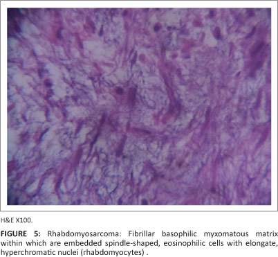
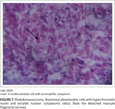
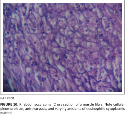
The growth was non-encapsulated and poorly demarcated (Figure 5). The ground substance was generally fibrillar and basophilic (myxoid matrix), (Figures 5 and 6) but with the areas of rhabdomyocytic differentiation appearing eosinophilic (Figures 7 and 8). The degree of cellularity varied in different parts of the tumour, being highest in the less differentiated areas. Isolated and interlacing bundles of muscle fibres exhibiting distinct cross striations were visible (Figure 8). The neoplastic cells were round to oval or spindle-shaped, and showed marked pleomorphism. The cytoplasm of these cells was uniformly eosinophilic, or contained eosinophilic material (Figures 9 and 10). Each cell contained a single, eccentric, round to elongated, hyperchromatic nucleus (Figure 9) with one or two fairly distinct nucleoli. There was remarkable anisokaryosis. Some mitotic cells were visible. A few binucleate and multinucleated giant cells were occasionally encountered (Figure 7). There were scattered inflammatory foci consisting of mononuclear cell infiltration. Some tissue necrosis was present, but haemorrhages were not observed. On the basis of these histopathological features, a diagnosis of rhabdomyosarcoma was made.


Discussion
The anaesthetic protocol adopted for the management of this case was determined by the condition of the animal. The tortoise's head was trapped by the tumour mass within the head orifice with only a small opening available for breathing (Figure 1). This precluded the need for sedation, or use of general anaesthesia, which would have precipitated life threatening hypoxia due to the low oxygen tension within the head orifice and the expected reduced ability of the animal to inhale an adequate amount of atmospheric oxygen under sedation or general anaesthesia (Hall, Clark & Trim 2001). The lignocaine local infiltration provided adequate intra-operative and immediate postoperative analgesia. The tortoise had an uneventful recovery, evidenced by the extension of its head to eat and drink following surgery (Figure 2).
Tortoises form part of the collections of most wildlife parks and zoological gardens. Maintenance of wild animals in captivity tends to be associated with many disease problems. Most of the reported disease conditions, especially tumours in reptiles, have been observed to occur in those animals in captivity (Cooper, Jackson & Harshbarger 1983; Cowan 1968). The relocation of the animals from their natural habitat to urban parks often encourages their continuous exposure to environmental pollutants, some of which are carcinogens. Inbreeding amongst animals in captivity also encourages the expression of lethal cancer genes in the offspring (Buckley, Hulse & Keep 1980).
Neoplasia is extremely rare in reptiles, especially in land tortoises (Sykes & Trupkiewicz 2006). Striated muscle tumours are quite infrequent in any animal species (Pulley & Stannard 1990). As far as the authors could determine, only one case of rhabdomyoma has been reported previously, in a male pine snake (Ball 1945).
Rhabdomyosarcomas are malignant tumours of striated muscle, and are thought to arise from embryonic mesenchyme (Cooper & Valentine 2002). They are commonly associated with a viral or chemical aetiology (Glaister 1981). The present tumour appeared to be an isolated and spontaneous case, as there were other tortoises of the same species but different ages living in the same environment. Besides, review of the management history of the tortoise did not implicate exposure of the animals to any known carcinogens in the captive environment.
Although the majority of neoplasms are seen in older animals, rhabdomyosarcomas occur most often in children (Ellison & Parham 2006) and younger animals (Van Vleet & Valentine 2006). Since tortoises only reach sexual maturity between 15 and 20 years of age (Raphael 2003), this 4-year-old tortoise is undoubtedly a juvenile. Moreover, its small size confirms that it is a young tortoise.
Rhabdomyosarcomas (RMS) in humans are often classified, based on histopathological features, as embryonal RMS (with botryoid RMS as a variant), alveolar RMS, and pleomorphic RMS. Embryonal rhabdomyosarcomas occur mostly in the head and neck regions of young animals, and the histological features include primitive cells that may be round, myoblastic, or myotubular ('strap cells'), with bi- or multinucleation; the tumour can, however, be composed of all these different cell types (Van Vleet & Valentine 2006). The age-old criterion for confirmation of a diagnosis is the identification of cross striations in tumour cells. Nevertheless, many confirmed rhabdomyosarcomas lack demonstrable cross striations (Cooper & Valentine 2002).
Conclusion
Tumours are rare in tortoises. This is a case of rhabdomyosarcoma, whose confirmatory diagnosis was based on the presence of cross striations, which to the knowledge of the authors is the first documentation of the tumour in a tortoise.
Acknowledgements
Competing interests
The authors declare that they have no financial or personal relationship(s) which may have inappropriately influenced them in writing this paper.
Authors' contributions
O.D.E. (University of Ibadan) was the surgeon, R.E.A. (University of Ibadan) was the chief pathologist who interpreted the slides from the processed tumour tissue, C.O.O. (University of Ibadan) was the anaesthetist; O.O.A. (Department of Veterinary Medicine, University of Ibadan) was the referring clinician, O.O.A. (Department of Veterinary Pathology, University of Ibadan) and J.O.O. (University of Ibadan) were the supporting pathologists who processed the tumour tissues, O.D.E. (University of Ibadan) wrote the manuscript and C.O.O. (University of Ibadan) is the corresponding author.
References
Balazs, G.H., Murakawa, S.K.K., Ellis, D.M. & Aguirre, A.A., 2000, 'Manifestation of fibropapillomatosis and rates of growth of green turtles at Kaneohe Bay in the Hawaiian Islands', in F.A. Abreu-Grobois, R. Briseno-Duenas, R. Márquez & L. Sarti (eds.), Proceedings of the 18th Annual Symposium on Sea Turtle Biology and Conservation, Mazatlán, Sinaloa, Mexico, U.S. Department of Commerce, NOAA Technical Memorandum NMFS-SEFSC 436, March 3-7, 1998, pp. 112-113. [ Links ]
Ball, H.A., 1945, 'Melanosarcoma and rhabdomyoma in two pine snakes (Pituophis melanoleucus)', Cancer Research, 134-138. [ Links ]
Bourn, D.M., Gibson, C., Augeri, D., Wilson, C.J., Church, J. & Hay, S.I., 1999, 'The rise and fall of the Aldabran giant tortoise population', Proceedings of the Royal Society London, Series B 266(1424), 1091-1100. http://dx.doi.org/10.1098/rspb.1999.0748, PMid:10406128 [ Links ]
Buckley, P., Hulse, E.V. & Keep, B.M., 1980, 'An inbred strain of rat with a high incidence of squamous-cell carcinomas of the mouth', British Journal of Cancer 41, 295-301. http://dx.doi.org/10.1038/bjc.1980.42, PMid:7370169 [ Links ]
Cooper, B.J. & Valentine, B.A., 2002, 'Tumors of muscle', in D.J. Meuten (ed.), Tumors in domestic animals, 4th edn., pp. 343-345, Blackwell, Ames. http://dx.doi.org/10.1002/9780470376928.ch6 [ Links ]
Cooper, J.E., Jackson, O.F. & Harshbarger, J.C., 1983, 'A neurilemmal sarcoma in a tortoise (Testudo hermanmy, Journal of Comparative Pathology 39, 541-545. http://dx.doi.org/10.1016/0021-9975(83)90060-9 [ Links ]
Cowan, D.F., 1968, 'Diseases of captive reptiles', Journal of the American Veterinary Medical Association 153, 848. PMid:5692920 [ Links ]
Donoghue, S., 2006, 'Nutrition', in D.R. Mader (ed.), Reptile medicine and surgery, 2nd edn., pp. 251-298, Saunders Elsevier, St. Louis, Missouri. http://dx.doi.org/10.1016/B0-72-169327-X/50022-5 [ Links ]
Ellison, D.A. & Parham, D.M., 2006, 'Rhabdomyosarcomas in adults and children: An update', Archives of Pathology and Laboratory Medicine 130, 1454-1465. PMid:17090187 [ Links ]
Garner, M.M., Hernandez-Divers, S.M. & Raymond, J.T., 2004, 'Reptile neoplasia: A retrospective study of case submission to a specialty diagnostic service', Veterinary Clinics of North America, Exotic Animal Practice 7, 653-671. http://dx.doi.org/10.1016/j.cvex.2004.04.002, PMid:15296868 [ Links ]
Glaister, J.R., 1981, 'Rhabdomyosarcoma in a young rat', Laboratory Animals 15, 145146. http://dx.doi.org/10.1258/002367781780959062, PMid:7278117 [ Links ]
Hall, L.W., Clark, K.W. & Trim, C.M., 2001, Veterinary anaesthesia, 10th edn., W.B. Saunders, London, New York, Toronto. [ Links ]
Hatt, J.M., 2008, 'Raising giant tortoises', in M.E. Fowler & R.E. Miller (eds.), Zoo and wild animal medicine current therapy, 6th edn., pp. 144-153, Saunders Elsevier, St. Louis. http://dx.doi.org/10.1016/B978-141604047-7.50021-X [ Links ]
Hauser, B., Mettler, F. & Honegger, R.E., 1977, 'Disorders of bone metabolism in Seychelles giant tortoises (Testudo [Geochelone] gigantea)', (German), in R. Ippen & H.-D. Schroder (eds.), Erkrankungen der Zootiere, Verhandlungsbericht der XIX. Internationalen Symposiums uber die Erkrankungen der Zootiere, Poznan, 18-22 May, 1977, pp. 121-125. [ Links ]
Herbst, L.H., 1994, 'Fibropapillomatosis of marine turtles', Annual Review of Fish Diseases 4, 389-425. http://dx.doi.org/10.1016/0959-8030(94)90037-X [ Links ]
McArthur, S., 2004, 'Infectious agents', in S. McArthur, R. Wilkinson & J. Meyer (eds.), Textbook of medicine and surgery of tortoises and turtles, pp. 307-377, Blackwell Science, Oxford. http://dx.doi.org/10.1002/9780470698877.ch2 [ Links ]
Murray, M.J., 1996, 'Pneumonia and normal respiratory function', in D.R. Mader (ed.), Reptile medicine and surgery, 1st edn., pp. 396-405, W.B. Saunders Company, Philadelphia. [ Links ]
O'Malley, B., 2005, Clinical anatomy and physiology of exotic species, Elsevier Saunders, London. [ Links ]
Pulley, L.T. & Stannard, A.A., 1990, 'Tumors of the skin and soft tissues', in J.F. Moulton (ed.) Tumors in domestic animals, p. 31, University of California Press, Berkeley. [ Links ]
Raphael, B.L., 2003, 'Chelonians (turtles, tortoises)', in M.E. Fowler (ed.), Zoo and wild animal medicine, 5th edn., pp. 48-58, Saunders Elsevier, St Louis. [ Links ]
Santoro, M., Stacy, B.A., Morales, J.A., Gastezzi-Arias, P., Landazuli, S. & Jacobson, E.R., 2008, 'Mast cell tumor in a Giant Galapagos tortoise (Geochelone nigra vicina)', Journal of Comparative Pathology 138, 156-159. http://dx.doi.org/10.1016/j.jcpa.2007.11.004, PMid:18308330 [ Links ]
Schramm, B., 1998, 'The Galapagos giant tortoise (Geochelone nigra) (German), Reptilia 3, 20-25. [ Links ]
Sykes, J.M. & Trupkiewicz, J.G., 2006, 'Reptile neoplasia at the Philadelphia Zoological Garden, 1901-2002', Journal of Zoo and Wildlife Medicine 37, 11-19. http://dx.doi.org/10.1638/04-112.1, PMid:17312806 [ Links ]
Van Vleet, J.F. & Valentine, B.A., 2006, 'Muscle and tendon', in M.G. Maxie (ed.), Jubb, Kennedy, and Palmer's pathology of domestic animals, 5th edn., vol. 1, pp. 273274, Saunders Elsevier, Philadelphia. [ Links ]
 Correspondence to:
Cecilia Oguntoye
Department of Veterinary Surgery and Reproduction
University of Ibadan, Nigeria
wumcel06@yahoo.com
Received: 15 May 2012
Accepted: 13 Oct. 2012
Published: 30 Nov. 2012

