


Services on Demand
Article
 English (pdf)
 Article in xml format
 Article references
Indicators

Related links
-
 Cited by Google
 -
 Similars in Google
Share

 Permalink
Journal of the South African Veterinary Association
On-line version ISSN 2224-9435
Print version ISSN 1019-9128
J. S. Afr. Vet. Assoc. vol.82 n.1 Pretoria Jan. 2011
CLINICAL COMMUNICATION KLINIESE MEDEDELING
Uterine adenocarcinoma with transcoelomic metastases in breeder hens (Gallus domesticus)
D G BwalaI; N M DuncanII,*; S P R BisschopI
IPoultry Reference Centre, Faculty of Veterinary Science, University of Pretoria, Private Bag X04, Onderstepoort, 0110 South Africa
IIPathology Department, Faculty of Veterinary Science, University of Pretoria, Private Bag X04, Onderstepoort, 0110 South Africa
ABSTRACT
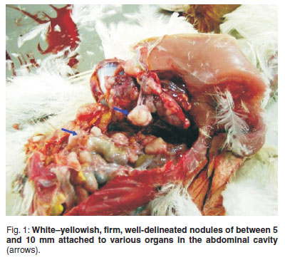
Hens involved in a Newcastle disease study were euthanased at regular intervals according to a designed protocol. Of these, 7.14 % (n = 42) of the 82-week-old specific pathogen-free breeder hens were found to have well-delineated firm white to yellowish nodules of varying sizes in the abdominal cavity. Histologically, the nodules were identified as an adenocarcinoma originating in the uterus. Transcoelomic spread was evidenced by the presence of similar neoplastic cells embedded in the serosa and outer longitudinal muscle layer of the intestines as well as the liver.
Keywords: adenocarcinoma, hens, uterine tumour.
INTRODUCTION
Tumours in commercial poultry are mostly caused by infection with avian herpes, leukosis and retroviruses3 and thus most studies on avian tumours have been focused on those with viral aetiology, probably due to their economic importance and also because they serve as a model for human cancer studies10.
Adenocarcinomas in poultry are among the groups of tumours of unknown aetiology, and have been reported by several veterinary pathologists and reviewed12.
The term adenocarcinoma9 or peritoneal carcinomatosis13 has been applied to tumours whose origin cannot be ascertained but that spread widely over the peritoneum and other visceral organs and are frequently observed in aged hens8. Spontaneous neoplasms or adenocarcinomas affecting various organs have been reported in specific pathogen-free (SPF) hens4, 5 and most of these tumours are thought to originate in the oviduct or ovary2,6,11. A study in Irish abattoirs reported 419 birds with nodules, 80.4 % of which were tumours, and 71.8 % of the tumours were adenocarcinoma of the intestine and reproductive tract15. A prevalence of 5-81 % for oviductal adenocarcinomas in end-of-lay hens7and 92.9 % for ovarian and oviduct adenocarcinomas1 in 305 4-year-old layers has been reported1.
Generally, information on the incidence of nonviral tumours in commercially raised chickens and turkeys is limited, probably due to their shorter life span than that required for the development of such tumours10,12. Also, there are few reports of tumours that are not virally induced because structured surveys are not conducted to assess the situation as they are generally perceived as incidental conditions. Nonviral tumours are also probably considered to be of less economic importance12. The present communication reports cases of uterine adecarcinoma in SPF breeder hens (Gallus domesticus ) encountered during an experimental virus challenge study.
MATERIALS AND METHODS
Forty-two commercial Hyline Brown layers (52 weeks old) and 42 SPF White Leghorn breeder hens (82 weeks old) selected for a Newcastle disease vaccine trial were randomly assigned into 8 groups. Birds were then vaccinated with La Sota vaccine after acclimatising for 2 days and subsequently challenged 10 days later with a virulent isolate of Newcastle disease virus belonging to lineage 5d/VIId. Eight birds (4 SPF and 4 commercial) were euthanased on days 2, 4, 6, 8 and 10 post-vaccination and post-challenge. The euthanased birds were necropsied and various organs required for the trial were collected. The organs with nodules encountered at necropsy were also sampled. Gross findings were recorded and samples of the organs including those with nodules were preserved in 10 % neutral buffered formalin, trimmed and then routinely processed and stained with standard haematoxylin and eosin (H&E) and then examined by light microscopy.
RESULTS
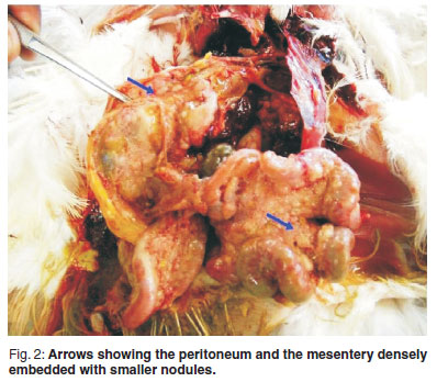
All the birds were in a relatively good bodily condition throughout the trial. Two of the hens euthanased on day 2 post-vaccination and another 1 euthanased on day 6 post-challenge, however, had prominent keel bones and shrunken breast muscles. The coelomic cavity of all 3 hens was filled with firm white to yellowish growths of varying sizes. The nodules were found on the oviduct, pancreas, liver, heart, spleen, proventriculus, intestines and the mesentery (Figs 1 & 2). There were adhesions between the various organs and especially between the intestinal loops. The intestinal mesentery was also extensively involved resulting in clumping of the intestine. The follicles were inactive with no mature or developing ova and the oviducts were small, flaccid and non-functional.
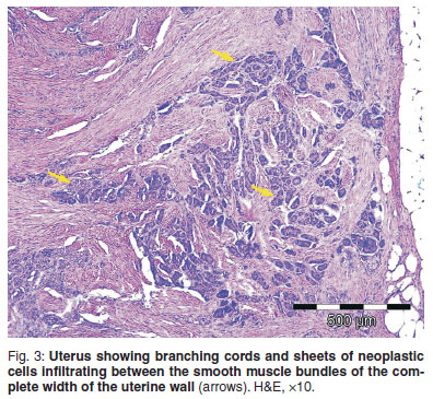
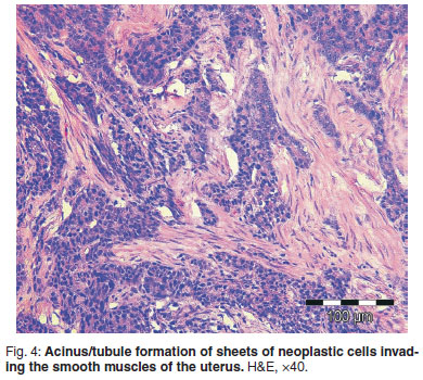
Microscopically, the uterine changes were characterised by transmural branching cords and sheets of neoplastic cells. Indications of acinus/tubule formation were also present within these aggregates of neoplastic cells (Figs 3 & 4). The neoplastic cells had medium to large vesicular nuclei with a single nucleolus and moderate amounts of grey, finely granular cytoplasm. The mitotic index was low. Marked fibroplasia (scirrhous reaction) was found surrounding the acinar structures (Figs 3 & 4). Transcoelomic spread was evidenced by the presence of similar neoplastic cells embedded in a prominent fibrous stroma abutting the serosa and outer longitudinal muscle layer of the intestines as well as on the capsule of the liver. The glandular origin of the neoplasm was much more evident in the metastatic foci, with tubule and acinus formation being prominent. There was no evidence of neoplasia in the ovarian and magnal tissues.
DISCUSSION
These cases were diagnosed as uterine adenocarcinoma with transcoelomic metastases based on the histological morphology of the uterine neoplasm. The neoplasm was characterised by branching cords and sheets of cells infiltrating between the smooth muscle bundles of the uterine wall. These changes were absent in the magnal tissue and ovary. Metastatic abdominal adenocarcinomas may originate from either the ovary or the oviduct and their differentiation may be difficult11. In these cases the absence of neoplastic cells in the ovarian and magnal tissues confirms the site of origin as uterine in nature. According to a previous report12, most adenocarcinomas of the oviduct originate in the upper magnal portion of the oviduct, but occasional cases of adenocarcinomas do occur in the shell gland (uterus) and infundibulum. Although this was not a structured research study, the prevalence of 7.14 % recorded agrees with previous findings of a prevalence rate of 5-81 % for oviductal adenocarcinoma7.
These adenocarcinomas were encountered in 82-week-old White Leghorn hens, which tallies with the reviewed published findings1that ovarian and oviductal adenocarcinomas are among the most frequently encountered tumors in older White Leghorn hens. The SPF birds used in the study had been laying eggs for approximately 62 weeks and prolonged reproductive activity and ageing has been suggested to have a direct relationship to the occurrence of oviductal adenocarcinomas in fowl4,15, presumably because the high egg production is associated with continuous gonadotrophin production, resulting in the excess stimulation of oestrogen-sensitive target organ epithelium14.
In conclusion, neoplastic diseases, irrespective of their aetiology or the organ affected, cause economic loss and therefore the recording and reporting of these diseases remain vital.
ACKNOWLEDGEMENT
We are grateful to the Poultry Reference Centre, Faculty of Veterinary Science, University of Pretoria, for funding the diagnostic bills and Dr Kerstin Junker for her assistance with the micrograph pictures.
REFERENCES
1. Alfonso M, Adochiles L, Hendrickson V M, Carver D K, Rodriguez G C, Barnes H J 2005 Metastatic adenocarcinoma in the lungs of older laying hens. Avian Diseases 49: 430-432 [ Links ]
2. Campbell J G 1969 Tumours of the fowl. Lippincott, Philadelphia, PA: 180-182 [ Links ]
3. Fadly A M 2003 Neoplastic diseases. In Saif Y M, Barnes H J, Glisson J R, Fadly A M, McDougald L R, Swayne D E (eds) Diseases of poultry (11th edn). Iowa State Press, Ames, IA: 405-407 [ Links ]
4. Fredrickson T N 1987 Ovarian tumors of the hen. Environmental Health Perspectives 73: 35-51 [ Links ]
5. Fredrickson T N, Helmboldt C F 1991 Tumors of unknown etiology. In Calnek B W, Barnes H J, Beard C W, Reid W M, Yoder H W (eds) Diseases of poultry (9th edn). Iowa State University Press, Ames, IA: 459-470 [ Links ]
6. Giles J R, Shivaprasad H L, Johnson P A 2004 Ovarian tumor expression of an oviductal protein in the hen: a model for human serous ovarian adenocarcinoma. Gynecologic Oncology 95: 530-533 [ Links ]
7. Goodchild W M 1969 Adenocarcinoma of the oviduct in laying hens. Veterinary Record 84: 122 [ Links ]
8. Haritani M, Kajigaya H, Akashi T, Kamemura M, Tanahara N, Umeda M, Sugiyama M, Isoda M, Kato C 1984 A study on the origin of adenocarcinoma in fowls using immunohistochemical technique. Avian Diseases 28: 1130-1134 [ Links ]
9. Ilchmann G, Bergmann V 1975 Histologische und elektronenmikroskopische Untersuchungen zur Adenokarzinomatose der Legehennen. Archiv für Experimentelle Veterinärmedizin 29: 897-907 [ Links ]
10. Kumar R, Nair M G, Lakkawar A W, Varshney K C 2004 Ovarian adenocarcinoma in a guinea fowl (Numida meleagris ) - a case report. Veterinarski Arhiv 74: 245-249 [ Links ]
11. Reece R L 1997 Tumours of unknown etiology. In Calnek B W (ed.) Diseases of poultry (10th edn). Iowa State University Press, Ames, Iowa: 459-463 [ Links ]
12. Reece R L 2003 Neoplastic diseases. Other tumours of unknown etiology. In Saif Y M, Barnes H J, Glisson J R, Fadly A M, McDougald L R, Swayne D E (eds) Diseases of poultry (11th edn). Iowa State Press, Ames, IA: 541-564 [ Links ]
13. Sokkar S M, Mohammed M A, Zubaidy A J, Mutalib A 1979 Study of some non-leukotic avian neoplasms. Avian Pathology 8: 69-75 [ Links ]
14. Swarbrick O, Campbell J G, Berry D M 1968 An outbreak of oviduct adenocarcinoma in laying hens. Veterinary Record 82: 57-59 [ Links ]
15. Talebi A, Collins J D, Dodd K 1993 Nodular lesions found in Irish poultry during veterinary inspection at poultry meat plants. Avian Pathology 22: 715-724 [ Links ]
Received: September 2009.
Accepted: March 2011.
* Author for correspondence. E-mail: neil.duncan@up.ac.za

