


Serviços Personalizados
Artigo
 Inglês (pdf)
 Artigo em XML
 Referências do artigo
Indicadores

Links relacionados
-
 Citado por Google
 -
 Similares em Google
Compartilhar

 Permalink
Journal of the South African Veterinary Association
versão On-line ISSN 2224-9435
versão impressa ISSN 1019-9128
J. S. Afr. Vet. Assoc. vol.81 no.1 Pretoria Jan. 2010
ARTICLE ARTIKEL
Seroprevalence of bovine brucellosis in trade cattle slaughtered in Ibadan, Nigeria, from 2004-2006
S I B CadmusI,*; H K AdesokanI; B O AdedokunII; J A StackIII
IDepartment of Veterinary Public Health and Preventive Medicine, University of Ibadan, Ibadan, Nigeria
IIDepartment of Epidemiology, Medical Statistics & Environmental Health, University of Ibadan, Nigeria
IIIDepartment of Statutory and Exotic Bacteria, Veterinary Laboratories Agency, New Haw, Addlestone, Surrey KT15 3NB, United Kingdom
ABSTRACT
A seroprevalence study was carried out among trade cattle slaughtered at Bodija Municipal Abattoir, Ibadan (southwestern Nigeria) over a period of 3 consecutive years from 2004 to 2006 with a view to determining the breed, sex and age distribution in the seropositivity of bovine brucellosis. In total, 1642 animals were examined for antibodies to Brucella abortus using the Rose Bengal test. Seroprevalences of 6.00 %, 6.17 % and 5.31 % were obtained in the years 2004, 2005 and 2006, respectively but a decrease in 2006 shows no significant difference (P > 0.05). The role of the breed (P > 0.05), sex (P > 0.05) and age (P > 0.05) in the occurrence of the infection was not statistically significant at 5 %, although higher rates were obtained for females and older animals. The trend in the disease over the 3-year period showed that it is endemic in trade cattle slaughtered in Ibadan and the public health implications of this are discussed.
Keywords: brucellosis, cattle, epidemiology, Nigeria, seroprevalence, zoonosis.
INTRODUCTION
Brucellosis is a disease that causes serious economic losses to the animal industry and extensive morbidity in humans and thus constitutes an important public health problem that is recognised worldwide. Bovine brucellosis is a zoonotic disease transmitted from cattle to humans by ingestion of infected food products, direct contact with an infected animal or inhalation of infected aerosol. Transmission via aerosol is very efficient given the relatively low concentrations of organisms (as few as 10-100 bacteria) needed to establish infection in humans and this route of infection has serious health and safety implications. This has brought renewed attention to this old disease14. As observed in most abattoirs in Nigeria, adequate personnel and meat inspectors are lacking and safety precautions are not adhered to by livestock workers and butchers. While the disease has been eradicated in many countries by implementing expensive long-term control programmes, the occurrence is increasing in developing countries21. Bovine brucellosis is widespread in Africa, where it remains one of the most important zoonotic diseases10. Recent investigations have shown that bovine brucellosis is endemic in Nigeria based on serological studies5,8,9,12,17.
Trade cattle constitute the majority of animals slaughtered in Ibadan, south-western Nigeria, and different studies have reported the seroprevalence of bovine brucellosis in these animals8,12. Data on the yearly trend of the disease in trade animals in Ibadan are lacking, and would provide valuable information for mapping out control and eradication strategies for the disease. We therefore designed a study to monitor the trend of bovine brucellosis in trade cattle slaughtered in Ibadan over a 3-year period with the aim of determining whether age, breed and sex play a role in the seroprevalence of the disease as measured by the Rose Bengal test (RBT).
MATERIALS AND METHODS
Location and duration of study
The animals screened were from the Bodija Municipal Abattoir which is the biggest in Ibadan, the capital of Oyo State (southwestern Nigeria). The study was conducted over a period of 4 months during each year from 2004 to 2006.
Animals screened and sample collection
Animals slaughtered in this abattoir were mostly from the northern parts of Nigeria and derived from herds that were extensively managed, unvaccinated and with limited or no veterinary care. Blood samples were collected at random from animals available at the abattoir. In addition, there were variations in the population of cattle slaughtered over the years due to gradual decentralisation of slaughtering in this abattoir so that varying numbers of animals were screened over the years. In total, 1642 cattle were screened, comprising 917 in 2004; 405 in 2005 and 320 in 2006. The breed, sex and age of all animals were recorded.
For each animal, approximately 10 m of blood was collected in 15 m sterile tubes during slaughter. The blood samples were allowed to clot and centrifuged at 3000 g for 5 minutes. Serum samples were decanted and stored at -20 ºC until they were assayed. The serum samples were examined by RBT3.
Statistical analysis
Chi-square (χ2) tests were used to analyse the data showing the observed data against the expected data calculated under the hypothesis that there was a difference between the age, sex, breed and year. Odds ratios were also calculated for the relative significance of infections among the major breeds of cattle screened.
RESULTS
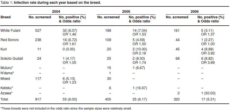
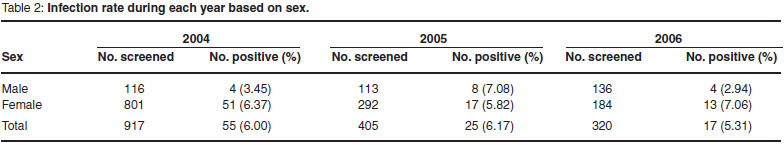
Over the study period, seroprevalence rates of 6.00 %, 6.17 % and 5.31 % were obtained in 2004, 2005, and 2006, respectively. The highest rates of 6.72 % (OR 1.61), 10.00 % (OR 2.18) and 8.89 % (OR 3.92) were obtained in the Red Bororo breed in 2004 and the Kuri in 2005 and 2006 (Table 1). The seroprevalence rate for each year based on sex (Table 2) revealed a higher rate in female animals. With reference to the age distribution, the most affected were the adult cattle with rates of 6.48 % in 2004, 6.96 % in 2005 and 5.54 % in 2006 (Table 3).
DISCUSSION
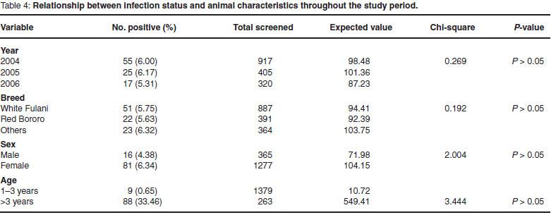
Seroprevalences of 6.00 %, 6.17 % and 5.31 % obtained in 2004, 2005 and 2006 respectively showed that brucellosis is endemic in trade cattle in Ibadan. The prevalence according to breed, sex, age and year of study (Table 4) revealed no significant differences in the infection rates over the period despite the decrease noticed in 2006 (χ2 for trend = 0.269, P > 0.05).
The steady nature of the infection rates over the 3-year period (Table 4) confirms that bovine brucellosis is endemic in trade cattle slaughtered in Ibadan and possibly widespread throughout Nigeria. Although some studies in Nigeria suggested an increasing trend in the prevalence of the disease18, rates obtained in this study may be due to the persistence of the disease in the cattle population resulting from the unchanging poor husbandry practices of the livestock owners and marketers as well as non-vaccination of the cattle coupled with little or no veterinary care. One such practice is the tradition of the nomadic Fulani pastoralists who manage about 95 % of the total animal population in Nigeria19. The Fulanis are accustomed to an extensive system of management, keeping both healthy and infected animals together under purely traditional systems and travelling over long distances with limited food and water supplies8.This exposes the animals to unfavourable physiological conditions which make them highly susceptible to diseases like brucellosis and tuberculosis before being transported to the abattoirs for slaughter.
Different breeds of cattle demonstrated different prevalence rates which were not statistically significant over the years (χ2 = 0.192, P > 0.05) (Tables 1 & 4), although in some studies6,8 the White Fulani breed was the most affected; however, they are the predominant breed in Nigeria4.
The male to female infection ratios recorded over the years were not statistically significant at 5 % (χ2 = 2.004, P > 0.05) (3.5:6.4 in 2004; 7.1:5.8 in 2005 and 2.9:7.1 in 2006) (Table 2) and do not support the conclusion that females were more prone to infection, although higher rates were obtained for female animals. Other studies carried out in the same abattoir12 also showed higher infection rates in females than males. The most plausible reason given for this was the inclusion of pregnant cows in the population studied. It must be noted that sexually mature pregnant cattle are more susceptible to infection than sexually immature cattle of either sex20.
Adult cattle over the age of 3 years had the highest seroprevalence (Table 3) but this was not statistically significant (χ2 = 3.444, P > 0.05). Ordinarily, young cattle (1-3 years of age) are said to be less susceptible to B. abortus than older, sexually mature animals; however, non-vaccinated young cattle are also at higher risk of brucellosis if exposed to pathogenic strains of the organism20.
Brucellosis is a worldwide zoonosis15,16 that causes serious economic losses in livestock and poses important human health hazards worldwide12. One of the major implications of the burden of this disease in the abattoir setting is the exposure of livestock traders, butchers and other meat processors as well as veterinarians/meat inspectors. The poor facilities and safety precautions in most abattoirs and slaughter slabs in Nigeria contribute to the likelihood of exposure. In most instances, these workers use their bare hands to handle infected organs and carcasses from diseased animals. Additionally, the consumption of unpasteurised milk and milk products by some members of society is complicated by direct consumption of unpasteurised milk from the udders of cows by some of the Fulani pastoralists7. This is made worse by the close contact and co-habitation of livestock with humans. The economic impact and public health significance of the uncontrolled prevalence of brucellosis in the Nigerian livestock population is undoubtedly high19 and the financial costs of the disease nationally have been estimated to be substantial1,2.
This study had some limitations, however. Firstly, sample collection was restricted to a few months of the year and this might not give the true picture of the prevalence of the disease. Secondly, the sample size of some of the breeds of cattle encountered during this study was relatively small compared with the more popular breeds at the abattoir. This may therefore create some bias with respect to the seroprevalence rates of breeds affected. Thirdly, only RBT was used in this study and no single serological test can be relied upon in all epidemiological situations because they all have their limitations. Moreover, it has been shown that although the low pH (+3.6) of the antigen enhances the specificity of the test, the temperature of the antigen and the ambient temperature at which the reaction takes place may influence the sensitivity and specificity13.
The above limitations notwithstanding, the study highlights the endemicity of the disease over the years in trade cattle slaughtered in Ibadan. It also shows that adult and female animals play a more important role in the epidemiology of the disease, athough this is not statistically significant.
In conclusion therefore, for the control and eradication of brucellosis in trade cattle in Ibadan and Nigeria as a whole, more emphasis should be directed towards early vaccination of young animals and separation of clean and infected animals/herds. This should be combined with more government intervention in the areas of regulations and policies concerning routine screening of all cattle populations, including trade animals destined for slaughter at abattoirs in Nigeria. Coupled with these should be awareness programmes involving stakeholders in the livestock industry as well as consumers to avert public health and economic losses associated with brucellosis in Nigeria.
ACKNOWLEDGEMENTS
We thank the Veterinary Laboratories Agency (VLA), United Kingdom, that provided the antigens used for this study.
REFERENCES
1. Ajogi I, Akinwunmi J A 2001 Cash-flow model of the cost of brucellosis in traditionally managed cattle herds in Nigeria. Bulletin of Animal Health and Production in Africa 49:169-173 [ Links ]
2. Ajogi I, Akinwunmi J A , Esuruoso G O, Lamorde G 1998 Settling the nomads in Wase-Zange grazing reserves in the Sudan Savannah zone of Nigeria. III. Estimated financial losses due to bovine brucellosis. Nigerian Veterinary Journal 19:86-94 [ Links ]
3. Alton G G, Jones L M, Angus R D, Verger J M 1988 Techniques for the brucellosis laboratory. Institut National de la Recherche Agronomique, Paris [ Links ]
4. Bourn D, Wint W, Blench R, Woolley E 1994 Nigerian livestock resources survey. World Animal Review 78:49-58 [ Links ]
5. Ate I U, Andrew P I R, Nok J, Tekdek L B 2007 Seroprevalence of brucellosis in puerperal cows and its public health implications in Zaria, northern Nigeria. Journal of Animal and Veterinary Advances 6:863-866 [ Links ]
6. Cadmus S I B, Adesokan H K, Stack J 2008 The use of the milk ring test and rose bengal test in brucellosis control and eradication in Nigeria. Journal of the South African Veterinary Association 79:113-115 [ Links ]
7. Cadmus S I B, Adesokan H K 2007 Phenotypic characterization and spoligotype profiles of Mycobacterium bovis isolated from unpasteurized cows' milk in Ibadan, Nigeria. Tropical Veterinarian 25:65-72 [ Links ]
8. Cadmus S I B, Ijagbone I F, Oputa H E, Adesokan H K, Stack J A 2006 Serological survey of brucellosis in livestock animals and workers in Ibadan, southwestern, Nigeria. African Journal of Biomedical Research 9:163-168 [ Links ]
9. Esuruoso G O, Ayanwale F O 1980 Bovine brucellosis in Lagos state of Nigeria. Bulletin of Animal Health and Production in Africa 28:11-15 [ Links ]
10. Gameel S E A M, Mohammed S O, Mustafa A A, Azwai S M 1993 Prevalence of camel brucellosis in Libya. Tropical Animal Health and Production 25:91-93 [ Links ]
11. Hamidy M E R, Amin A S 2002 Detection of Brucella sp. in the milk of infected cattle, sheep, goats and camels by PCR. Veterinary Journal 163:299-305 [ Links ]
12. Ishola O O, Ogundipe G A T 2000 Seroprevalence of brucellosis in trade cattle slaughtered in Ibadan, Nigeria. Bulletin of Animal Health and Production in Africa 48:53-55 [ Links ]
13. MacMillan A 1990 Conventional serological tests. Animal Brucellosis 206:153-197mm [ Links ]
14. Maloney G E 2001 CBRNE brucellosis. eMedicine.com, Inc. Online at: http://www.emedicine.com/abortus.shtml (accessed April 2009) [ Links ]
15. Maloney G E, Fraser W R, 2001 CBRNE-brucellosis. Online at: http://www.emedicine.com/aboutus.shtml (accessed October 2006). [ Links ]
16. Nicoletti P 1993 Brucellosis. Current veterinary therapy. In Howard J L (ed.) Food animal practice (3rd edn). W B Saunders, Philadelphia: 551-555 [ Links ]
17. Ocholi R A 1990 Prevalence of Brucella antibodies in Fulani cattle herds in Kaduna State. M.Sc. thesis, Ahmadu Bello University, Zaria [ Links ]
18. Ocholi R A, Kwaga J K P, Ajogi I, Bale J O O 2004 Phenotypic characterization of Brucella strains isolated from livestock in Nigeria. Veterinary Microbiology 103:47-53 [ Links ]
19. Oladosu L A, Falade S, Akpokodje U 1986 Equine brucellosis in Nigeria. Zariya Veterinarian 1:129-133 [ Links ]
20. Radostits O M, Blood D C, Gay C C 1995 Veterinary medicine - A textbook of the diseases of cattle, sheep, pigs and horses (8th edn). Baillière Tindall, London [ Links ]
21. Seifert H S H 1996 Diseases caused by aerobic rods. I. Brucellosis. In Bokma B H, Blouin E E, Bechara G H (eds) Tropical animal health. Kluwer Academic, Dordrecht, 356-367 [ Links ]
Received: December 2009.
Accepted: March: 2010.
* Author for correspondence. E-mail: sibcadmus@yahoo.com

