


Services on Demand
Article
 English (pdf)
 Article in xml format
 Article references
Indicators

Related links
-
 Cited by Google
 -
 Similars in Google
Share

 Permalink
South African Journal of Animal Science
On-line version ISSN 2221-4062
Print version ISSN 0375-1589
S. Afr. j. anim. sci. vol.53 n.6 Pretoria 2023
http://dx.doi.org/10.4314/sajas.v53i6.03
Marek's disease in backyard chickens of Pakistan: A study of pathological lesions in correlation to viral load
M. AzeemI; M. ur Rehman KhanI, #; G. SaleemI; M. Hassan MushtaqII
IDepartment of Pathology, University of Veterinary and Animal Sciences, 54000 Lahore, Pakistan
IIDepartment of Epidemiology & Public Health, University of Veterinary and Animal Sciences, 54000 Lahore, Pakistan
ABSTRACT
Marek's disease (MD) is an infectious, lymphoproliferative disorder caused by the alpha herpesvirus genus, Mardivirus, serotype 1 (Gallid Herpesvirus 2, or GaHV-2). There is no current information on Marek's disease virus (MDV) pathotypes and serotypes circulating in backyard poultry in Pakistan. This study aimed to characterize GaHV-2 in the backyard poultry of Rawalpindi division based on glycoprotein C and to investigate the correlation of a viral oncogene (meq) with the histological, lymphoproliferative infiltration in the visceral organs of naturally-infected birds. The study was performed from May 2019 to April 2020 in the Rawalpindi, Jhelum, Chakwal, and Attock districts of the Rawalpindi division. An overall 13.96% prevalence of MDV infection based on gross morphology and 18.43% using polymerase chain reaction (PCR) was recorded. The Pakistani GaHV-2 isolates could be divided into two clades, based on sequencing and phylogenetic analysis of the gC gene, closely related to the US virulent isolates and mildly virulent vaccine strain, CVI988, respectively. Of total PCR-positive cases, 25 (75.76%) samples revealed gross tumours. Median histopathological scores of lymphoproliferative infiltrations in the spleen and liver tissues were recorded as moderate (++), whereas in the heart and gonads, mild (+) changes were observed. The mean viral load quantified using real-time PCR of 14.72 copies of meq gene/ß-actin revealed a strong (R2 = 0.88) correlation with histopathology scores of spleen tissues. Differences in the meq gene/ß-actin ratio were observed between non-tumorous and tumorous birds, which is indicative of pathological changes in individuals of the present study.
Keywords: alpha herpesvirus, glycoprotein C, histopathology scores, meq-oncogene, Rawalpindi division, real-time polymerase chain reaction, Gallid herpesvirus-2
Introduction
Marek's disease (MD) is caused by Marek's disease virus (MDV), which consists of three serotypes; MDV-1 is the pathogenic form, whereas MDV-2 and MDV-3 (herpesvirus of turkeys; HVT) are non-pathogenic and primarily used as protective immunizations against MDV-1. MD is a cell-associated, alpha herpesvirus demonstrated as the fifth most common infectious disease in chickens in a backyard poultry disorder survey (Mete et al., 2016). The oncogenic MDV-1 virus can cause infection in 1-day-old chicks. The first phase of this process is the cytolytic phase, followed by latency. The second cytolytic phase occurs next, transforming the T lymphocytes of the affected host, leading to the development of lymphoproliferative disease or lymphomas (Payne & Nair, 2012).
Vaccination is the primary method of preventing MD, and strategies such as the widespread usage of vaccines within the poultry industry have been relatively successful in lowering economic losses (Mete et al., 2016). The most effective way to administer vaccines created from serotypes MDV-2 (such as SB-1), HVT, and attenuated MDV-1 (such as CVI988; Rispens) is through in-ovo vaccination with Rispens (Gimeno, 2008). Vaccine failures can be caused by prior virus exposure, inadequate handling, and administration of the vaccine, or weakened immunity (due to insufficient maternal antibodies or concurrent infections) in the chicken (Baigent et al., 2006; Dunn et al., 2014). Furthermore, the emergence of more virulent strains (virulent pathotypes) of MDV-1 have been linked to the failure of some vaccines (Witter, 1997; Nair, 2005), such that the most commonly-used HVT vaccine does not provide adequate immunization against very virulent MDV-1 (Gimeno, 2008).
Surveys and on-field experience indicate that backyard chicken farmers do not understand vaccination protocols for MD (Lister & Houghton-Wallace, 2012; Elkhoraibi et al., 2014; Whitehead & Roberts, 2014). MDV-1 pathotype has been studied in chickens, primarily in commercial poultry lines under laboratory conditions. Such settings often use genetically susceptible or resistant chickens that lack antibodies and are referred to as specific-pathogen-free or antibody-free chickens to observe the progress of the disease, its viral pathogenesis, and the efficacy of vaccines (Bacon et al., 2001a).
Diagnosing MD in backyard chickens is complex due to the wide variety of host lineages and infective MDV pathotypes. Additionally, the clinical signs and lesions of MD may be similar to those of other lymphoproliferative diseases or even peripheral neuropathy, an autoimmune disorder (Bacon et al., 2001b; Witter et al., 2010). Researchers have identified several criteria that can help distinguish MD in chickens, including the age of the chicken, changes observed under a microscope, immunohistochemistry tests, and molecular techniques. Nevertheless, multiple combined methods are often necessary to properly diagnose the condition (Witter et al., 2010). Glycoprotein C (gC) is a valuable diagnostic tool in MD, an essential avian virus known to cause severe neurological disease and death in poultry. Further, analysis of the glycoprotein profile can be used to differentiate between the genotypes of each MD virus strain, ultimately leading to improved targeted strategies to combat this essential avian pathogen (Jarosinski & Osterrieder, 2012; Hassanin et al., 2013).
Real-time quantitative polymerase chain reaction (qPCR) analysis is suggested as a definitive diagnostic tool for tumours where the MDV load is substantially higher than in nontumorous tissues. As a result, diagnosing MD in birds of varying or unknown ages and vaccination histories with an extensive overlap of lesions poses a unique challenge for diagnosticians in the field, who lack the necessary resources. This need is especially notable in settings where individual poultry health monitoring is prioritized over the flock approach.
The present study aimed to study the phylogenetic analysis of MDV tumorous and non-tumorous backyard chickens of Rawalpindi division, and to correlate the histopathologic scores and MDV-1 viral loads.
Materials and Methods
The study was conducted after approval from the research ethical guidelines committee, University of Veterinary and Animal Sciences, Lahore (DR/209, 24th April, 2019).
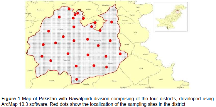
The samples were collected from May 2019 to April 2020 from 40 backyard chicken flocks (179 birds) from four districts in Rawalpindi Division: Rawalpindi, Jhelum, Chakwal, and Attock districts (Figure 1). The samples were taken from two to three birds per flock. Samples were taken from each bird's visceral organs with diffuse or localized tumours. The samples were kept at -20 °C until the DNA was extracted. Information regarding age, sex, breed, clinical signs, and vaccination status were also collected. Based on the owners' information, the vaccinated birds had a history of inoculation on day 1 at the hatchery level with commercially-available, live cell-associated vaccine (Rispens+HVT, Merial, Inc., USA).
Total cellular DNA was extracted using the ThermoScientific® GeneJET Genomic DNA Purification kit, according to the manufacturer's instructions. PCR was conducted to detect a 686-bp glycoprotein C (gC) amplicon from Gallid alpha herpes virus type 1, with primers and conditions described previously by Becker et al. (1993). The forward: 5'-ATACCACGCCAACGAAAAGAATGT-3' and reverse primers: 5'-CTATAGTACATATTGCATACCAT-3' were as described by Becker et al. (1993). The PCR amplification was carried out in a 25 μl volume containing 3 μl of DNA, 1 μl (10 μmol) of each forward and reverse primer, 12.5 μl of Master Mix (Thermo Scientific Dream TaqTM Green PCR), and 7.5 μl distilled water.
Viral genomic DNA was subjected to thermal cycling that at 95 °C for 120 s, followed by 35 cycles at 94 °C for 60 s, 55 °C for 60 s, and 72 °C for 180 s, and a final extension at 72 °C for 300 s. PCR product (5 ul) was separated at 100 V for 20 min on a 1% agarose gel using the 100-bp DNA marker (ThermoScientific). PCR products were visualized in a gel documentation system (Cleaver Scientific, Ltd).
The amplified gC gene segments were sequenced using gene-specific primers by a commercial sequencing service (AB13730XL; Macrogen Inc., Seoul, Korea). Reference sequences and sequences of MDV gC under various accession numbers from different parts of the world were downloaded from https://www.ncbi.nlm.nih.gov/nucleotide/ and aligned with the sequences of the present study using BIOEDIT (version 7.2.5). The phylogenetic trees were constructed using the neighbour-joining (N-J) tree method, and the validity of each branch was assessed using 1,000 bootstrap replications. Branches corresponding to partitions reproduced in less than 50% of bootstrap replicates were collapsed. The evolutionary distances were computed using the Maximum Composite Likelihood method (Tamura et al., 2004) and are presented in the units of the number of base substitutions per site. The differences in the composition bias among sequences were considered in evolutionary comparisons (Tamura & Kumar, 2002). This analysis involved 37 nucleotide sequences. Codon positions included were 1st+2nd+3rd+Noncoding. All ambiguous positions were removed for each sequence pair (pairwise deletion option). There were a total of 1547 positions in the final dataset. Evolutionary analyses were conducted in MEGA software (version 11.0.13) (Kumar et al., 2016). The nucleotide identity percentage among the sequences was calculated using the sequence identity matrix tool in BIOEDIT (version 7.2.5).
The absolute MDV genome loads in splenocytes were quantified using real-time PCR with previously published primers specific to the meq gene of MDV using the extracted DNA (Abdul-Careem et al., 2006). The Meq primer sequences were F-5'-GTCCCCCCTCGATCTTTCTC-3' and R-5'-CGTCTGCTTCCTGCGTCTTC-3'. The DNA template was added to a total volume of 20 μl containing PCR buffer, oligonucleotide primers at 0.2 μM each of primer, and 2 μl of Fast SYBR™ Green Master Mix (Applied-Biosystems, Singapore). The chicken ß-actin gene in each sample was amplified using a primer pair (F-5'-GAGAAATTGTGCGTGACATCA-3', R-5'-CCTGAACCTCTCATTGCCA-3'). To describe the ratio between the two genes, the amplification of the meq gene was compared to the ß-actin in each sample. The results were averaged across all samples tested in triplicate.
The gross examination findings and tumour lesions were documented using the criteria indicated by Burgess et al. (2001). The scoring of the lymphocytic infiltrations was conducted on formalin-fixed, paraffin-embedded (FFPE), 5-μm thick sections of the liver, spleen, heart, and gonads, stained with Haematoxylin and Eosin. The following histologic scores were designed for this study as scattered lymphocytic infiltrations (+), moderate multifocal lymphoid cells clusters (++), and large multifocal to coalescing sheets of lymphocytes obliterating the tissue architecture (+++), as described by Kano et al. (2009). Each part was scored after a comparison with the established reference, and the overall scores were recorded.
The association of risk factors with MDV infection was analysed using a Chi-squared test. The relative fractions were expressed in percentages and 95% confidence intervals (95% CI) using GraphPad Prism 8 for windows 10 (GraphPad Software Inc., La Jolla, CA). Adjusted odds ratios related to dichotomous and polychotomous variables were assessed using Binary Logistic Regression Model using Minitab® 21.3.1 (64-bit). An independent t-test was used to analyse the difference between the mean viral loads of birds revealing gross tumorous lesions and those that did not. Spearman's ranked correlation was used to evaluate the correlation between the viral loads of the spleen samples and histological lymphocytic infiltration scores in the tissues. Statistical significance was defined at P < 0.05.
Results
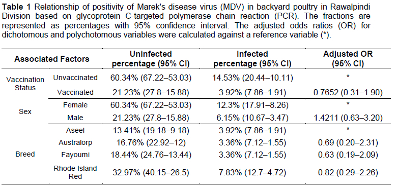
An overall 13.96% (95% CI; 19.80-9.64) presence of MDV infection based on gross morphology and 18.43% (95% CI; 24.75-13.43) with PCR was recorded in the chickens from the four districts of Rawalpindi division. The chi-squared test results did not reveal an association of disease positivity with the analysed risk factors (P >0.05). The birds with a vaccination history against MDV showed 23.48% fewer odds of infection than the unvaccinated individuals. Moreover, males showed almost 1.4-times more chance of infection than female birds. However, different backyard poultry breeds, including Aseel, Australorp, Fayoumi, and Rhode Island Red (RIR), had almost similar odds of infection with MDV. Table 1 shows the details of the infected and healthy birds with the associated factors.
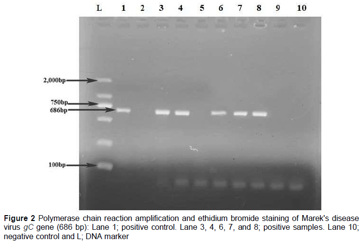
A positive PCR amplicon (686 bp) of the gC gene for MDV was identified using agarose gel electrophoresis. DNA extracted from different samples yielded a clear band in the gel electrophoresis after amplifying the target gene (Figure 2). The PCR amplified gC gene sequences were submitted to the GenBank database under accession numbers MN923520.1, MN923519.1, MN923518.1, MN923517.1, MN727368.1, MN727367.1, MN727366.1, and MN727365.1.
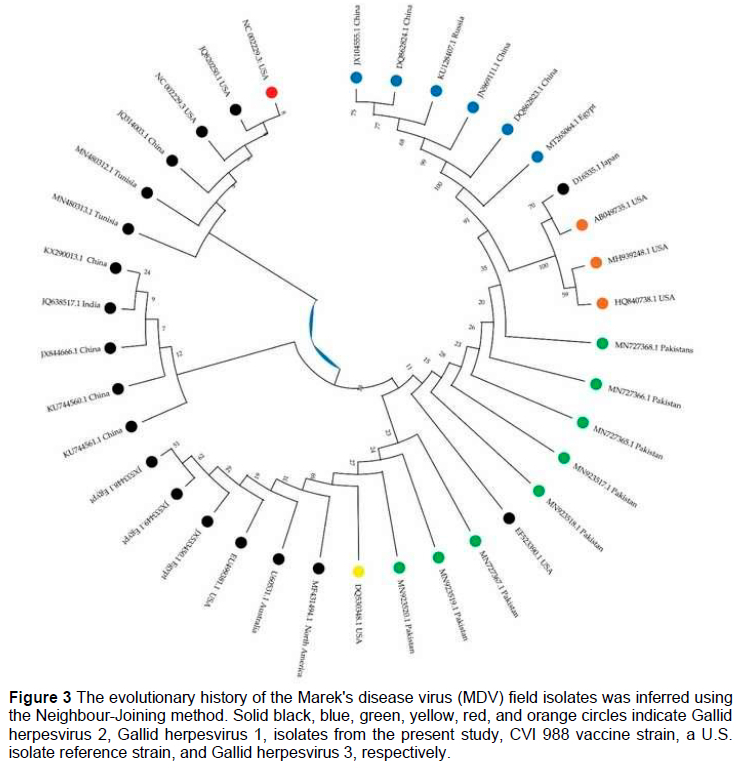
The nucleotide sequence identity matrix revealed maximum similarity of MN727366.1 of 47.3% each with virulent strain virus isolates, KX290013.1, KU744561.1, KU744561.1, JX844666.1 (China) and EF523390.1 (USA). Isolate MN727365.1 showed 47.2% similarity each with isolates KX290013.1, KU744561.1, KU744561.1, JX844666.1 (China), and EF523390.1 (USA). Moreover, the isolate MN727368.1 had 47.1% similarity with KX290013.1, KU744561.1, KU744561.1, JX844666.1 (China), and EF523390.1 (USA). The isolates, MN727367.1, MN923520.1, and MN923519.1 showed 45.8%, 46.2%, and 46.2% similarity with JX533448.1, the mildly virulent Egyptian isolate. The phylogenetic analysis of nucleotide sequences revealed the three major clades. Isolates with accession numbers MN923518.1, MN923517.1, MN727368.1, MN727366.1, and MN727365.1 were placed with virulent US isolate (EF523390.1). MN727367.1, MN923520.1, and MN923519.1 were placed with mildly virulent vaccine strain virus CVI988 from the USA (DQ530348.1). The phylogenetic analysis of nucleotide sequences is shown in Figure 3.
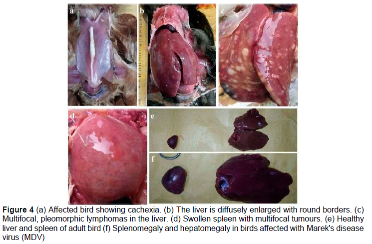
Out of the total PCR-positive cases, 25 (75.76%) samples revealed gross tumours. In comparison, 8 (24.25%) did not reveal gross lesions on the visceral organs. In many cases, the peripheral nerves did not display any macroscopic alterations. The gross examination of the visceral organs indicated that the organs (liver & spleen) had substantial enlargement. The surface had elevated white foci and the lesion's distribution may have been either diffuse or multifocal. The sliced parts of the organs revealed nodules embedded deeper inside the parenchyma of the organs. There was a wide range of sizes among the nodules, ranging from pinpoint to enormous nodules with a diameter of 1 -2 cm. There was diffuse expansion and no noticeable nodular focal lesions in some situations (Figure 4).
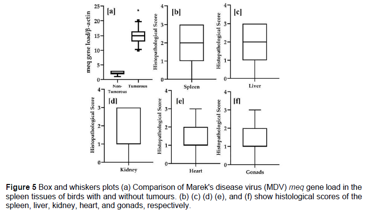
Total genomic DNA was isolated from the splenocytes of chickens, and MDV genome loads were quantified using real-time PCR. The MDV viral loads ranged between 10 and 20 copies of meq/ß-actin in fresh spleens of the 25 tumorous chickens. The mean viral load quantified was 14.72 copies of meq/ß-actin (95% CI; 13.69-15.75). The 5% and 95% percentile individuals lay at 10.3 and 19.7 copies of meq/ß-actin, respectively. In the eight non-tumorous chickens, including the PCR-positive cases, MDV loads were at very low levels, revealing marked differences between tumorous and non-tumorous spleen samples. Figure 5 shows the meq gene load in the spleen tissues of both tumorous and non-tumorous individuals, along with the infiltrating lymphocytic score in the visceral organs of affected birds.
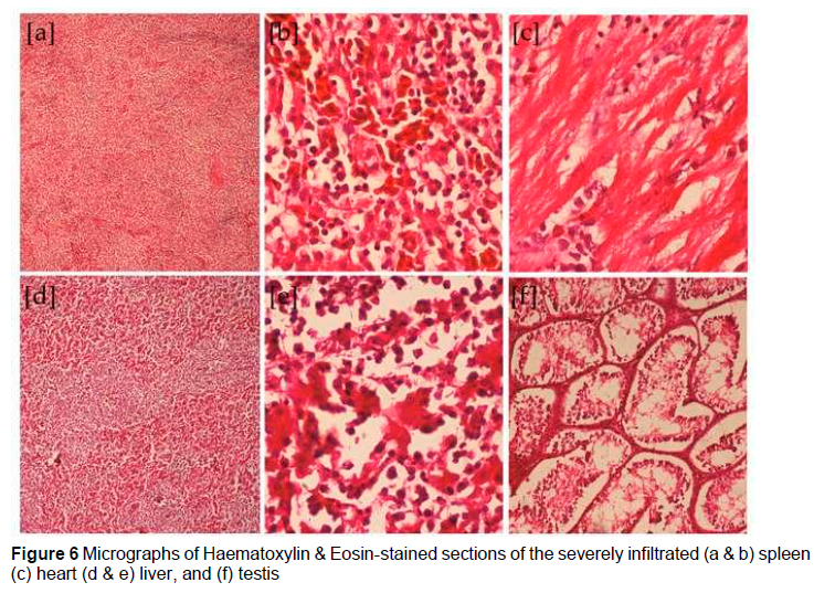
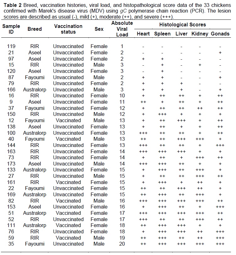
A histopathological examination of the visceral organs revealed lymphoproliferative abnormalities in the heart, liver, spleen, kidney, and gonads. These lymphoproliferative changes varied from mild (+) and moderate (++) to severe (+++). Figure 6 describes histopathological lesions of varying severity in the various visceral organs of infected birds. Lymphoid tissue entirely replaced the parenchyma in several of the spleen tissues. There was a marked uniformity in the size of the neoplastic lymphocytes, ranging from tiny to medium. These lymphocytes were replacing segments of the parenchyma in the liver and spleen that ranged from multifocal to dispersed. The standard consistency of the organ was disrupted due to the presence of pleomorphic lymphoblastic and lymphocytic cells, comprising the majority of the cells in the organ. Median histopathological scores of spleen tissues were moderate (++). Moreover, 95 percentile of individuals lay in severe (+++) histopathological scores (Figure 5b). In the liver, there was unequivocal evidence of both focal and diffuse pleomorphic cell infiltration. These cells were a combination of lymphoblasts, lymphocytes of small to medium size, plasma cells, and macrophages. Additionally, some macrophages with a few plasma cells were present. The infiltrating lymphoblasts were found to have a substantial number of mitotic figures. There was cellular infiltration of lymphocytes with replacement of normal parenchyma. Perivascular lymphoblast infiltration was also observed. Median histological scores of liver tissue were moderate (++); 95 percentile individuals revealed severe (+++) changes in the hepatic parenchyma. In heart tissue, observed median histological lymphocytic infiltration score was mild (+), (5 and 95 percentile of individuals lay between normal to moderate (++) scores). Gonads revealed median or mild (+) histological changes (5 and 95 percentile individuals shown normal and severe (+++) histological scores, respectively). Individual details of infected chickens are described in Table 2.
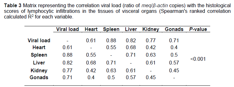
Spearman's ranked correlation regarding the quantification of MDV viral load (ratio of meq/ß-actin copies) revealed a strong correlation with histological scores of various organs (P <0.001). The highest correlation was observed with the spleen (88%), followed by the kidney (77%), liver (82%), gonads (71%), and heart (61%). Table 3 shows the correlation matrix of the histological scores of various organs and viral loads in the spleen.
Discussion
MDV has become more virulent over the last few decades, which has invited researchers globally to develop vaccines with many recent viral isolates. Although there are a substantial number of underlying connected elements, the cause of the continual increase in virulence is not known precisely (Jarosinski et al., 2006). Lower levels of immune titre can result from a sub-adequate dose of vaccines, as compared to the recommended dosage. This could have been done for the sake of economic benefit, or it could have resulted from improper storage and handling (Witter, 1997). There are several different processes that might be due to mistakes while handling vaccinations, such as combining antibiotics with vaccines or performing the thawing process incorrectly. The higher occurrence rates can also be caused by other factors, including weak biosecurity (Dunn & Gimeno, 2013). The overall 13.96% (95% CI; 19.80-9.64) presence of MDV infection based on gross morphology and 18.43% (95% CI; 24.75-13.43) using PCR was recorded in chickens from the four districts of Rawalpindi division. This higher prevalence rate in the present study might be attributed to the fact that the sampling was done from the area where the field veterinarians were suspecting incidence of MDV in the backyard poultry.
It is well established that several structural proteins found in MDV are substantially involved in these processes. MDV glycoproteins found in the virus's envelope play a role in virus attachment and entrance or in virus maturation, emigration, and direct cell-to-cell dissemination, which have not been identified (Elliott et al., 1995). In vitro and in vivo examinations of gC-like proteins, which are found in alpha herpes viruses and are the critical components of the viral envelope, reveal that these proteins have various roles in both conditions. In a broad sense, gC orthologues play an integral role in the first interaction between the virion and the cell by facilitating the attachment of the free virus to heparin- and chondroitin-like glycosaminoglycans present on the surface of the plasma membrane (Roizman & Knipe, 2001). In addition to their capacities to facilitate viral attachment and release, gC homologs are thought to be able to escape the immune system. It is believed that these functions are mediated by complement-component C3 binding, which then blocks the component's activity (Huemer et al., 1993; Huemer et al., 1995), in addition to playing a part in the process of leukocyte migration mediated by chemokines (González-Motos et al., 2017). Several studies have demonstrated the effectiveness of PCR amplification of the gC gene for diagnosing MDV. One study, for example, used a PCR-based method to differentiate virulent from avirulent MDV strains based on gC gene amplification (Jarosinski & Osterrieder, 2012).
The phylogenetic analysis of gC gene nucleotide sequences revealed that present study isolates with accession numbers MN923518.1, MN923517.1, MN727368.1, MN727366.1, and MN727365.1 were placed with the virulent US isolate. MN727367.1, MN923520.1, and MN923519.1 were placed with the mildly virulent vaccine strain virus, CVI988, from the USA. The spleen tissue revealed higher viral load quantitation in the samples infected with the virulent MDV strain. In contrast, the birds infected with mild strain had substantially lower viral loads in the spleen tissue. Moreover, the birds also revealed marked differences in the gross and histopathological changes between both strains. The findings of phylogenetic analysis also concur with the meq gene-based quantitation of the virus in the splenic tissue. Similarly, Spearman's ranked correlation revealed a strong correlation (88%) of viral load with histological lymphocytic infiltration scores in the spleen (P <0.001).
The real-time PCR is a reliable technique to assess the severity of an MDV infection and the potential for developing MD lymphoma by measuring the meq gene load in infected tissues (Abdul-Careem et al., 2006). The lesions in the present study were well-matched to those described by Hayajenh et al. (2021), Zelnik et al. (2004), and Fodor et al. (2001). Vieira-Pinto et al. (2003) described that histopathology could be used to confirm the presence of MD. Neoplastic foci and pleomorphic cells were found during the histological analysis of liver and spleen tissues for the subclinical type of MD. There was evidence of lymphomatous lesions in certain areas containing lymphoblasts and lymphocytes. These lesions were usually elongated and ranged in size from small to medium. Although the tumour microenvironment is the same across organs, the extent of organ involvement can often vary (Stamilla et al., 2020; Khan et al., 2021). Wen et al. (2018) identified the presence of MD in the nucleus of neoplastic lymphoid cells found in the liver. Zelnik et al. (2004) reported the incidence of MD in vaccinated birds. It was observed by Khan et al. (2021) that infected liver cells proliferated around the small blood vesicles, and the lesions in the tumour consisted of proliferated tumour cells.
The capacity of the host to endure minimal detrimental consequences as a result of infection is what is meant by the term "tolerance," which refers to the term "genetic resistance to infection" (Râberg et al., 2007; JovanoviC et al., 2009). Disease resistance mechanisms are complicated and incompletely understood at the molecular level. Genetic resistance to infectious diseases is controlled by a number of variables, including both biological and environmental factors (Zekarias et al., 2002; Zeleke et al., 2005). The majority of research that has been conducted on disease resistance mechanisms has focused on investigating the function of innate immune responses. This is because it is generally acknowledged that innate immune responses are the factors that determine whether or not an individual is resistant or susceptible to infectious pathogens (Glass et al., 2012; Iwasaki & Medzhitov, 2015). There is a correlation between the major histocompatibility complex, also known as MHC haplotypes, and the variation in disease susceptibility and resistance (Janeway, 2001; Leveque et al., 2003; Kapczynski et al., 2013).
To a great extent, antigen processing and presentation rely on the MHC molecules. Because each gene has several variations in the population, the MHC can process and display any antigens that might exist (Janeway Jr et al., 2001). The MHC genotype limits T-cell antigen recognition. The specific T-cells might not recognize the antigen because of the defect in the MHC molecule. Consequently, the MHC molecules are responsible for controlling the antigen specificity of T-cells (Janeway Jr et al., 2001). According to specific reports, there is a connection between the MHC haplotypes and the varying predispositions of chickens to infectious diseases (Goto et al., 2009). However, in the present study, different backyard poultry breeds, including Aseel, Australorp, Fayoumi, and Rhode Island Red had almost similar odds of infection with MDV.
Conclusion
In conclusion, it is suggested that virulent and mild pathotypes of MDV-1 strains are prevalent in the backyard poultry of the Rawalpindi division, which is a continuous source of threat for commercial poultry in the study area.
Acknowledgement
The authors would like to thank all the facilitators for their support in sample collection and processing, and Higher Education Commission (HEC) Pakistan for their financial funding of the research project through the HEC-NRPU program.
Authors' contribution
MA and MURK conceived the idea and facilitated the execution of research activities. MA, MURK, GS, and MHS performed the sampling and testing. MA and MURK facilitated in the statistical analysis, manuscript write-up and reviewing it.
Conflict of Interest
The authors declare that there is no conflict of interest related to this article.
References
Abdul-Careem, M.F., Hunter, B.D., Nagy, É., Read, L.R., Sanei, B., Spencer, J.L., & Sharif, S., 2006. Development of a real-time PCR assay using SYBR Green chemistry for monitoring Marek's disease virus genome load in feather tips. J. Virol. Methods. 133, 34-40, https://doi.org/10.1016/j.jviromet.2005.10.018 [ Links ]
Bacon, L., Hunt, H., & Cheng, H., 2001a. Genetic resistance to Marek's disease. Marek's Dis. 121-141, https://doi.org/10.1007/978-3-642-56863-3_5 [ Links ]
Bacon, L., Witter, R., & Silva, R., 2001b. Characterization and experimental reproduction of peripheral neuropathy in White Leghorn chickens. Avian Pathol. 30, 487-499, https://doi.org/10.1080/03079450120078680 [ Links ]
Baigent, S., Nair, V., & Currie, R., 2006. Real-time quantitative PCR for Marek's disease vaccine virus in feather samples: Applications and opportunities. Dev. Biol. 126, 271. [ Links ]
Becker, Y., Tabor, E., Asher, Y., Davidson, I., Malkinson, M., & Witter, R., 1993. PCR detection of amplified 132 bp repeats in Marek's disease virus type 1 (MDV-1) DNA can serve as an indicator for critical genomic rearrangement leading to the attenuation of virus virulence. Virus genes. 7, 277-287, https://doi.org/10.1007/BF01702588 [ Links ]
Burgess, S., Basaran, B., & Davison, T., 2001. Resistance to Marek's disease herpesvirus-induced lymphoma is multiphasic and dependent on host genotype. Vet. Pathol. 38, 129-142, https://doi.org/10.1354/vp.38-2-129 [ Links ]
Dunn, J.R., Auten, K., Heidari, M., & Buscaglia, C., 2014. Correlation between Marek's disease virus pathotype and replication. Avian Dis. 58, 287-292, https://doi.org/10.1637/10678-092513-Reg.1 [ Links ]
Dunn, J.R., & Gimeno, I.M., 2013. Current status of Marek's disease in the United States and worldwide based on a questionnaire survey. Avian Dis. 57, 483-490, https://doi.org/10.1637/10373-091412-ResNote.1 [ Links ]
Elkhoraibi, C., Blatchford, R., Pitesky, M., & Mench, J., 2014. Backyard chickens in the United States: A survey of flock owners. Poult. Sci. 93, 2920-2931, https://doi.org/10.3382/ps.2014-04154 [ Links ]
Elliott, G., Mouzakitis, G., & O'Hare, P., 1995. VP16 interacts via its activation domain with VP22, a tegument protein of herpes simplex virus, and is relocated to a novel macromolecular assembly in coexpressing cells. J. Virol. 69, 7932-7941, https://doi.org/10.1128/jvi.69.12.7932-7941.1995 [ Links ]
Fodor, I., Coman, M., & Catana, N., 2001. An outbreak of Marek's disease in broiler chickens: Epidemiological, clinical and anatomopathological aspects. Lucr. ştiint Med. Vet. 42, 129-142. [ Links ]
Gimeno, I.M., 2008. Marek's disease vaccines: A solution for today but a worry for tomorrow? Vaccine. 26, C31-C41, https://doi.org/10.1016/j.vaccine.2008.04.009 [ Links ]
Glass, E.J., Baxter, R., Leach, R.J., & Jann, O.C., 2012. Genes controlling vaccine responses and disease resistance to respiratory viral pathogens in cattle. Vet. Immunol. Immunopathol. 148, 90-99, https://doi.org/10.1016/j.vetimm.2011.05.009 [ Links ]
González-Motos, V., Jürgens, C., Ritter, B., Kropp, K.A., Durán, V., Larsen, O., Binz, A., Ouwendijk, W.J., Lenac Rovis, T., & Jonjic, S., 2017. Varicella zoster virus glycoprotein C increases chemokine-mediated leukocyte migration. PLOS Pathog. 13, e1006346, https://doi.org/10.1371/journal.ppat.1006346 [ Links ]
Goto, R.M., Wang, Y., Taylor Jr, R.L., Wakenell, P.S., Hosomichi, K., Shiina, T., Blackmore, C.S., Briles, W.E., & Miller, M.M., 2009. BG1 has a major role in MHC-linked resistance to malignant lymphoma in the chicken. Proc. Natl. Acad. Sci. U.S.A. 106, 16740-16745, https://doi.org/10.1073/pnas.0906776106 [ Links ]
Hassanin, O., Abdallah, F., & El-Araby, I.E., 2013. Molecular characterization and phylogenetic analysis of Marek's disease virus from clinical cases of Marek's disease in Egypt. Avian Dis. 57, 555-561, https://doi.org/10.1637/10337-082912-Reg.1 [ Links ]
Hayajenh, F., Zakaria, H., & Alkhazaleh, J., 2021. Histological and ELISA investigations to uncover subclinical and clinical Marek's disease in broiler chickens in Jordan. Agrocienc. [ Links ]
Huemer, H., Larcher, C., & Babiuk, L., 1993. Species selective interaction of Alpha herpesvirinae with the "unspecific" immune system of the host. Arch. Virol. 130, 353-364, https://doi.org/10.1007/BF01309666 [ Links ]
Huemer, H.P., Nowotny, N., Crabb, B.S., Meyer, H., & Hübert, P.H., 1995. gp13 (EHV-gC): A complement receptor induced by equine herpesviruses. Virus Res. 37, 113-126, https://doi.org/10.1016/0168-1702(95)00027-N [ Links ]
Iwasaki, A., & Medzhitov, R., 2015. Control of adaptive immunity by the innate immune system. Nat. Immunol. 16, 343-353, https://doi.org/10.1038/ni.3123 [ Links ]
Janeway, C.A., 2001. The immune system in health and disease. http://www.garlandscience.com. [ Links ]
Janeway Jr, C.A., Travers, P., Walport, M., & Shlomchik, M.J., 2001. The major histocompatibility complex and its functions. Immunobiology: The Immune System in Health and Disease. 5th edition. Garland Science. [ Links ]
Jarosinski, K.W., & Osterrieder, N., 2012. Marek's disease virus expresses multiple UL44 (gC) variants through mRNA splicing that are all required for efficient horizontal transmission. J. Virol. 86, 7896-7906, https://doi.org/10.1128/JVI.00908-12 [ Links ]
Jarosinski, K.W., Tischer, B.K., Trapp, S., & Osterrieder, N., 2006. Marek's disease virus: Lytic replication, oncogenesis, and control. Expert Rev. Vaccines. 5, 761-772, https://doi.org/10.1586/14760584.5.6.761 [ Links ]
Jovanović, S., Savić, M., & Živković, D., 2009. Genetic variation in disease resistance among farm animals. Biotechnol. Anim. Husb. 25, 339-347, https://doi.org/10.2298/BAH0906339J [ Links ]
Kano, R., Konnai, S., Onuma, M., & Ohashi, K., 2009. Cytokine profiles in chickens infected with virulent and avirulent Marek's disease viruses: Interferon-gamma is a key factor in the protection of Marek's disease by vaccination. Microbiol. Immunol. 53, 224-232, https://doi.org/10.1111/j.1348-0421.2009.00109.x [ Links ]
Kapczynski, D.R., Afonso, C.L., & Miller, P.J., 2013. Immune responses of poultry to Newcastle disease virus. Dev. Comp. Immunol. 41, 447-453, https://doi.org/10.1016/j.dci.2013.04.012 [ Links ]
Khan, A., Akram, M., Noor-un-Nisa, N.K., Waheed, S., & Khan, A., 2021. 01. Histological study of broiler chicken liver infected with Marek's Disease (MDV). Pure Appl. Biol. 10, 1028-1032, https://doi.org/10.19045/bspab.2021.100107 [ Links ]
Kumar, S., Stecher, G., & Tamura, K., 2016. MEGA7: Molecular evolutionary genetics analysis version 7.0 for bigger datasets. Mol. Biol. Evol. 33, 1870-1874, https://doi.org/10.1093/molbev/msw054 [ Links ]
Leveque, G., Forgetta, V., Morroll, S., Smith, A.L., Bumstead, N., Barrow, P., Loredo-Osti, J., Morgan, K., & Malo, D., 2003. Allelic variation in TLR4 is linked to susceptibility to Salmonella enterica serovar Typhimurium infection in chickens. Infect. Immun. 71, 1116-1124, https://doi.org/10.1128/IAI.71.3.1116-1124.2003 [ Links ]
Lister, S., & Houghton-Wallace, J., 2012. Backyard poultry 2. Veterinary care and disease control. In Pract. 34, 214-225, https://doi.org/10.1136/inp.e1187 [ Links ]
Mete, A., Gharpure, R., Pitesky, M.E., Famini, D., Sverlow, K., & Dunn, J., 2016. Marek's disease in backyard chickens, a study of pathologic findings and viral loads in tumourous and nontumourous birds. Avian Dis. 60, 826-836, https://doi.org/10.1637/11458-062216-Reg [ Links ]
Nair, V., 2005. Evolution of Marek's disease-a paradigm for incessant race between the pathogen and the host. Vet. J. 170, 175-183, https://doi.org/10.1016/j.tvjl.2004.05.009 [ Links ]
Payne, L., & Nair, V., 2012. The long view: 40 years of avian leukosis research. Avian Pathol. 41, 11-19, https://doi.org/10.1080/03079457.2011.646237 [ Links ]
Râberg, L., Sim, D., & Read, A.F., 2007. Disentangling genetic variation for resistance and tolerance to infectious diseases in animals. Sci. 318, 812-814, https://doi.org/10.1126/science.1148526 [ Links ]
Roizman, B., & Knipe, D.M., 2001. Herpes simplex viruses and their replication, In D. M. Knipe and P. M. Howley (ed.), Fields Virology, Lippincott-Raven Publishers, New York, N.Y. [ Links ]
Stamilla, A., Messina, A., Condorelli, L., Licitra, F., Antoci, F., Lanza, M., Loria, G.R., Cascone, G., & Puleio, R., 2020. Morphological and immunohistochemical examination of lymphoproliferative lesions caused by Marek's disease virus in breeder chickens. Animals. 10, 1280, https://doi.org/10.3390/ani10081280 [ Links ]
Tamura, K., & Kumar, S., 2002. Evolutionary distance estimation under heterogeneous substitution pattern among lineages. Mol. Biol. Evol. 19, 1727-1736, https://doi.org/10.1093/oxfordjournals.molbev.a003995 [ Links ]
Tamura, K., Nei, M., & Kumar, S., 2004. Prospects for inferring very large phylogenies by using the neighbor-joining method. Proc. Natl. Acad. Sci. U.S.A. 101, 11030-11035, https://doi.org/10.1073/pnas.0404206101 [ Links ]
Vieira-Pinto, M., Mateus, T., Seixas, F., Fontes, M., & Martins, C., 2003. O papel da inspeção sanitária post mortem em matadouro na detecção de lesões e processos patológicos em aves. Quatro casos de lesões compatíveis com a doença de Marek em carcaças de aves rejeitadas. Rev. Port. Cienc. Vet. 98, 145-148. [ Links ]
Wen, Y., Huang, Q., Yang, C., Pan, L., Wang, G., Qi, K., & Liu, H., 2018. Characterizing the histopathology of natural co-infection with Marek's disease virus and subgroup J avian leucosis virus in egg-laying hens. Avian Pathol. 47, 83-89, https://doi.org/10.1080/03079457.2017.1375079 [ Links ]
Whitehead, M.L., & Roberts, V., 2014. Backyard poultry: legislation, zoonoses and disease prevention. J. Small Anim. Pract. 55, 487-496, https://doi.org/10.1111/jsap.12254 [ Links ]
Witter, R., 1997. Increased virulence of Marek's disease virus field isolates. Avian Dis., 149-163, https://doi.org/10.2307/1592455 [ Links ]
Witter, R.L., Gimeno, I., Pandiri, A., & Fadly, A., 2010. Tumour diagnosis manual: The differential diagnosis of lymphoid and myeloid tumours in the chicken. Avian Dis. 1, 7-84, https://doi.org/10.2307/1592455 [ Links ]
Zekarias, B., Ter Huurne, A.A., Landman, W.J., Rebel, J.M., Pol, J.M., & Gruys, E., 2002. Immunological basis of differences in disease resistance in the chicken. Vet. Res. 33, 109-125, https://doi.org/10.1051/vetres:2002001 [ Links ]
Zeleke, A., Gelaye, E., Sori, T., Ayelet, G., Sirak, A., & Zekarias, B., 2005. Investigation on infectious bursal disease outbreak in Debre Zeit, Ethiopia. Int. J. Poult. Sci. 4, 504-506, https://doi.org/10.3923/ijps.2005.504.506 [ Links ]
Zelnik, V., Harlin, O., Fehler, F., Kaspers, B., Göbel, T., Nair, V., & Osterrieder, N., 2004. An enzyme-linked immunosorbent assay (ELISA) for detection of Marek's disease virus-specific antibodies and its application in an experimental vaccine trial. J. Vet. Med. B Infect. Dis. Vet. Public Health. 51, 61-67, https://doi.org/10.1111/j.1439-0450.2004.00728.x [ Links ]
Submitted 8 May 2023
Accepted 10 July 2023
Published 23 November 2023
# Corresponding author: muhammad.azeem@uvas.edu.pk

