


Serviços Personalizados
Artigo
 Inglês (pdf)
 Artigo em XML
 Referências do artigo
Indicadores

Links relacionados
-
 Citado por Google
 -
 Similares em Google
Compartilhar

 Permalink
South African Journal of Animal Science
versão On-line ISSN 2221-4062
versão impressa ISSN 0375-1589
S. Afr. j. anim. sci. vol.53 no.5 Pretoria 2023
http://dx.doi.org/10.4314/sajas.v53i5.02
Diagnosis and prognosis of bovine mastitis using ultrasonography and the associated risk factors on dairy farms
O. M. AbdullahI, *; S. AslamI; M. A. KhanI; H. MushtaqII; M. HassanIII; H. AkbarI; N. HussainI; M. IjazIV
IDepartment of Veterinary Surgery and Pet Sciences, Faculty of Veterinary Sciences, University of Veterinary and Animal Sciences, Lahore 54000, Pakistan
IIDepartment of Epidemiology and Public Health, Faculty of Veterinary Sciences, Faculty of Veterinary Sciences, University of Veterinary and Animal Sciences, Lahore 54000, Pakistan
IIIDepartment of Clinical Sciences, College of Veterinary and Animal Sciences (Sub-Campus University of Veterinary and Animal Sciences, Lahore), Jhang 35200, Pakistan
IVDepartment of Animal Sciences, College of Veterinary and Animal Sciences (Sub-Campus University of Veterinary and Animal Sciences, Lahore), Jhang 35200, Pakistan
ABSTRACT
The study aimed to test whether the ultrasonographic morphometry and characteristic traits of udders and teats could be used to identify clinical mastitis in forty-eight adult Holstein-Friesian dairy cows. The inclusion and exclusion criteria were also declared with the help of the California Mastitis Test and Somatic Cell Count. Clinically, dairy cows (n = 48) were in their third lactation in order to undergo both ultrasonographic and phenotypic morphometry. Udder parenchyma as echotexture, teat width, teat width at the rosette of Furstenberg, teat cistern width, teat wall thickness, teat canal length, and ratios of TC/TWT, TWT/TW and TC/TW, as well as supramammary lymph node size, were evaluated, whereas phenotypic morphometric traits, i.e., udder circumference, udder depth, udder length, teat length, teat circumference, teat diameter, and the shortest distance from teat ends to floor were measured. There was substantial difference between morphological parameters of the teat such as, teat width, teat width at the rosette of Furstenberg, teat wall thickness, ratio of TC/TWT, ratio of TWT/TW, ratio TC/TW, teat canal length, echogenicity variables, and phenotypic morphometric traits, except for the teat cistern width. Pearson correlation indicated a strong correlation between lymph node length and lymph node width. Clinical mastitis in cattle is related to udder and teat morphometric traits. Incorporating these characteristics into the selection and breeding programme may aid in the selection of animals for mastitis resistance. Ultrasound is an effective imaging and morphological assessment tool for the diagnosis and prognosis of clinical mastitis in dairy cows.
Keywords: ultrasound, udder, teat, traits, mastitis
Introduction
Mastitis in cattle is a contagious and inflammatory disease of the mammary gland. It is the most common and expensive disease of dairy cattle. Early diagnosis and early treatment of the disease is an essential part of its treatment. Mastitis has an impact on labour and replacement expenses, lower lactation persistence, early culling, and milk quantity and quality (Cheng & Han 2020). In order to produce milk of the highest quality, contemporary animal husbandry places a high priority on maintaining ruminant mammary gland health. At dairy farms, disturbances that reduce milk output are a big issue. Various mastitis forms result in losses and poor modifications to milk quality. The adverse economic impact also includes higher healthcare costs and the early euthanasia of animals (Council, 1988). These factors make prompt and precise diagnosis and prognosis crucial. This requires the use of current, precise, and quick examination techniques, such as ultrasonography.
Numerous studies explain the significance of mastitis, particularly in terms of its potential to harm public health and cause financial losses. The most prevalent and expensive illness affecting dairy cattle is mastitis (Gaviglio et al., 2021). A crucial component of treatment is a timely and correct identification of the illness. Mastitis quarters are classified as either clinical or subclinical mastitis (Bajwa et al., 2004). Establishing an affordable, precise, and quick approach for diagnosing mastitis on the farm is essential due to the importance of early diagnosis and early treatment. Long-term sub-chronic and chronic mastitis can cause atrophy of the udder lobes, which are the primary source of milk supply (Martins et al., 2019), resulting in the early culling of animals with high milk output. This circumstance makes it difficult to choose research targeted at reducing somatic cell count (SCC) in milk. As a result, the study of correlations between SCC in milk and various udder and teat features, as well as the creation of novel selection criteria for reducing mastitis occurrence, are beginning to be important (Alhussien & Dang, 2018). Mastitis resistance and milk SCC were discovered to have a close relationship with udder and teat conformation features. As a result, it has been proposed that selecting cows with ideal udder and teat shape may aid in reducing the occurrence of mastitis and improving milk quality (Bharti et al., 2015).
With the help of ultrasonography, it is possible to look for haematomas, abscesses, inflammation, tissue growth, congenital malformations, mucosal lesions, and foreign objects in the udder (Szencziová & Strapák, 2012). Caruolo & Mochrie (1967) used an A-mode ultrasonic device and a 1 MHz probe to examine nursing cows' teats in the first time that ultrasound was used to examine the mammary gland of ruminants. For the quick identification of mastitis, ultrasound technology is thought to be a beneficial technique. Ultrasound has demonstrated a relationship between the severity of clinical mastitis and changes in supramammary lymph node size (length and width), as well as other morphological characteristics (Hussein et al., 2015). These criteria comprised teat width (TW), teat cistern width (TC), teat wall thickness (TWT), teat canal length (TCL), and TC/TW. In addition to pathogenic factors, udder and teat morphometry has been reported to affect the prevalence of mastitis. Udder and teat traits are crucial elements of dairy animal selection in herds and herd growth programmes (Schauer et al., 2021). Udder features make up ~40% of the total score on the dairy cow scorecard, which indicates optimal dairy conformation. Udder type traits may be identified early in an animal's life and are moderately to highly heritable, facilitating effective selection (Getu & Campus, 2015). There is a strong genetic correlation between milk output and udder type features. It is possible to boost production and reduce udder-related disorders like mastitis by selecting cows based on optimum udder type attributes. Milk yield is an important selection criterion in dairy cattle breeding. The udder of the cow is one of the most important criteria used to predict performance. Type traits are recorded relatively early in life and are heritable (Brito et al., 2021).
In dairy cattle breeding, milk output is an essential selection factor. Some anatomical characteristics are thought to be associated with milk production (Miglior et al., 2017). One of the most important parameters used to predict production performance is the cow's udder. Udder dimensions have been known to be inherited (Rehman & Khan, 2012a). Udder height is a more accurate predictor of lactation performance. A big udder with a high proportion of glandular tissue and a symmetrical form is advantageous in a milk animal. Milk output was shown to be substantially linked with udder length. Teat measurements and milk production have strong phenotypic relationships (Khan & Khan, 2016). Where production data are insufficient, udder features are found to be crucial in connection to milk output.
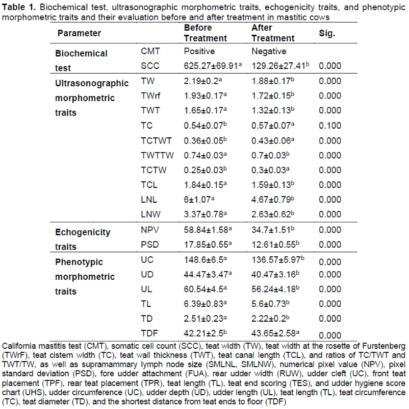
Udder and teat measurements (cm) were taken before and after treatment, including udder circumference (cm), udder depth (cm), udder length (cm), teat length (cm), teat circumference (cm), teat diameter (cm), and the shortest distance from teat ends to the floor (cm). The following phenotypic traits of cattle were recorded: fore udder attachment (FUA), rear udder width (RUW), udder cleft (UC), front teat placement (TPF), rear teat placement (TPR), teat length (TL), teat end scoring (TES), and udder hygiene score chart (UHS). Real-time, two-dimensional views of tissues were obtained using ultrasound. By establishing the correlation between the somatic cell count, the morphometric properties of the teats, and the supramammary lymph nodes in the dairy cows, we succeeded in our objective in this study by providing a quick approach to identify mastitis.
Material and Methods
This study was approved by the Advance Studies and Research Board as a non -invasive technique (DAS/440). The experiments were conducted on private livestock farms that were committed to the production of milk under a semi-intensive production system. These were located in Lahore, in the province of Punjab (31.5204° N, 74.3587° E). There is an average temperature of 12.1-33.2 °C and a rainfall of 823.8 mm per year. The livestock farms had an average of 500 Holstein milking cows, with an average production of 22.3 L per cow/per day. Data on 48 cows was analysed for the test. A total of 48 teats from 48 clinically-affected, mastitic Holstein-Friesian cows in the third lactation stage, kept under the same conditions, fed by identical ration twice a day, and milked twice using an automatic milking system, were used. Lactating cows and cattle that passed the 60-day voluntary waiting period and were in a position to be inseminated were considered. Cows that had subclinical mastitis identified using CMT and SCC tests were not considered in this study because they received anti-inflammatory and antibiotic treatment to control udder infection. The diagnosis of clinical mastitis was made by the Veterinary Physician of the livestock farm, using the field test CMT and SCC. For negative, weak positive, distinct positive, and strong positive, CMT scores were 1 (no coagulation or gel formation), 2 (a coagulation begins but disappears after rotation, no gel formation), 3 (mixture coagulates, does not stick, and distinctly forms a gel), and 4 (mixture coagulates and sticks, tends to form jelly). Inclusion criteria were: one clinically affected quarter, third lactation, CMT (+), and SCC >450,000 cells/mL.
Ultrasonographic scanning of the teats was carried out in a water-filled plastic cup for 30 min before milking with a 7.5 MHz linear array transducer. Each examination and measurement was performed by the same clinician. The water in the cup was renewed after each use. The teats were carefully and slowly submerged in the water. To acquire clear images, contact gel (Konix®, Turkuaz Medikal Kozmetik, Istanbul) was applied to both transducer and cup.
Ultra-sonographic morphometry was done in 48 clinically-affected teats and 48 supramammary lymph nodes from 48 dairy cows in their third lactation. Teat width (TW), teat width at the rosette of Furstenberg (TWrF), teat cistern width (TC), teat wall thickness (TWT), teat canal length (TCL), and ratios of TC/TWT and TWT/TW, as well as supramammary lymph node size (SMLNL, SMLNW), were determined.
Any abnormalities in secretion, size, consistency, or temperature of all milking cows in the selected farms were thoroughly evaluated. Clinical mastitis was characterized by palpatory pain, milk alterations (blood-mixed milk, watery discharges, flakes, pus), and udder integrity variation. Udder health was assessed by clinical examination of the udder; California Mastitis Test (CMT; Kepro Cmt Solution, The Netherlands) and somatic cell count (Lactoscan SCC, Milkotronic Ltd, Bulgaria) were performed. Milk samples were collected before ultrasonographic examination. Clinically-affected quarters (mastitis) of the udder in the present study had a minimum of 500 000 cell count/mL. The farms were surveyed twice a year, once during the dry season and once during the wet season. Farmers were visited in order to gather information about their farming system. To detect clinical mastitis, udders and milk were analysed. The udder and CMT scores were recorded in accordance with the National Mastitis Council method (1999). Udder palpation findings were rated as 1, 2, 3, and 4 for no swelling or discomfort in the udder, swollen ventral quarter, generalised swollen quarter, and swollen and painful udder, respectively.
Risk factors associated with the incidence of teat and udder affections were observed and analysed using phenotypic morphometry. The identification of risk variables is critical for the design of mastitis management programmes in cows. During a farm tour, information regarding farms and management was gathered.
Statistical analysis was performed using SPSS 21.0 software (SPSS Inc., Chicago, IL, USA). The data was analysed using analysis of variance (ANOVA); the significance level was set at 5%. Pearson correlations were performed using the same software to investigate the relationship of different parameters. Chi-square tests were used to determine the association between the morphometric traits of the udder and teats. The data are shown as mean ± standard deviation (SD).
Results and Discussion
A total of 48 clinically-affected cows were subjected to the present study for morphometric evaluation (n = 48) in which clinical mastitis was evident from seven different dairy farms in the Lahore district and its surroundings. Results of the measurements of all teat parameters are shown in Table 1. Ultrasonography of udder parenchyma, teats, and supramammary lymph nodes in cows is an optimal choice for research as it is non-invasive, with minimal restraint of the animals. Ultrasonographic examination is simple to perform and has the advantage of facilitating maintenance and sequential examination of the experimental animals, with increased welfare (Szencziová & Strapák, 2012).
Ultrasonographic images of the parenchyma of the healthy mammary gland appear as a homogenous structure of average echogenicity, filled with anechoic content (milk) (Khoramian, 2015). This distinctive appearance is due to the uniform distribution of glands and connective tissue (Yano & Rudner, 2022). It is possible to see where the big lactiferous ducts enter the gland cistern. The amount of fullness in the milk gland affects how echogenic it is. This distinctive appearance is due to the uniform distribution of gland parenchyma, which has a lower echoic density, and connective tissue, which has a higher echoic density.
Ultrasonographic observations of clinical mastitis include an aberrant shape of the teat canal and sinus, the lack of the three-layered appearance of the teat, an overlapping papillary duct and rosette of Furstenberg, and a clear picture of the udder parenchyma and gland sinus. This is similar in the case of subclinical mastitis, but the intensity increases (Cartee et al., 1986). The typical teat walls were seen as having three distinct layers: an outside layer that was hyperechoic, a middle layer that was thicker and hypoechoic, and an inner layer with dilated anechoic walls (Amin et al., 2017). The teat wall histology showed three distinct layers: the teat skin layer (outer), the fibro-muscular vascularized layer (middle), and the boundary mucous membrane layer (inner).
The teat width (TW) in clinically affected quarters was larger (2.19 ± 0.2 cm) (P <0.05) than teat width (1.88 ± 0.17 cm) when dairy animals were declared healthy with CMT (negative) and SCC <150 cells/mL (Table 1). The exterior, middle, and inner hyperechoic layers of the teat wall could be observed clearly. The outside, middle, and interior hyperechoic layers of the teat wall were observable. An annular fold existed between the gland cistern and the teat. There were additional blood arteries coming from the Furstenberg's venous ring. The use of the transducer in the water bath technique was simple. The teat canal and sinus both displayed regular outlines (Vidal et al., 2018).
The teat width at the Furstenberg rosette (TWrF) in clinically-affected quarters was greater (P <0.05) than in healthy cows at 1.93 ± 0.17 cm and 1.72 ± 0.15 cm, respectively treatment (Table 1).The mean teat wall thickness (TWT) was 1.62 ± 0.17 cm in mastitic cows and 1.32 ± 0.13 cm in the control (P <0.05); teat cistern (TC) measurements were similar (0.54 ± 0.07 cm and 0.57 ± 0.07 cm, respectively; P >0.05) (Table 1).
Mastitic cows exhibited a partly-visible teat cistern, which was anechoic due to the presence of watery milk. The presence of hypoechoic, caseated material completely obstructed the teat cistern, and the three-layered pattern of the teat wall was disrupted. There was also thelitis, as evidenced by thicker teat wall and increased diameter, as well as Furstenberg blockage with hyperechoic, caseated materials in the teat cistern (More et al., 2022). The afflicted teat of a cow with mastitis includes hyperechoic flakes, and the teat canal and Furstenberg disappear. A thick, hyperechoic teat wall, as well as many hyperechoic structures in the teat cistern and udder parenchyma, and complete replacement of milk alveoli with hyperechoic fibrous tissue have been reported (Youssef, 2012; Abd-El-Hady, 2015). Similarly, the ratio between teat cistern diameter and teat wall thickness (TC/TWT) was lower before treatment (0.36 ± 0.05) than after treatment (0.43 ± 0.06) (P <0.05) (Table 1). The ratio between teat wall thickness and teat width (TWT/TW) before treatment was higher (P <0.05) (0.74 ± 0.03) than after treatment (0.7 ± 0.03). The ratio between teat cistern width and teat width (TC/TW) before treatment was higher (P <0.05) (0.25 ± 0.03) than after treatment (0.43 ± 0.06). Teat canal length (TCL) before and after treatment was 1.84 ± 0.15 cm and 1.59 ± 0.13 cm, respectively (P <0.05) (Table 1).
A total of 48 supramammary lymph nodes were evaluated on both sides. The supramammary lymph node is situated 1.5-2 cm below the udder surface, caudal and dorsal to each hindquarter. It is possible to recognize a typical lymph node as an oval-shaped object with a thin echogenic capsule. The parenchyma of the node is hypoechoic, and the hilar region with its arteries is represented by a central, linear, echogenic structure. This finding is consistent with that found in sheep (Hussein et al., 2015) and cattle (Amin et al., 2017). A portable ultrasound machine with a 3.5-5 MHz convex transducer was used to identify the supramammary lymph node size (Kaixin 5600, VET Portable Ultrasound). The length (dorsoventral dimension) and depth (caudocranial dimension) were measured (Khoramian et al., 2015).
The length of the lymph node before treatment was higher (P <0.05) at 6 ± 1.07 cm than the mean after treatment of 4.67 ± 0.79 cm (Table 1). The mean width of the lymph node before treatment was 3.37 ± 0.78 cm 2.63 ± 0.62 cm after treatment (P <0.05) (Table 1). Somatic cell count (SCC) indicates a protective effect in the animal body to fight infectious organisms and is therefore used to evaluate the incidence of tissue damage due to mastitis. An elevated SCC in milk has a negative influence on the quality of raw milk. The ultrasound imaging of a subclinical mastitic udder reveals a considerable amount of turbid echoic content (milk) in the cistern, with normal teat wall thickness and teat canal thickness (Scholten, 2022); chronic, severely-fibrosed udder tissue reveals hyperechoic tissue without acini or milk in the cistern, as well as hyperechoic teat canal thickness. The ultrasound picture of the udder parenchyma reveals a hypoechoic visible capsule containing hypoechoic and hyperechoic secretion (a calcified pus abscess), as well as a hyperechoic teat wall thickness with a limited lumen. As a result, the use of ultrasonography can discern probable alterations, prognosis, and monitoring in response to therapy (Ebtsam et al., 2020).
Typical alterations that occur throughout the mastitis stages in the mammary glandular parenchyma, teat, and milk exhibit varying echogenicity. The parenchyma of the mammary glands is shown as a homogeneous, hypoechoic structure; blood arteries are anechoic; and lobes and alveoli are anechoic globular structures (Nissan et al., 2022). According to some accounts, the mammary gland is homogeneous and hyperechoic with anechoic alveoli. The udder cistern is echogenic due to the presence of milk, and its wall is uneven due to the opening of numerous lactiferous ducts. The lactiferous ducts and arteries of the bovine mammary gland are shown as anechoic structures. The glandular parenchyma of cows with acute mastitis are non-homogenous and low to hyperechoic. The glandular parenchyma of cows with chronic mastitis is hyperechoic, with few lactiferous ducts (Meinecke-Tillmann & Meinecke, 2007).
With a small teat canal, diagnostic teat-wall thickness is also indicative. The normal animal mammary gland examined by ultrasonography exhibits parenchyma that are homogenous and hypoechogenic with interspersed anechoic blood arteries, milk alveoli, lactiferous ducts, and gland cisterns (Nishimura et al., 2011). When filled with milk, the teat cistern is defined as a dilated anechoic structure with few hypoechogenic regions. The papillary duct is viewed as a single hypoechoic zone, and the structures that separate it from the teat cistern resembled the Furstenberg venous ring (Abu-Seida, 2016).
Udder circumference decreased after treatment, 148.6 ± 6.5 cm vs 136.57 ± 5.97 cm, respectively (P <0.05). Udder diameter decreased from clinically-affected to healthy cattle (44.47 ± 3.47 cm vs 40.47 ± 3.16 cm; P <0.05); similarly for udder length (60.54 ± 4.5 cm vs 56.24 ± 4.18 cm; P <0.05) (Table 1). The overall teat length decreased after treatment (6.39 ± 0.83 cm vs 5.6 ± 0.73 cm; P <0.05). The teat diameter decreased after treatment (2.51 ± 0.23 cm vs 2.22 ± 0.2 cm; P <0.05). The shortest distance from the tip of teat to floor increased (42.21 ± 2.5 cm vs 43.65 ± 2.58 cm; P <0.05).
Pearson's correlations indicated that TW was different (P <0.01) and positively correlated with TWrf (r = 0.989), TWT (r = 0.958), TC (0.327), TCL (r = 0.523), and SCC (r = 0.620) (Table 2). TC/TWT and TWT/TW showed significant (P <0.05) but negative ipsilateral correlations (r = -0.239) and positive ipsilateral correlation (r = 0.233) with TW. TW had a negative correlation with TC/TW (r = -0.551) and with treatments (r = -0.651). SCC was different between treatments (P <0.01) and positively correlated with TW (r = 0.620), TWrf (r = 0.508), TWT (r = 0.709), TWT/TW (r = 0.551), and TCL (r = 0.649). TC/TWT and TC/TW were negatively correlated (r = -0.546) and (r = -0.684), respectively. SCC had a non-significant (P >0.05) negative correlation with teat cistern width (TC) (r = -0.178) (Table 2).
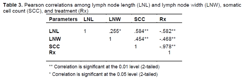
With Pearson's correlation, it was statistically observed that LNL was different between treatments (P <0.01) and positively correlated with SCC (r = 0.584) and negatively correlation with Rx (r = -0.582), whereas, LNW was affected by treatment and had a positive ipsilateral correlation (r = 0.255) (Table 3).
The nodes were distinct from the surrounding tissue in the majority of cows. The parenchyma of the nodes was hypoechoic to anechoic with a bright hyperechoic region in the centre and a narrow hyperechoic line around the nodes. Although the node sizes fluctuated, their internal design was similar. The lymph nodes were clearly distinguished because they had an oval appearance and a thin echogenic capsule (Bhargava and Bhargava, 2018). Within the hypoechoic parenchyma of lymph nodes, there was a focused hyperechoic region. The teat canal was seen as a narrow, bright white line that was surrounded by parallel, dark grey bands using different frequencies (7.5-18 MHz) of the linear array transducer in the vertical plane. The clinical mastitis lymph node was clearly visible, enlarged, and almost totally lost the highly echogenic hilus. The entire structure looked to be completely hypoechogenic (Samreen et al., 2020). Accordingly, considerable alterations were discovered in morphometric parameters and the ultrasonographic configuration of supramammary lymph nodes in cattle with clinical mastitis.
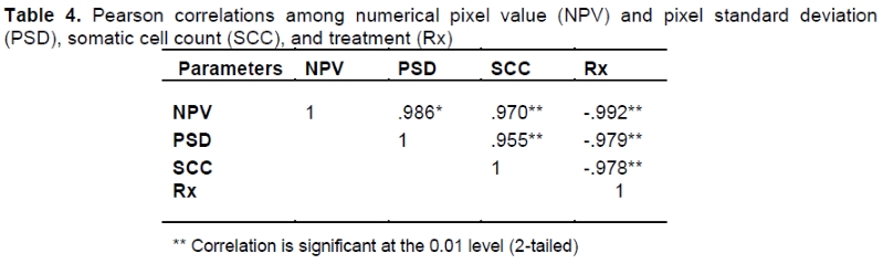
NPV was different between treatments (P <0.01)and positively correlated with PSD (r = 0.986) and SCC (r = 0.970) and negative correlated with Rx (r = -0.992). This density disparity allowed for the ultrasonographic separation of the wall layer. The teat cistern was visible as a small, hypoechogenic dot-filled region with dilated anechoic walls. The normal Furstenberg rosette was a bright and thin white line composed of two to three parallel, thin, hypoechogenic lines at the end of the teat. The teat canal and cistern lacked the distinctive three-layered appearance of clinical mastitis and revealed an uneven contour lining, homogeneous and hypoechogenic contents, a smaller lumen, a somewhat thicker wall (Abu-Seida, 2016). During clinical mastitis, the Furstenberg rosette was not clear (Plummer & Plummer, 2012).
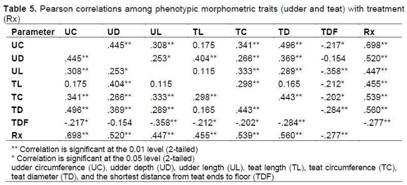
Mastitis is known to be associated with teat-end condition, cow dirtiness, breed, production system, and stage of lactation, all of which are substantially connected to management and rearing environment. Mastitis, according to Iraguha et al. (2015) is a complex condition that is linked to practically every imaginable management and environmental component. Pearson's correlations indicated 17 positive and three negative correlations (P <0.01), and one positive and three negative correlations (P < 0.05) (Table 5).
Udder and teat measurements (cm) were taken before and after treatment. There was an effect of treatment on UC (P <0.01), which was positively correlated with UD (r = 0.445), UL (r = 0.308), TC (r = 0.341) and negatively correlated with TDF (r = -0.217). There was a treatment effect on UD (P <0.01) and a positive correlation with UC (r = 0.445), TL (r = 404), TC (r = 0.266), TD (r = 0.369) and UL (r = 0.253). There was a treatment effect on UL (P <0.01) and a positive correlation with TC (r = 0.333), TD (r = 0.289) and a negative correlation with TDF (r =- 0.358). There was a treatment effect on TL (P <0.01) and a positive correlation with TC (r = 0.298) and groups (r = 0.455), and negative ipsilateral (P <0.05) correlation with TDF (r = -0.212). There was a treatment effect on TC (P <0.01) and a positive correlation with TD (r = 0.443) and groups (r = 0.539) and negative ipsilateral correlation with TDF (r = -0.202). There was a treatment effect on TD (P <0.01) a negative correlation with TDF (r = -0.284), and positive correlation with group (r = 0.560). There was a treatment effect on TDF (P <0.01) and a negative correlation with groups (r = -0.277).
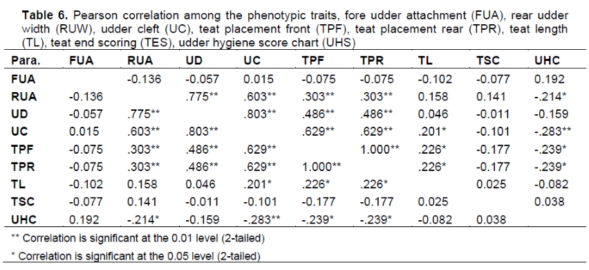
Pearson's correlations indicated 10 positive correlations and one negative correlation (P <0.01), and three positive and four negative correlations (P <0.05) (Table 6). Phenotypic morphometric evaluation of the udder and teat parameters of clinically-affected mastitic cows showed that 22.9% of the cows had extremely broken front udder base attachment, 41.7% had intermediate attachment, and 31.9% had extremely tight attachment. A proportion of 20.1% had extremely narrow rear udder attachment, 56.2% had intermediate attachment, and 29.9% had extremely wide attachment. Of the cows, 22.9% had a deep udder below the hock, 62.5% were below the hock, and 14.6% were above the hock (P <0.05).
A proportion of 33.2% had a weak udder cleft, 62.5% had an intermediate cleft, and 6.2% had a strong cleft (P <0.05). Phenotypically, front and rear teat placement indicated 54.2% with outside-pointed teat placement, 45.8% had middle placement (P <0.05). A total of 33.3% of the cows had small teats, 29.2% had intermediate teats, and 37.5% had large teats (P <0.05). A total of 6.2% of the cows had no ring teat ends, 27.1% had smooth teat ends with a slight ring, 47.9% had a rough ring, and 18.8% had very rough teat end. A total of 4.2% of the cows had a dirt-free udder, 37.5% had slight dirt, 41.7% had mild dirt, and 16.7% had udder covered with dirt (Table 6).
The length and size of the udder were shown to be related to milk production. Teats that were longer, wider, and closer to the ground were shown to be more prone to mastitis in Holstein-crossbred cows and riverine buffaloes. While larger teat diameter provides for quicker milk production, it also allows more irritants or germs to enter the mammary system, raising the risk of mastitis development. Teat infection can arise as a result of numerous circumstances. The teat and teat connective tissue act as natural defence against udder infections. There was no discernible effect of teat canal length on any new invading infection of the udder, but in different studies, it has been concluded that the increase in length increases the chances of infection, A comparatively longer teat canal leads to more udder infections and may be related to chronic or subclinical mastitis, according to a theory that suggests the length of the teat canal is not related to acute mastitis. According to certain reports, both large and short teat canals encourage bacterial colonization of the mammary gland. But in the current study, the diagnostic effect of ultrasound was discussed. Future work on teat canal length and diameter from the prospective of infection will be pursued (Zigo et al., 2021). It has been established that the udder and teat morphology are strongly related to the development of mastitis in dairy cattle and buffalo. Clinically-infected animals showed noticeably higher RUW, loose FUA, and lower RUH. Additionally, it was discovered that the mastitic animals had deeper udders than the healthy ones (Sharma et al., 2017)
While larger teat diameter provides for quicker milk production, it also allows more irritants or germs to enter the mammary system, raising the risk of mastitis development. Teat infection can arise as a result of numerous circumstances (Bharti et al., 2015). Mastitis prevalence increases quantitatively with teat-end injury and keratinization increased. Furthermore, badly injured, ulcerated teat-ends with scabs or open lesions were the most vulnerable to mastitis. Mastitis prevalence rose as cow dirtiness increased, with the extensive production system having a greater prevalence of mastitis than semi-intensive systems. Mastitis is more common among pure breeds. It was also greater in the late and early phases of lactation than in the middle of lactation. The teat and teat connective tissue act as natural defence against udder infections. There was no discernible effect of teat canal length on any new invading infection of the udder, but in different studies, it has been concluded that the increase in length increases the chance of infection, A comparatively longer teat canal leads to more udder infections and may be related to chronic or subclinical mastitis, according to a theory that suggests the length of the teat canal is not related to acute mastitis (Ndihokubwayo et al., 2019). According to certain reports, both large and short teat canals encourage bacterial colonization of the mammary gland.
Because of their proximity to the ground, deep udders are more likely to be contaminated with environmental infections due to liner slips and sores. Only when animals with mastitis had loose central ligaments was the length of the central ligament proven to be relevant in Sahiwal cattle as stated by Underwood et al. (2015). Teat morphometric features relating to length and diameter were shown to be of substantial value. The teat canal not only serves as a physical barrier against invading microorganisms, but it also serves as a source of antibacterial compounds to help prevent new infections. Cows and buffaloes with longer teats had a greater prevalence of clinical and subclinical mastitis, according to previous research. Mastitis-affected cows also had considerably longer rear teats than healthy animals. Teat circumference and teat diameter were also measured in addition to length (Sarba & Tola 2017). Mastitic cattle had larger teat diameters than healthy animals. This is consistent with previous research that indicated thicker teats to be a risk factor for mastitis. Mastitic udders have a greater gap between their rear teats and a significant positive correlation with length of udder ligament.
In terms of UD, the cows that had an udder base that was lower than the hock joint had greater mastitis susceptibility (Rehman & Khan, 2012b). The deeper udders were shown to have a greater risk of developing mastitis due to their increased propensity to become dirty and subsequently infected with environmental microorganisms. A deeper udder may also make it difficult for the cow to lie down comfortably and raises the possibility of teat sores. Additionally, lower udders needed longer milking times and have more liner slides, which are detrimental to udder health. The increased roughness of the teat end caused by the prolonged machine-on time is linked to a higher risk of developing new intramammary infections (Tančin et al., 2018).
Conclusion
Ultrasound provides real-time, two-dimensional insights of tissues. We achieved the study's objective of offering a rapid method to diagnose mastitis by demonstrating a link between the somatic cell count, the morphometric features of the teats, and the supramammary lymph nodes in the dairy cow and different associated risk factors.
Acknowledgements
We acknowledge Dr. Sadaf Aslam and Dr. Hamid Akbar for their guidance.
Author Contributions
SA, MAK, HM, and MH conceptualized the hypothesis of this manuscript. OMA, SA, and MH conducted the research. OMA and MI statistically analysed the data, wrote, and edited the manuscript. SA, HA, NH, and MI reviewed the manuscript.
Conflict of Interest
The authors have declared that there is no conflict of interests regarding the publication of this article.
References
Abd-El-Hady, A., 2015. Clinical observations on some surgical udder and teat affections in cattle and buffaloes. Scholars J Agri. Vet. Sci., 2(4A), 270-281. [ Links ]
Abu-Seida, A. M., 2016. Current status and prospect of ultrasonographic application in buffaloes. Asian J Anim Vet Adv., 11, 144-157. DOI:10.3923/ajava.2016.144.157 [ Links ]
Alhussien, M. N., Dang A. K., 2018. Milk somatic cells, factors influencing their release, future prospects, and practical utility in dairy animals: An overview. Vet. World. 11(5), 562. https://doi.org/10.14202/vetworld.2018.562-577. [ Links ]
Amin, N., Patil D., Kelawala D., Parikh P., Mer D., Gameti K., Gohil K., 2017. Ultrasonography of udder and teat in dairy animals. Rumin. Sci. 6(1), 173-177. [ Links ]
Bajwa, I., Khan M., Khan M., Gondal K., 2004. Environmental factors affecting milk yield and lactation length in Sahiwal cattle. Pakistan Vet. J. 24(1), 23-27. [ Links ]
Bhargava, S., Bhargava S. K., 2018. Differential Diagnosis in Ultrasound. Jaypee Brothers Medical Publishers. [ Links ]
Bharti, P., Bhakat C., Pankaj P. K., Bhat S. A., Prakash, M.A., Thul, M.R., Japheth, K.P., 2015. Relationship of udder and teat conformation with intra-mammary infection in crossbred cows under hot-humid climate. Vet. World, 8(7), 898. DOI:10.14202/vetworld.2015.898-901 [ Links ]
Brito, L., Bedere N., Douhard F., Oliveira H., Arnal M., Penagaricano F., Schinckel A., Baes C., Miglior F., 2021. Review: Genetic selection of high-yielding dairy cattle toward sustainable farming systems in a rapidly changing world. Anim. 15, 100292. DOI:10.1016/j.animal.2021.100292 [ Links ]
Cartee, R., Ibrahim A., McLeary D., 1986. B-mode ultrasonography of the bovine udder and teat. J. Am. Vet. Med Assoc. 188(11), 1284-1287. DOI:10.1016/j.cvfa.2009.07.007. [ Links ]
Caruolo, E., Mochrie R., 1967. Ultrasonograms of lactating mammary glands. J. Dairy Sci. 50(2), 225-230. DOI:10.3168/jds.S0022-0302(67)87392-2 [ Links ]
Cheng, W. N., Han S. G., 2020. Bovine mastitis: risk factors, therapeutic strategies, and alternative treatments: A review. Asian-Australas. J. Anim. Sci. 33(11), 1699-1713. DOI:10.5713/ajas.20.0156 [ Links ]
Council, N. R., 1988. Lactation biology and methods of increasing efficiency. In. Designing Foods: Animal Product Options in the Marketplace. National Academies Press (US). [ Links ]
Flöck, M., Winter P., 2006. Diagnostic ultrasonography in cattle with diseases of the mammary gland. Vet. J. 171(2), 314-321. DOI:10.1016/j.tvjl.2004.11.002 [ Links ]
Gaviglio, A., Corradini A., Marescotti M. E., Demartini E., Filippini R., 2021. A theoretical framework to assess the impact of flooding on dairy cattle farms: Identification of direct damage from an animal welfare perspective. Anim. 11(6), 1586. DOI:10.3390/ani11061586. [ Links ]
Getu, A., Campus S., 2015. The role of conformational traits on dairy cattle production and their longevities. Open Access Library Journal. 2(03), 1. DOI:10.4236/oalib.1101342 [ Links ]
Hussein, H. A., EL-Khabaz K. A., Malek S. S., 2015. Is udder ultrasonography a diagnostic tool for subclinical mastitis in sheep? Small Rumin. Res. 129, 121-128. DOI:10.1016/j.smallrumres.2015.05.010 [ Links ]
Iraguha, B., Hamudikuwanda H., Mushonga B., 2015. Bovine mastitis prevalence and associated risk factors in dairy cows in Nyagatare District, Rwanda. J. South African Vet. Assoc. 86(1), 1 -6. DOI:10.4102/jsava.v86i1.1228 [ Links ]
Khan, M., Khan M., 2016., Genetic parameters of udder traits and their relationship with milk yield in Sahiwal cows of Pakistan. J. Anim. Plant Sci. 26(4). [ Links ]
Khoramian, B., Vajhi, A., Ghasemzadeh-Nava, H., Ahrari-Khafi, M.S., Bahonar, A., MS A. K., 2015. Ultrasonography of the supramammary lymph nodes for diagnosis of bovine chronic subclinical mastitis. Iran J Vet Res. 16(1), 75-77. [ Links ]
Martins, S. A., Martins, V. C., Cardoso, F. A., Germano, J., Rodrigues, M., Duarte, C., Bexiga, R., Cardoso, S., Freitas, P. P., 2019. Biosensors for on-farm diagnosis of mastitis. Front. Bioengineer. and Biotech. 7, 186. DOI:10.3389/fbioe.2019.00186 [ Links ]
Meinecke-Tillmann, S., Meinecke, B., 2007. Ultrasonography in small ruminant reproduction. Comp. Reprod. Biol. 349-376. DOI: 10.1002/9780470390290.ch 14 [ Links ]
Miglior, F., Fleming, A., Malchiodi, F., Brito, L. F., Martin, P., Baes, C. F., 2017. A 100-Year Review: Identification and genetic selection of economically important traits in dairy cattle. J. Dairy Sci. 100(12), 10251-10271. DOI:10.3168/jds.2017-12968. [ Links ]
More, S. J., McAloon, C., Boloña P. S., O'Grady, L., O'Sullivan, F., McGrath, M., Buckley, W., Downing, K., Kelly, P., Ryan, E. G., 2022. Mastitis control and intramammary antimicrobial stewardship in Ireland: Challenges and opportunities. Front. Vet. Sci. 310. DOI:10.3389/fvets.2022.748353 [ Links ]
Ndihokubwayo, F., Koç, A., Uzun, N., 2019. Teat profile and its importance in dairy cows. Cappadocia, Turkey. pp. 325. [ Links ]
Nishimura, M., Yoshida, T., El-Khodery, S., Miyoshi, M., Furuoka, H., Yasuda, J., Miyahara, K., 2011. Ultrasound imaging of mammary glands in dairy heifers at different stages of growth. J. Vet. Med. Sci. 73(1), 19-24. DOI:10.1292/jvms.09-0503. [ Links ]
Nissan, N., Bauer E., Massasa, E. E., Sklair-Levy, M., 2022. Breast MRI during pregnancy and lactation: Clinical challenges and technical advances. Insights into Imaging. 13(1), 1-21. [ Links ]
Plummer, P. J., Plummer C., 2012. Diseases of the Mammary. Sheep & Goat Medicine-E-Book. pp442. [ Links ]
Rehman, Z., Khan, M., 2012a. Environmental factors affecting performance traits of Sahiwal cattle in Pakistan. Pak. Vet. J. 32(2), 229-233. [ Links ]
Rehman, Z., Khan, M., 2012b. Genetic factors affecting performance traits of Sahiwal cattle in Pakistan. Pak. Vet. J. 32(3), 329-333. [ Links ]
Samreen, N., Dhage, S., Gerber, N. K., Chacko, C., Lee, C. S., 2020. Imaging and management of internal mammary lymph nodes. J. Breast Imag. 2(6), 530-540. DOI:10.1093/jbi/wbaa046 [ Links ]
Sarba, E. J., Tola, G. K., 2017. Cross-sectional study on bovine mastitis and its associated risk factors in Ambo district of West Shewa zone, Oromia, Ethiopia. Vet. World. 10(4), 398. DOI:10.14202/vetworld.2017.398-402 [ Links ]
Schauer, B., Wald, R., Urbantke, V., Loncaric, I., Baumgartner, M., 2021. Tracing mastitis pathogens: Epidemiological investigations of a Pseudomonas aeruginosa mastitis outbreak in an Austrian dairy herd. Anim. 11(2) 279. DOI:10.3390/ani11020279. [ Links ]
Scholten, B. A., 2022. Dairy Farming in the 21st Century: Global Ethics, Environment and Politics. Bloomsbury Publishing. [ Links ]
Sharma, T., Das, P.K., Ghosh, P.R., Banerjee, D., Mukherjee, J. 2017. Association between udder morphology and in vitro activity of milk leukocytes in high yielding crossbred cows. Vet. World. 10(3): 342. DOI:10.14202/vetworld.2017.342-347 [ Links ]
Szencziová, I., Strapák, P., 2012. Ultrasonography of the udder and teat in cattle: Perspective measuring technique. Slovak J. Anim. Sci. 45(3), 96-104. [ Links ]
Tančin, V., Mikláš, Š., Mačuhová, L., 2018. Possible physiological and environmental factors affecting milk production and udder health of dairy cows: A review. Slovak J. Anim. Sci. 51(1), 32-40. [ Links ]
Ebtsam, E. Z. Kotb, Fadel, M., Ola, A., Abd El-Fattah, A. M., Azab, A. Z., Leil., A., 2020. Ultrasonography, histopathological udder alterations and bacteriological investigations for diagnosis of mastitic goats. J. Appl. Vet. Sci. 5(2), 77-86. DOI:10.21608/javs.2020.85593 [ Links ]
Underwood W. J., Blauwiekel, R., Delano, M. L., Gillesby, R., Mischler, S. A., Schoell, A., 2015. Biology and diseases of ruminants (sheep, goats, and cattle). In. Laboratory Animal Medicine. Elsevier. pp. 623-694. DOI:10.1016/B978-0-12-409527-4.00015-8 [ Links ]
Vidal, J. D., Wood, C. E., Colman, K., Whitney, K. M., Creasy, D. M., 2018. Reproductive system and mammary gland. In. Toxicologic Pathology. CRC Press. pp. 889-1020. [ Links ]
Yano, L. M., Rudner, M. A., 2022. Postoperative breast. In. Modern Breast Cancer Imaging. Springer. pp. 331-414. [ Links ]
Youssef, A. H. K., 2012. Diagnostic imaging of the uro-genital system in sheep and goat. Benha University. [ Links ]
Zigo, F., Vasil, M., Ondrašovičová, S., Výrostková, J., Bujok, J., Pecka-Kielb, E., 2021. Maintaining optimal mammary gland health and prevention of mastitis. Front. Vet. Sci. 8, 607311. DOI:10.3389/fvets.2021.607311 [ Links ]
Submitted 31 October 2022
Accepted 10 August January 2023
Published 27 September 2023
* Corresponding author: obaid.abdullah@uvas.edu.pk

