


Services on Demand
Article
 English (pdf)
 Article in xml format
 Article references
Indicators

Related links
-
 Cited by Google
 -
 Similars in Google
Share

 Permalink
South African Journal of Animal Science
On-line version ISSN 2221-4062
Print version ISSN 0375-1589
S. Afr. j. anim. sci. vol.53 n.2 Pretoria 2023
http://dx.doi.org/10.4314/sajas.v53i2.08
Healthy, sub-clinical, and clinical mastitis in Holstein-Friesian cattle: A comparative echotextural and electrical conductivity study
Obaid Muhammad AbdullahI, #; Sadaf AslamI; Muhammad Arif KhanI; Hassan MushtaqII; Mubbashar HassanIII; Muawuz IjazIV
IDepartment of Veterinary Surgery and Pet Sciences, Faculty of Veterinary Sciences, University of Veterinary and Animal Sciences, Lahore 54000, Pakistan
IIDepartment of Epidemiology and Public Health, Faculty of Veterinary Sciences, Faculty of Veterinary Sciences, University of Veterinary and Animal Sciences, Lahore 54000, Pakistan
IIIDepartment of Clinical Sciences, College of Veterinary and Animal Sciences (Sub-Campus University of Veterinary and Animal Sciences, Lahore), Jhang 35200, Pakistan
IVDepartment of Animal Sciences, College of Veterinary and Animal Sciences (Sub-Campus University of Veterinary and Animal Sciences, Lahore), Jhang 35200, Pakistan
ABSTRACT
The goal of this research was to investigate the echotextural parameters, numerical pixel values (NPVs), and pixel heterogeneity (PSDs) of the lymph node and udder parenchyma, as well as the correlation between echotextural parameters and electrical conductivity (EC), in order to develop it as an alternative to laboratory testing for mastitic animal diagnosis and prognosis. The ultrasonographic images of each quarter were processed using digital image analyses to obtain mean NPVs and mean PSDs of mammary gland and lymph node parenchyma. The mean EC increased with the progression of infection. The mean NPVs of lymph node parenchyma decreased, whereas the mean NPVs of udder parenchyma increased from healthy to subclinical and clinical cows, respectively. The mean PSDs of lymph node and udder parenchyma increased with the progression and severity of infection from healthy to subclinical and clinical cows, respectively. It was concluded that, in dairy cattle, the variation in echotextural variables (NPVs and PSDs) of mammary gland and supramammary lymph node parenchyma and electrical conductivity appeared to be good indicators for the diagnostic and prognostic evaluation of sub-clinical and clinical mastitis in animals and simultaneously assists in the evaluation of udder health status.
Keywords: lymph node, udder, numerical pixel values, pixel heterogeneity
Introduction
Mastitis is one of the most serious diseases to afflict dairy herds and has an influence on the sector's profitability and production (Capper and Cady, 2020). Worldwide, mastitis is caused by a wide variety of microbes, including bacteria, viruses, and fungi. There are many contributing risk factors for bovine mastitis. Mastitis is a collaborative effort between pathogens, hosts, and the surrounding environment. These considerations are integral to the creation of mastitis control programmes. Bovine mastitis is an expensive disease because it causes losses (due to a drop in production) and expenses (due to the treatment of the disease), which are cumulative, with additional inputs to reduce the level of mastitis. Treatment, production losses, culling, variations in product quality, and the risk of contracting other diseases all contribute to the monetary burden of mastitis (clinical or subclinical). Costs associated with diagnostic tests, other disease expenses, animal culling, and lost milk production (Halasa et al., 2007; Hogeveen et al., 2011) due to drug rejection all contribute to the final price of the product.
Mastitis is classified into three types based on the severity of the inflammation: sub-clinical, chronic, and clinical. Visual abnormalities such as red and swollen teats and fever are easily detectable in dairy cows with clinical mastitis. In contrast to clinical mastitis, subclinical mastitis does not show observable changes in the udder or milk, although milk supply does decrease as the somatic cell count rises (Bhumarkar et al., 2021). Although it is impossible to quantify the financial losses caused by subclinical mastitis, experts agree that it causes herds to incur more enormous financial losses than clinical instances (Motaung et al., 2017). Chronic mastitis is an inflammatory illness that advances over several weeks with occasional clinical outbursts. The entire economic loss due to dairy mastitis is estimated to be on average $147 per cow per year. Production losses and culling have a substantial impact on this cost, accounting for 11-18% of the gross margin per cow per year. Mammary tissue injury, which impairs milk production, accounts for 70% of total losses (Puerto et al., 2021).
Technologies are making it easier to gather, analyse, and share information and data on animal health. In the field of animal health, these kinds of technologies (e.g., sonography) have not yet been used to their full potential in terms of early disease detection and quick responses to natural or planned disease outbreaks. Globally, there is a growing demand for better and more advanced mHealth and eHealth technologies that can collect and analyse data in real time. Animal tracing systems and supply chain management are crucial to ensuring the safety of animals and humans and in facilitating commerce (Holmstrom and Beckham, 2017; Özmen et al., 2022).
Ultrasound is commonly used in human medicine to check the mammary glands. Cystic and solid lesions are differentiated using 5-13 MHz scanners. These scanners can investigate areas that traditional mammography cannot (for example, metastases in the axillary lymph nodes). Ultrasound is a non-invasive and non-destructive diagnostic tool mainly used by veterinarians and places minimal stress on the animals. In recent decades, it has been used for the evaluation of morphometric traits of the udder and teats in dairy animals (Schwarz et al., 2020). Ultrasound provides real time images of the healthy and mastitic organ, which can then be used for accurate diagnosis and prognosis. In addition to digital ultrasonographic images, there is computer-assisted echotextural analysis (e.g., ImageJ) software, which can be used to classify the udder health status of healthy and affected udder quarters based on the mean numerical pixel values (NPV) and pixel heterogeneity (PSD) of udder parenchyma and lymph nodes (Zhang et al., 2022). This method has been tried on a range of animal species of veterinary relevance (Santos et al., 2015). Ultrasonographic imaging allows for the frequent and safe evaluation of a wide range of internal organ areas in animals.
Numerous studies have been published on the ultrasonographic examination of the ruminant mammary gland, but there appears to be a scarcity of data on the diagnostic utility of mammary gland and supramammary lymph node ultrasonography in cattle over the course of a longitudinal study using healthy and mastitic cattle. As a result, the purpose of this research was to determine if ultrasonography could aid in the diagnosis and prognosis of subclinical and clinical mastitis in Holstein-Friesian cows (Schwarz et al., 2020). The Holstein-Friesian was chosen due to their superlative milk production and the availability of Holstein-Friesians globally (Straczek et al., 2021).
Only digital image analysis allows for objective examination of echogenicity based on measurements of pixel intensity. The standard deviation of pixel intensity has previously been used to quantitatively evaluate ultrasonic homogeneity/heterogeneity. Pixel heterogeneity in udder ultrasonography has been linked to tissue biochemical composition. Numerical pixel values (NPVs), a quantitative measure of pixel brightness, can be calculated using a variety of computer-assisted image analysis applications. The NPVs and PSDs are both unitless numbers; the NPVs range from 0 (black) to 255 (white), and the PSD is the standard deviation of the NPVs in the research area (Murawski et al., 2019). Both are regarded as reliable evaluations of echogenic properties of tissue and effective predictors of concomitant histophysiological changes.
Over the last few decades, the EC of milk has been used as an indication of mastitis. The presence of ions is used to calculate EC (Ilie et al., 2010). When cows and buffaloes suffer from mastitis as a result of udder inflammation, the concentration of Na, K, and Cl ions rises. Mastitis alters the chemical makeup and nutritional content of animal milk (Neculai-Valeanu & Ariton, 2022).
Our study's initial goal was to measure the echotextural features of the mammary gland and supramammary lymph nodes of healthy, sub-clinically affected, and clinically-affected dairy cows in their third lactation. The second objective of this study was to evaluate the electrical conductivity of udder and lymph node parenchyma of dairy cows in healthy, sub-clinically affected, and clinically-affected mastitic cows.
Materials and Methods
Ethical approval was waived by the Advanced Studies and Research Board (ASRB) as handling and measurements of the animals were done by qualified veterinarians (University of Veterinary and Animal Sciences 54000, Lahore, Pakistan, DAS/440, 03-02-2022).
This study was conducted on seven different dairy farms in Lahore district and its surroundings in Punjab. The study compared the outcomes in three different categories of dairy cow: healthy, sub-clinical, and clinically mastitic cows. Forty-eight udder quarters and 96 supramammary lymph nodes of adult dairy cows were evaluated in each group. Animals were kept under the same climatic and environmental conditions, fed a total mioxed ration (TMR) twice a day, and milked twice a day using an automatic milking system (DeLaval VMS milking system, V300). Inclusion criteria: All animals were enrolled on the basis of (1) California mastitis test (CMT) and somatic cell count (SCC) data, (2) adult dairy cows (3rd lactation), (3) the animal must be in mid-lactation (60 days after parturition and before drying off), and (4) animals included in the sub-clinical and clinical study must have only one affected quarter.
For clinical evaluation of the udder, animals were cast and restrained. The mammary glands and teats were then palpated. The shape, size, temperature, and consistency of each gland were assessed and recorded. Any discomfort response and the development of abnormal structures within the gland were noted during the investigation (Haskell et al., 2022).
The CMT is the most popular field test in dairy cattle for the detection of subclinical mastitis; it does not produce numerical, but rather, categorical results. The test involves introducing a detergent to the milk, linear alkylbenzene sulphonate, which causes the release of DNA from leukocytes in the udder, which are then transformed into a gelatinous complex in association with protein agents in the milk. The findings are classified in different ways, such as negative when the reagent and blended milk are still watery. When the cell count rises, the reagent-milk combination practically hardens (CMT test, KEPRO CMT Solution, Deventer, The Netherlands). The somatic cell count is another traditional procedure used to diagnose the occurrence of mastitis in herds and to assess the hygienic quality of milk. A high somatic cell count number in raw milk indicates not only that the cows have mastitis, but also provides information on metabolic changes in the milk, up to and including production losses (LACTOSCAN SCC, Milkotronic Ltd, Nova Zagora, Bulgaria).
In a pioneering study for the detection of mastitis using non-traditional technology, a model was developed in which data variables were acquired, online and automatically, every 5 seconds, including electrical conductivity per mammary quarter, milk temperature, and milk production per cow. The combination of these parameters was determined to improve the detection of subclinical mastitis (Shoshani and Berman, 1992). The concentration of cations and anions in milk is used to calculate the EC. The most frequent ions in milk are Na+, Cl-, and K+. Mastitis causes changes in the concentrations of Na+, Cl-, and K+ in milk, which causes an increase in EC. The increased concentration of these ions allows these ions to enter the alveolar lumen. Electrical conductivity was measured using a conductometer (Metrohm 660; Herisau, Switzerland) equipped with a conductivity cell (cell constant = 1.24/cm) and automatic correction to the reference temperature (20 °C) using a Pt100 probe.
Ultrasonography is useful for detecting and monitoring changes in the teat(s) and mammary gland(s); it provides information on udder anatomy while being non-invasive, non-ionizing, fast, and painless. A low frequency (Kaixin 5600, VET Portable Ultrasound, BIOVET COMPANY LLC, Bila Tserkva, Ukraine) linear-array transducer was used to acquire ultrasonographic images of healthy, sub-clinical, and clinically-afflicted cows. The same operator examined each cow while it was standing up and without anaesthetic. To get high-quality ultrasonographic pictures, the udder hair was clipped as needed. Coupling gel was applied to the transducer probe prior to ultrasonography. By putting the transducer directly on the udder surface, the mammary parenchyma was scanned ultrasonographically. Images were taken either horizontally or vertically. To evaluate the parenchyma, the probe was placed on the caudal surface of each mammary gland along its longitudinal axis and moved from left to right; scanning depths of 80 and 120 mm were available.
It is possible to quantify the echogenicity of lymph nodes and udder parenchyma by using specialised software (Image J, Picture J, NACL Co. Ltd, Tokyo, Japan) to examine image brightness and calculate the mean grey value of the ultrasonographic image (El-Husseiny, 2020). Quantitative ultrasonographic assessment of the lymph node, udder parenchyma, and content echogenicity was done using ImageJ measures and image brightness analysis. Previously, Meiburger et al. (2018), Wijntjes & van Alfen (2021), and Mannelli et al. (2016) used a similar method to accurately interpret soft tissue ultrasonographic images based on quantitative assessment rather than qualitative evaluation, which is more descriptive but less accurate. Heterogeneity is therefore defined as the standard deviation in echogenicity pixels; echogenicity is defined as pixel intensity ranging from 0 (absolute black) to 255 (absolute white) (Lapuente et al., 2020).
Multivariate analysis of variance was used to compare the echotextural variables (NPV and PSD) using analysis of variance. SPSS (Version 23.0) was used to examine the correlation between echotextural variables of the udder and lymph nodes. A P <0.05 level was declared as statistically significant. All findings are given as means ± standard error.
Results
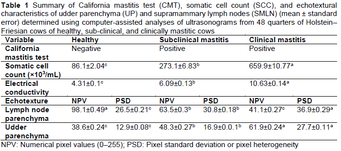
A total of 144 quarters of the udders and 192 pairs of supramammary lymph nodes were evaluated using a low frequency (3.5 MHz) linear probe ultrasound (Bustos et al., 2020). The animals were grouped into healthy, sub-clinical, and clinically mastitic ones. The scanned data were then quantified on the basis of echogenicity using a digital image analysis process with ImageJ software. An oval-shaped area of interest inside the parenchyma of the lymph node and a polygonal area of interest within the parenchyma of the mammary gland were chosen from the ultrasonogram and quantified. Data were quantified using numerical pixel values (NPV) and pixel heterogeneity (PSD). NPV describes the echogenicity of pixel intensity in the ultrasonogram, whereas PSD describes the heterogeneity.
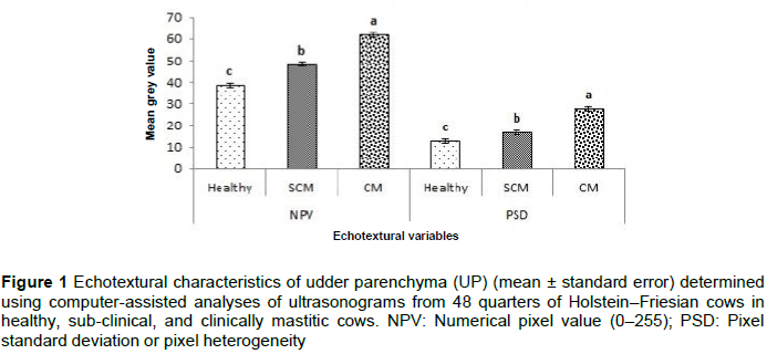
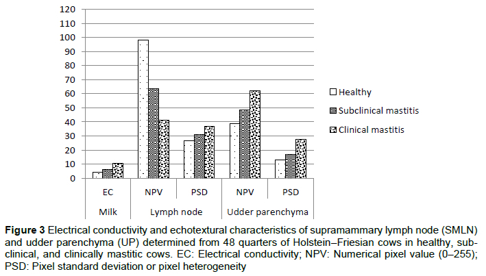
There were marked effects for both NPV and PSD during scanning of healthy, subclinical, and clinical udder quarters and supramammary lymph node parenchyma (Table 1). In the case of udder parenchyma, the NPV increased as infection in the udder progressed from subclinical to clinical cases. The NPV of the healthy udder was used as a control. In the case of healthy cows, the NPV of udder parenchyma was 38.6 ± 0.24, which increased in subclinical mastitic udder parenchyma (48.3 ± 0.27), and in clinical cases (61.9 ± 0.24). When compared to PSDs, there was a similar pattern because the PSD defines the level of heterogeneity and this increased as the infection progressed in parenchyma (12.9 ± 0.08 vs 16.9 ± 0.1 vs 27.7 ± 0.11) in healthy vs subclinical vs clinical mastitic cows, respectively (Table 1; Figure 1).
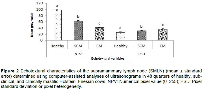
In the case of lymph node parenchyma, the NPV (means ± standard deviation) decreased as infection in the udder progressed from subclinical to clinical cases (NPV of healthy lymph nodes acted as the control). In case of healthy cows, the lymph node parenchyma was found to be 98.1 ± 0.49, which was higher than in subclinical mastitis (63.5 ± 0.3), and clinical cases (41.1 ± 0.27). When compared to PSD, the reverse pattern was evident. PSD defines the level of heterogeneity, which increases as the infection progresses in the parenchyma (26.5 ± 0.21 vs 30.8 ± 0.18 vs 36.9 ± 0.29) in healthy, subclinical, and clinically mastitic cows, respectively (Table 1, Figure 2). The mean EC increased (P <0.05) with the progression of infection (4.31 ± 0.1 vs. 6.09 ± 0.13 vs. 10.63 ± 0.14 in healthy, subclinical, and clinically mastitic cows, respectively (Table 1).
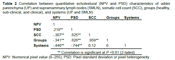
There were eight significant correlations among the echotextural variables of groups (healthy, subclinical, and clinical), systems (udder parenchyma and lymph node), and somatic cell count (Table 2). A positive linear correlation was noted only between NPV and PSD (r = 0.219), PSD and SCC (r = 0.625), PSD and between groups (healthy, subclinical, and clinical) (r = 0.626), and lastly, between SCC and groups (healthy, subclinical, and clinical) (r = 0.959). NPV was negatively correlated with SCC (r = -0.307) among all groups (r = -0.341), and between systems (lymph node and udder parenchyma; r = -0.446). Similarly, PSD also showed a significant but negative correlation with systems (lymph node and udder parenchyma) (r = -0.744) (Table 2).
Discussion
The main objective of this study was to examine the echotextural variables of udder and lymph node parenchyma, and at the same time, evaluate the non-traditional method of electrical conductivity in healthy cows, sub-clinical, and clinically mastitic cows to standardize the quantitative data. The data were evaluated using digital analysis in the form of NPVs and PSDs to make the diagnostic and prognostic power more effective (Zhang et al., 2022). It is plausible to recommend using NPV and PSD to determine the animal's disease state (healthy, subclinical, and clinical) using an ultrasonographic approach and digital image processing. In the current study, the effect of parenchymal echogenicity in healthy, sub-clinical, and clinically affected cows was evaluated. In the case of the mammary gland, NPVs were lower in healthy cows and higher in subclinical and clinical cases due to inflammation and secondly, due to the negative impact of infection on milk let-down. This is why the mean parenchymal echogenicity increases as the infection progresses. NPVs of the mammary gland decrease with the progression of infection. The PSD increases from healthy to clinical cases (McNally et al., 2017). In lymph node parenchyma, the NPVs decreased with the progression of infection from subclinical to clinical states. The increase in NPV is due to the loss of an echogenic fatty hilus and the misshapen organ (Belotta et al., 2019). PSD (mean heterogeneity) increases in the case of the mammary gland because there is quantifiable data to show the extent of the difference in echogenicity of that organ with the progression of infection. Utilizing various image analysis software programs, NPV, a quantitative measure of pixel brightness, was established. As they travel through tissues, ultrasound rays can be dispersed or deflected, or they might be reduced. Both echotextural variables objectively quantified tissue echogenicity. It is therefore plausible to infer that the ultrasonographic approach combined with computerized image processing may potentially determine milk composition despite the excretory product compartment's minor contribution to total udder content and echotexture.
Previously, echotextural variables of the mammary gland and their relationship to the physiochemical properties of milk in lactating cows were examined using ultrasound. Together with digital image analysis to determine tissue composition, Belotta et al. (2019) quantified the echotextural relationship in testicular tissue. As evidenced by the fatty content of the hilar part of the lymph node parenchyma, testicular NPVs were inversely correlated with protein content, whereas PSDs were favourably correlated with lipid content (Risk, 2011). Therefore, it is plausible to argue that ultrasonographic methods and digital image processing can assist in the detection of minute changes in the content and echotexture of udder and lymph node tissue. A key point for clinical application of quantitative pixel intensity measurement and computation of pixel intensity change is that these are measurements are at the level of the parenchyma, not a gross or overall evaluation at the organ level, which is a fundamentally different notion.
According to the authors, this is the first effort to link the quantitative echotextural features of lymph node parenchyma and mammary glands in healthy, subclinical, and clinically mastitic cows. There is a wealth of data to back up the use of computerized image processing and ultrasonic imaging to forecast the state of various interior organs and tissues. Some experimental and clinical research has also shown a link between echogenicity and tissue physiochemical features, e.g., in ram testes (Ahmadi et al., 2012), the chicken pectoralis major muscle (Harris-Love et al., 2016), and human dystrophic muscles (Schwarz et al., 2021). Image processing can be utilized to estimate intramuscular fat levels in live beef cattle following ultrasonography (Fabbri et al., 2021). Various echotextural properties of the mammary gland parenchyma in nursing ewes may be induced by differences in milk production and chemical composition in different genotypes of sheep (Kurowska et al., 2019). In mastitis, the degree of structural abnormality in the tissue dictates the sonographic image of the udder. In animals that have been unwell for a shorter amount of time, a hyperechoic parenchyma with only a few lactiferous ducts was also discovered. In the case of acute mastitis, there is a non-homogeneous (predominantly hypoechoic pattern), as well as an enhanced echogenicity as a result of fibrosis in chronic mastitis.
The ultrasonograms of the mammary glands were evaluated using commercially available image analysis software. Polygon measures were utilised to highlight the areas of interest (sinuses, alveoli, and parenchyma). For each region of interest, the mean numerical pixel value (NPV), pixel heterogeneity (standard deviation of NPV), and maximum and lowest pixel values were computed. Greyscale ultrasonograms indicated hyperechoic mammary parenchyma with coarse echotexture, as well as the presence of non-echogenic content (milk) in the cistern area, which appeared as echogenic patches in healthy animals. With the exception of milk, which was homogenous in H and SCM and heterogeneous in CM, the echotexture of the mammary parenchyma was heterogeneous in all three groups. The occurrence of flocculation and hyperechogenicity of the mammary parenchyma in clinically-ill buffalo and local Bulgarian goats are similar. An inflammatory process that leads to tissue fibrosis with a greater density than normal parenchyma might explain the hyperechogenicity. Because inflammation alters the echogenicity of afflicted organs, quantitative measurement can be utilised to quantify inflammation. To the best of the authors' knowledge, this new collection of data may pave the way for the development of ultrasonography as a diagnostic tool in clinical assessments of mammary glands (Murawski et al., 2019).
Ultrasonography has long been used as a diagnostic tool in dairy cows (Buckrell, 1988; Gürbulak et al., 2009). Transcutaneous udder and teat examinations are commonly performed to detect irregularities in milk flow, measure the size of distinct udder compartments, and evaluate the presence of mastitis (Schwarz et al., 2020). Elevated SSC levels in composite milk samples indicate preclinical or severe mastitis. When compared to animals with subclinical illness and clinically healthy controls, the variability of the mammary parenchyma in goats with clinical mastitis was much greater (Janik, 2013). The udder parenchyma and lymph nodes of dairy calves were assessed for soundness using computer-assisted analysis of a B-mode ultrasonogram as a non-invasive approach (Ribadu & Nakao 1999; Bobe et al., 2008). A normal cow mammary gland ultrasonic image is echoic and coarse. The anechoic antrums are usually poorly defined. They might be blood vessels or lactiferous ducts. Echoic particles increase in milk flow from inflamed lactiferous ducts. The huge lactiferous ducts are clearly visible entering the gland cistern. The fullness level of the lactiferous gland also influences its echogenicity.
The physicochemical and bacteriological properties of milk are affected by mastitis (Ruegg & Reinemann, 2002; Hadimli et al., 2013). During infection, the EC of milk increased as Na+ and Cl- ion concentrations increased. EC is considered a suitable characteristic for identifying mastitis in milk since it is highly sensitive to charged compounds. It can be used to detect subclinical mastitis, hence reducing economic losses in the early stages of infection. For a long time, EC was measured frequently during milking on a large number of cows. EC values in the current study in healthy, subclinical, and clinically infected quarters concur with those of Ferraboschi et al. (2021). The EC of normal milk at 25 °C is generally between 4.0 and 5.0 mS. Mastitis raises milk EC as a result of bacterial damage to udder tissue; the concentration of ions is increased (primarily Na+ and Cl-), while K+ and lactose concentrations in milk decrease. EC levels in milk from infected quarters have been observed to be higher in several investigations. The mean EC values for clinically infected quarters in this study correlate well with values of 5.0 to 9.0 mS reported in multiple experiments. The infected quarter in a clinically ill cow will have a higher EC during the bulk of the milking, particularly at the beginning and end. The mean EC values from clinically infected cows vary more than the mean EC values from healthy cows. Our findings for subclinically infected cows are consistent with other investigations, which reported absolute EC values of 6.45-6.85 mS and 4.83-7.03 mS. Substantial differences between healthy and subclinically infected quarters are more difficult to explain than differences between healthy and clinically infected quarters. Because milk from subclinically infected cows is only slightly physically altered, higher variation in EC measurements within milkings is unlikely. Clots, on the other hand, can be identified in a chronically diseased cow. Milk is conductive due to the high concentration of mineral salts such as sodium, chloride, potassium, calcium, magnesium. Mastitis caused by breast inflammation increases the concentrations of Na+ and Cl- ions. Somatic cell count is a good method for detecting subclinical mastitis, but the EC test is now commonly used to identify subclinical mastitis (Adkins & Middleton, 2018). Mastitis changes the chemical composition and nutritional value of the milk. A high SSC lowers milk and milk product quality, as well as shelf life, taste, and physiochemical properties of milk (Crotty, 2020). Mastitic milk contains both pathogens and bacterial toxins. The use of such milk may increase the risk of food-borne illnesses, both directly and indirectly.
Conclusion
The fluctuation in echotextural parameters of the parenchyma of the mammary glands and the parenchyma of the supramammary lymph nodes in dairy cattle was found to be a good indicator of the diagnostic and prognostic assessment of sub-clinical and clinically mastitic animals. The current study was conducted to establish the EC as an optimal method for identifying mastitis (particularly subclinical mastitis) in order to prevent economic losses in the early stages of infection and allow for earlier treatment. The selection of animals for future breeding purposes on the basis of their soundness, tested using non-invasive ultrasonography technology requires further study in a larger number of animals.
Acknowledgements
We acknowledge Dr Sadaf Aslam and Dr Muhammad Arif Khan for their guidance.
Author Contributions
SA, MAK, HM, and MH conceptualized the hypothesis of this manuscript. OMA, SA, and MH conducted the research. OMA and MI statistically analysed the data and wrote and edited the manuscript. SA, MH, and MI reviewed the manuscript.
Conflict of Interest
The authors declare that there are no conflict of interests regarding the publication of this article.
References
Ahmadi, B., Lau, C. P.-S., Giffin, J., Santos, N., Hahnel, A., Raeside, J., Christie, H., & Bartlewski, P., 2012. Suitability of epididymal and testicular ultrasonography and computerized image analysis for assessment of current and future semen quality in the ram. Exp. Biol. Med. 237 (2),186-193. DOI: 10.1258/ebm.2011.011050 [ Links ]
Adkins, P.R., Middleton J.R., 2018. Methods for diagnosing mastitis. Veterinary Clinics: Food Anim. Pract. 34(3), 479-491. [ Links ]
Belotta, A. F., Gomes, M. C., Rocha, N. S., Melchert, A., Giuffrida, R., Silva, J. P. & Mamprim, M. J., 2019. Sonography and sonoelastography in the detection of malignancy in superficial lymph nodes of dogs. J. Vet. Intern. Med. 33 (3),1403-1413. DOI: 10.1111/jvim.15469. [ Links ]
Bhumarkar, R., Mahajan, G., & Kumar, A., 2021. Doubling farmers income and attaining resilience in agriculture through crop diversification. Agriculture & Food: E-Newsletter. [ Links ]
Bobe, G., Amin, V. R., Hippen, A. R., She, P., Young, J. W., & Beitz, D. C., 2008. Non-invasive detection of fatty liver in dairy cows by digital analyses of hepatic ultrasonograms. J. Dairy Res. 75 (1),84-89. DOI: 10.1017/S002202990700297X [ Links ]
Buckrell, B., 1988. Applications of ultrasonography in reproduction in sheep and goats. Theriogenology 29 (1),71-84. DOI: 10.1016/0093-691X(88)90032-5 [ Links ]
Bustos, S. S., Kapoor, T., Schechter, L. S., Forte, A. J., Del Corral, G., & Manrique, O. J., 2020. Impact of social media presence on online reviews among plastic surgeons who perform gender confirming surgeries. J. Plast. Reconstr. Aesthet. Surg. 73 (4),783-808. DOI: 10.1097/GOX.0000000000003478 [ Links ]
Crotty A., 2020. A comparison of the use of whole milk and fat-filled milk powders for production of heat-stable long-life beverages. E-Thesis. https://cora.ucc.ie/items/cee6dfad-778a-4d36-bea5-335b9bf59793. [ Links ]
Capper, J. L., & Cady, R. A., 2020. The effects of improved performance in the US dairy cattle industry on environmental impacts between 2007 and 2017. J. Anim. Sci. 98 (1), skz291. DOI: 10.1093/jas/skz291 [ Links ]
El-Husseiny, M., 2020. Platelet-rich fibrin augmented versus non-augmented glycerolized bovine pericardium and polypropylene mesh for repairing of large abdominal wall defects. Eur. J. Natural Sci. Med. 3 (2), 7-28. [ Links ]
Fabbri, G., Gianesella, M., Gallo, L., Morgante, M., Contiero, B., Muraro, M., Boso, M., & Fiore, E., 2021. Application of ultrasound images texture analysis for the estimation of intramuscular fat content in the longissimus thoracis muscle of beef cattle after slaughter: A methodological study. Anim. 11 (4),1117. DOI: 10.3390/ani11041117 [ Links ]
Gürbulak, K., Canoglu, E., Abay, M., Atabay, Ö., Bekyürek, T., 2009. ineklerde subklinik mastitisin farkli yöntemlerle saptanmasi. Kafkas Univ. Vet. Fak. Derg. 15(5), 765-770. DOI: 10.30607/kvj.801954 [ Links ]
Haskell, S. R., Loghry, B. L., BAS, M., Wegner-Fowley, K., Gilsenan, V., Johnson, A. L., 2022. Physical Exam. Large Animal Medicine for Veterinary Technicians. Eds: Loly, S., Hopkinson, H. John Wiley and Sons, Inc. pp 67-103. [ Links ]
Holmstrom, L., Beckham, T., 2017. Technologies for capturing and analysing animal health data in near real time. Rev. Sci. Tech. 36, 525-538. [ Links ]
Hadimli, H. H., Sayin, Z., Erganis, O., Kürsat, K., 2013. Identification and antibiotic susceptibility of catalase-negative, gram-positive cocci isolated from dairy cows with subclinical mastitis. Eurasian J. Vet. Sci. 29(3), 153-158. [ Links ]
Halasa, T., Huijps, K., 0steras, O., Hogeveen, H., 2007. Economic effects of bovine mastitis and mastitis management: A review. Vet. Quart. 29(1), 18-31. DOI: 10.1080/01652176.2007.9695224 [ Links ]
Hogeveen, H., Huijps, K., Lam, T., 2011. Economic aspects of mastitis: New developments. New Zealand Vet. J. 59(1), 16-23. DOI: 10.1080/00480169.2011.547165 [ Links ]
Harris-Love, M. O., Seamon, B. A., Teixeira, C., & Ismail, C., 2016. Ultrasound estimates of muscle quality in older adults: Reliability and comparison of Photoshop and ImageJ for the grayscale analysis of muscle echogenicity. PeerJ. 4,e1721. DOI: 10.7717/peerj.1721. eCollection 2016 [ Links ]
Ilie, L., Tudor, L., Galis, A. M., 2010. The electrical conductivity of cattle milk and the possibility of mastitis diagnosis in Romania. Medicina Veterinara 43(2), 220-227. [ Links ]
Janik, I. A., 2013. The detection and prediction of mastitis in dairy cows by particle analysis. Doctoral Thesis, Coventry University. https://pureportal.coventry.ac.uk/en/studentTheses/the-detection-and-prediction-of-mastitis-in-dairy-cows-by-particl [ Links ]
Kurowska, P., Dawid, M., Mlyczynska, E., Tworzydlo, W., Dupont, J., & Rak, A., 2019. Expression of vaspin and GRP78 in corpus luteum are dependent on estrous phase in Large White pigs. Paper read at 23rd Annual Conference of the European Society for Domestic Animal Reproduction (ESDAR). [ Links ]
Lapuente, C., Merlo, M. L., Barbeito, C., & Gobello, C., 2020. Feline testicular ultrasonogram differentiates pre vs. post-pubertal and normal vs. disrupted spermatogenesis. Theriogenology 157, 503-507. DOI: 10.1016/j.theriogenology.2020.08.012. [ Links ]
Mannelli, G., Cecconi, L. & Gallo, O., 2016. Laryngeal preneoplastic lesions and cancer: Challenging diagnosis. Qualitative literature review and meta-analysis. Crit. Rev. Oncol. Hematol. 106, 64-90. DOI: 10.1016/j.critrevonc.2016.07.004. [ Links ]
McNally, L., Bernardy, E., Thomas, J., Kalziqi, A., Pentz, J., Brown, S. P., Hammer, B. K., Yunker, P. J., & Ratcliff, W. C., 2017. Killing by Type VI secretion drives genetic phase separation and correlates with increased cooperation. Nat. Commun. 8 (1), 1-11. [ Links ]
Meiburger, K. M., Salvi, M., Giacchino, M., Acharya, U. R., Minetto, M. A., Caresio, C., & Molinari, F., 2018. Quantitative analysis of patellar tendon abnormality in asymptomatic professional "Pallapugno" players: A texture-based ultrasound approach. Appl. Sci. 8 (5), 660. DOI: 10.3390/app8050660 [ Links ]
Motaung, T. E., Petrovski, K. R., Petzer, I.-M., Thekisoe, O., & Tsilo, T. J., 2017. Importance of bovine mastitis in Africa. Anim. Health Res. Rev. 18 (1), 58-69. DOI: 10.1017/S1466252317000032. [ Links ]
Murawski, M., Schwarz, T., Jamieson, M., Ahmadi, B., & Bartlewski, P. M., 2019. Echotextural characteristics of the mammary gland during early lactation in two breeds of sheep varying in milk yields. Anim. Reprod. 16, 853-858. DOI: 10.21451/1984-3143-AR2019-0025 [ Links ]
Neculai-Valeanu, A.-S., Ariton, A.-M., 2022. Udder health monitoring for prevention of bovine mastitis and improvement of milk quality. Bioengineer. 9(11), 608. DOI: 10.3390/bioengineering9110608 [ Links ]
Özmen, G., Ozkan, iA., Seref, I., Tasdemir, S., Mustafa, ¢., Arslan, E., 2022. Sound analysis to recognize cattle vocalization in a semi-open barn. Gazi Mühendislik Bilimleri Dergisi. 8(1), 158-167. [ Links ]
Puerto, M., Shepley, E., Cue, R., Warner, D., Dubuc, J., & Vasseur, E., 2021. The hidden cost of disease: I. Impact of the first incidence of mastitis on production and economic indicators of primiparous dairy cows. J. Dairy Sci. 104 (7), 7932-7943. DOI: 10.3168/jds.2020-19584 [ Links ]
Ribadu, A. Y., & Nakao, T., 1999. Bovine reproductive ultrasonography: A review. J. Reprod. Dev. 45 (1),13-28. DOI: 10.1262/jrd.45.13 [ Links ]
Ruegg, P. L., Reinemann, D. J., 2002. Milk quality and mastitis tests. The Bov. Pract. 41-54. [ Links ]
Risk, L., 2011. Open clinical uro-oncology trials in Canada. Can. J. Urol. 18 (2), A10. [ Links ]
Schwarz, T., Scheeres, N., Malopolska, M. M., Murawski, M., Agustin, T. D., Ahmadi, B., Strzalkowska, N., Rajtar, P., Micek, P., & Bartlewski, P. M., 2020. Associations between mammary gland echotexture and milk composition in cows. Anim. 10 (11), 2005. DOI: 10.3390/ani10112005 [ Links ]
Schwarz, T., Weglarz, A., Andres, K., Wojtysiak, D., Murawski, M., Ahmadi, B., Bartlewski, P. M., & Ahmadi, B., 2021. Correlations among ultrasonographic, physicochemical and sensory characteristics of pectoralis major muscles in turkeys reared in a sustainable farming system. Anim. 12(1), 5. DOI: 10.3390/ani12010005 [ Links ]
Strqczek, I., Mlynek, K., & Danielewicz, A., 2021. The capacity of Holstein-Friesian and Simmental cows to correct a negative energy balance in relation to their performance parameters, course of lactation, and selected milk components. Anim. 11 (6),1674. DOI: 10.3390/ani11061674 [ Links ]
Shoshani E, Berman A., 1992. Composite milk electrical resistance as a means for monitoring mastitis. european Assoc. For Anim. Prod. 65, 126-126. DOI: 10.3168/jds.S0022-0302(82)82245-5 [ Links ]
Santos, V., Simplício, K., Sanchez, D., Coutinho, L., Teixeira, P., Barros, F., Almeida, V., Rodrigues, L., Bartlewski, P., Oliveira, M., 2015. B-Mode and doppler sonography of the mammary glands in dairy goats for mastitis diagnosis. Repro. Domest. Anim. 50(2), 251-255. DOI: 10.1111/rda.12479 [ Links ]
Wijntjes, J., & van Alfen, N., 2021. Muscle ultrasound: Present state and future opportunities. Muscle & Nerve, 63, (4), 455-466. DOI: 10.1002/mus.27081 [ Links ]
Zhang, X., Ahmad, M. J., An, Z., Niu, K., Wang, W., Nie, P., Gao, S., & Yang, L., 2022. Relationship between somatic cell counts and mammary gland parenchyma ultrasonography in buffaloes. Front. Vet. Sci. 9. DOI: 10.3389/fvets.2022.842105 [ Links ]
Submitted 28 October 2022
Accepted 16 February 2023
Published 15 May 2023
# Corresponding author: obaid.abdullah@uvas.edu.pk

