


Serviços Personalizados
Artigo
 Inglês (pdf)
 Artigo em XML
 Referências do artigo
Indicadores

Links relacionados
-
 Citado por Google
 -
 Similares em Google
Compartilhar

 Permalink
South African Journal of Animal Science
versão On-line ISSN 2221-4062
versão impressa ISSN 0375-1589
S. Afr. j. anim. sci. vol.52 no.4 Pretoria 2022
http://dx.doi.org/10.4314/sajas.v52i4.07
Intestinal morphology and mucin composition in Japanese quails fed on olive cake diet
Z. KarakogI; C. OzcanII, #
IDepartment of Laboratory Animals, Faculty of Veterinary Medicine, Dicle University, Diyarbakir, Turkey
IIDepartment of Animal Nutrition and Nutritional Diseases, Faculty of Veterinary Medicine, Siirt University, Siirt, Turkey
ABSTRACT
Olive cake, sometimes also called spent olive, is a low-energy nutrition source. It possesses various biological properties that are antioxidant, anti-inflammatory, antimicrobial, and antiviral in property due to its rich polyphenolic compound content. Mucins can be found in the composition of the mucus that cover the surface of the gastrointestinal tract. Feed additives can influence the mucin composition, as well as the height and width of the villi, which are biological appendages tasked to increase the absorption surface in the small intestinal mucosa. The aim of the present study was to determine the histology of the small intestine in Japanese quails fed with olive cake. In the study, mixed-sex quails fed with rations containing different amounts of olive cake were used as the live material. Morphological measurements, Alcian blue/periodic acid-Schiff, and aldehyde fuchsin/Alcian blue histochemical stains were performed on stomach and small intestine tissues taken from quails. Significant increases in villus height, villus width, and crypt depth in duodenum, jejunum, and ileal tissues were determined in control and experimental groups. The increase in crypt depth was greatest in the ileum. In the jejunal tissue, it was determined that the width of the villus decreased in groups 1 and 2 but increased in group 3. In the histochemical evaluation, it was determined that sulphate mucins were intense in the proventriculus, while carboxylic mucins were intense in all three parts of the small intestine. Considering the physiological functions of mucins, olive cake is thought to play an important role in the protection of the mucosa in quails.
Keywords: intestine, morphology, mucin, olive pulp, quails
Introduction
The use of aromatic plants in poultry nutrition is gradually increasing due to their antioxidant, antimicrobial, antiviral, antiprotozoal, antidepressive, immunomodulatory, and anti-radiation effects. In addition to these features, many studies are also reporting the performance enhancing properties of aromatic plants for farm animals (Bolacali & Irak, 2017; Tufan & Bolacali, 2017).
In Turkey, the use of vegetable production oil industry byproducts (olive cake, hazelnut pulp, sunflower cake, cottonseed cake, safflower cake, corn gluten meal) to remedy the vegetable protein deficit in poultry nutrition is becoming ever more prominent (Kutlu & §ahin, 2017). Olive cake is one of the most used aromatic plant-based feed additives. It is a by-product consisting of the core, skin, and pulp of the olive that remain after the oil is extracted from it.
Olive cake is a low-energy nutritional source. High oil content after the sieving of the kernel, the polyphenolic compound-rich structure of the oil, and versatile biological properties of it such as its antioxidant, antithrombotic, anti-inflammation, hypocholesterolaemic, antimicrobial, and antiviral effects, have all been demonstrated by various scientific studies (Basmacioglu-Malayoglu & Akta§, 2011). Olive cake has also become a feed source that has a great potential in increasing the quality of animal products, a property that has been emphasized in recent years (Molina-Alcaide & Ruiz, 2008; Cibik, 2014; Ozcan et al., 2020; Ozcan et al., 2021).
The gastrointestinal tract develops defense mechanisms as it fights against harmful agents such as pathogenic microorganisms, chemical agents entering from the external environment, along with food and beverages, and antigens consisting of nutrients, in addition to the normal gastrointestinal flora. One of these mechanisms is the mucus layer, which has a viscoelastic structure and covers the entirety of the lumen-facing surface of the system. This layer acts as a barrier against harmful factors in the luminal environment, makes the surface slick, and also tends to mask some surface antigens (Karakoc et al., 2016). Mucus contains approximately 95% water, fats (such as phospholipids and cholesterol), salts, lysozyme, and proteins that serve the purpose of defense (such as immunoglobulin and growth factors). High molecular weight mucins are responsible for the viscoelastic property of mucus. Mucins are secreted by goblet cells in the epithelium covering the gastrointestinal tract, and by the mucosal glands in the lamina propria of the system (Smirnov et al., 2006; Karakoc et al., 2019). Mucins are divided into two groups as neutral and acid mucins. Acid mucins are also divided into two subgroups as sulfated (sulfomucin) and carboxylated (sialomucin) mucins (Erdogan & Sagsöz, 2018).
The efficiency of the barrier function of the gastrointestinal epithelium is determined by the genetic characteristics of the host, and by the microbiota composition of the environment. A normal microbiota is based on a balance between bacterial populations. The dynamic between the mucus layer, epithelial cells, microbiota, and the immune system plays a key role for the barrier function of the intestinal mucosa (Kuter at al., 2020). Feeds and feed additives have an effect on the mucin composition, as well as on the height and width of the villi, which are formed to increase the absorption surface in the small intestinal mucosa. Feeds and additives also influence the crypt depths (Sur et al., 2017).
The aim of the present study was to determine the histology of the small intestine in Japanese quails fed with olive cake, as well as to investigate the presence and possible changes in mucin in their stomach and intestines. This investigation is based on the hypothesis that antioxidant, anti-inflammatory, antimicrobial, and antiviral properties of olive cake (which is frequently used as a feed additive in quails) might influence their mucin composition.
Material and Methods
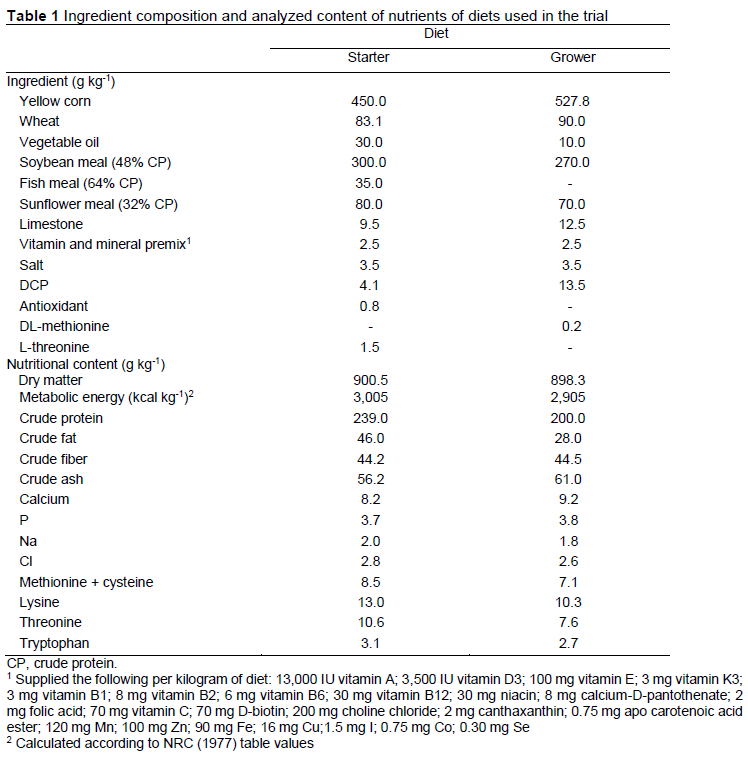
The study was carried out according to the animal experiments manual of the Siirt University Animal Experiments Local Ethics Committee (Protocol no: 2020-05). In the study, 40 one-day-old, mixed sex Japanese quail (Coturnix japonica) chicks obtained from Siirt University Wildlife Research and Application Center were used as animal material. Chicks were grouped with 10 members in each group as follows: the control group, 0.25% olive cake (Group 1), 0.50% olive cake (Group 2) and 0.75% olive cake (Group 3). The olive cake used as a feed additive in the research was obtained from a commercial company selling ground olive cake. Quails were fed with a ration (control group) whose nutrient contents were prepared in accordance with NRC (1994) between days 1-42 (Table 1). No feed additives were added to the ration of the control group, while 0.25%, 0.50% and 0.75% olive cake was added to the other three groups, respectively. Quails were placed in cages with dimensions of 96 x 46 x 25 cm. During the first week the coop temperature was kept at 32-34 °C. The temperature was gradually reduced every day beginning from the fourth day of the study and was stabilized at 21 °C. A 24-hour lighting program was implemented by daylight and artificial lighting systems in the coop environment. The research was continued for 42 days. Feed and water were provided ad libitum.
After 42 days, proventriculus, duodenum, jejunum, and ileum tissue samples were taken from decapitated animals. Tissue samples were fixed with alcoholic formalin for 18 h, passed through a graded series of ethanol, methyl benzoate, and benzene, and then embedded in paraffin wax. Serial sections of 5 µm thickness were taken from the prepared paraffin blocks. Crossman's Triple staining was applied to determine the general structure and the presence of any pathological conditions in the gastrointestinal tract. Periodic acid-Schiff-Alcian Blue (PAS-AB) (pH 2.5) was applied to the preparations to determine neutral and acid mucin in glands, and aldehyde fuchsin-Alcian blue (pH 2.5) (AF-AB) stains were applied to determine carboxylated and sulfated acid mucins (Bancroft & Cook 1984).
Once stained, the preparations were examined, evaluated, and photographed under a research microscope with a Nikon-Eclipse 400 DSRI Nikon digital camera (NIS Elements Imaging Software-version 3.10) attachment.
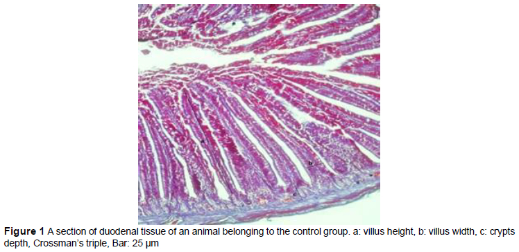
Measurements were made on the villi from different parts of each animal. Villus length, villus thickness, and crypt depth were measured in the intestines. The villi heights were measured as the length from the apex of the villi to the starting point of the crypts, while the crypt depths were evaluated as the distance from the point where the invagination began in the region between the villi to the end of the Lieberkuhn glands. Villus widths were obtained by measurements made from the widest region of the villi (Figure 1) (Sur et al., 2017).
Histochemical staining was performed semi-quantitatively using the intensity score method. In the intensity score, staining intensities in the cells were evaluated as no staining (-), weak staining (+), moderate staining (++), and strong staining (+++) (Erdogan et al., 2012; Sagsoz et al., 2013).
The data were subjected to a completely randomized design of the analysis of variance using the General Linear Models procedure of the SPSS 20.0 statistical program. Significant differences were determined using a Duncan's multiple range test at the level of P <0.05. A Kruskal-Wallis variance analysis test was applied to the intestinal measurements. The raw data were analyzed using the Bonferroni-corrected Mann Whitney U-test thereafter. P-values <0.05 were considered statistically significant.
Results and Discussion
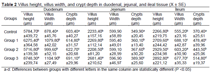
The villus heights, villus widths, and crypt depths of the small intestine histologies of Japanese quails fed with rations supplemented with different proportions of olive cake are given in Table 2. Increases occurred in villus height in duodenal, jejunal, and ileal tissue of the animals in the experimental groups, depending on the ratio of olive cake added to the ration (P <0.05).
Increases occurred in villus width in duodenal, jejunal, and ileal tissue of the animals in the groups depending on the ratio of olive cake added to the ration (P <0.05). In the jejunal tissue, it was determined that the width of the villus decreased in groups 1 and 2 but increased in group 3. However, this increase was found to be statistically insignificant (P >0.05).
It was determined that increases were caused in villus height in duodenal, jejunal, and ileal tissues of the animals in the groups depending on the ratio of olive cake added to the ration (P <0.05). It was determined that the increase in the crypt depth occurred mostly in the ileal tissue.
In the histological evaluation, it was determined that mucins were regionally expressed in the proventriculus and all parts of the small intestine, in both control and experimental groups. Expression in the proventriculus was determined to be located in the cells especially in luminal gastric glands (gll. Proventricularis superficialis), and in the deep proventriculus glands of the submucosa and lamina propria (gll. Proventricularis profundum). In the small intestines, mucin expression was determined in goblet cells in the mucosa, and in the deep tubular glands and ducts.
In PAS/AB (pH 2.5) staining, both PAS (+) and mixed staining cells were determined in the corpus glandule and ducts of the luminal and deep glands in the proventriculus. It was revealed that the glands on the surface showed strong AB (+), while the deep glands showed a weak reaction. It was also revealed that PAS (+) reacting cells showed an intense reaction in luminal glands and a weak reaction in deep glands. Another revelation was that, especially goblet cells in the duodenum showed strong AB (+) and weak PAS (+) reactions. In the jejunum and ileum, although goblet cells gave a strong AB (+) reaction as in the duodenum, the PAS (+) reaction was found to be moderate. Findings were similar in both control and experimental groups (Table 3, Figure 2).
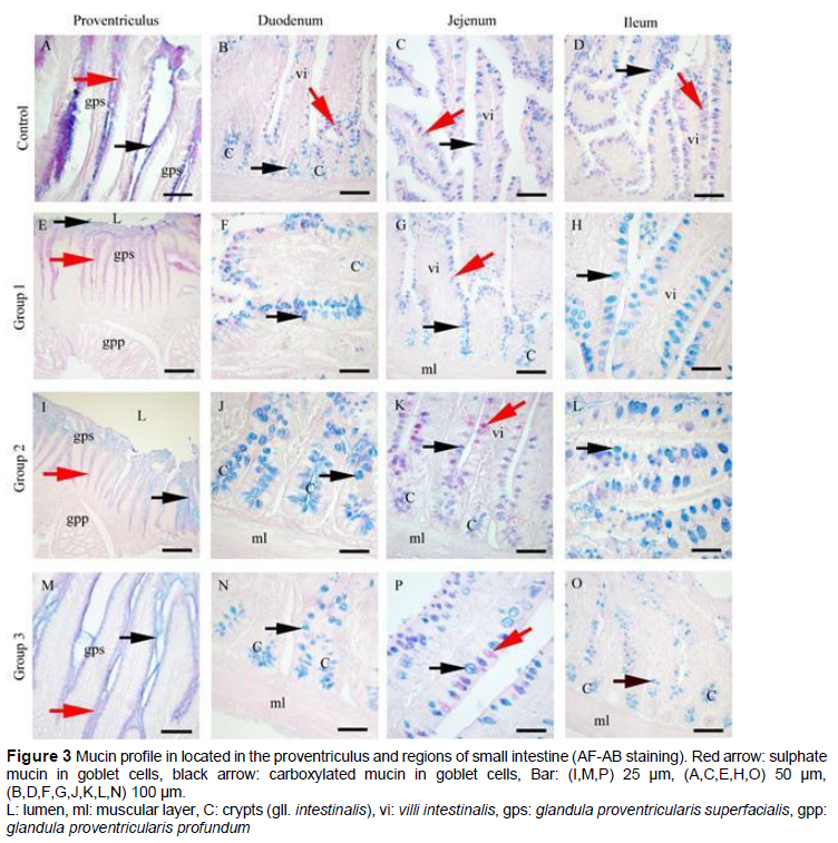
It was determined by AF/AB (pH 2.5) staining that the AF reaction in the proventriculus was localized in both luminal and deep glandular epithelial cells and the reaction was strong. AB (+) reaction was found to be weak. In contrast to the proventriculus, the AB (+) reaction was strong, and the AF (+) reaction was weak in goblet cells in the duodenum, jejunum, and ileum. Findings were similar in both control and experimental groups (Table 3, Figure 3).
The development of intestinal villi is dependent on the intake of nutrients. The absorption of ingested nutrients depends on the functional capacity of the villi. Crypts are sites where epithelial cell proliferation occurs. Consequently, crypt development directly affects villi development and the intestinal absorptive surface. Studies have shown that feed additives introduced to the ration of poultry animals in order to increase feed efficiency can result in increases in the villi heights and crypt depths in the small intestines (Shafey et al., 2013; Hassan et al., 2014; Shiraze et al., 2017; Sur et al., 2017).
In a study investigating the effects of organic mixtures containing acid and essential oils in broiler nutrition, it was reported that significant differences were observed in villus height and crypt depth in other groups compared to the control group (Stamilla et al., 2020). Shiraze et al. (2017) reported in their study on broiler chickens that olive leaves added to the ration at different rates caused an increase in villus height, villus width, and crypt depth in the small intestines. In the present study, it was determined that, depending on the ratio of olive pulp added to the rations, significant increases occurred in villus height in duodenum, jejunum, and ileum tissues of the animals in the experimental groups (Table 2, P <0.05).
Saki et al. (2017) reported that another property that influences the absorptive level in the intestines is the width of the villi. In the present study, it was determined that depending on the ratios of olive pulp added to the rations, significant increases in villus width in duodenal and ileal tissues were observed in the birds in the groups (Table 2, P <0.05). It was determined that there was no significant increase in villus width in the jejunal region (P >0.05).
Sur et al. (2017) reported in their study that the increase in the depth of the crypts is an indicator of the cell renewal rate in the intestinal epithelium, and deep crypts also mean long villi. The ileal histomorphometry showed that crypt depth and the villus height to crypt depth ratio in groups that received 2.5% and 5% olive oil "decreased" and "increased" respectively, compared to the control group (P <0.05) (Soroush et al., 2020). In the current study, it was determined that, depending on the ratio of olive cake added to the ration, a significant increase in crypt depth occurred in all three parts of the small intestine (Table 2, P <0.05). It was determined that the most affected part of the intestine was the ileum, the magnitude of change being depending on the amount of olive cake added.
Many studies have been conducted on the composition and expression of mucins located in the stomach and intestines of humans, mammals, and other species (Schumacher et al., 2004; Senol et al., 2014; Godwin et al., 2016; Karakoç et al., 2016; Vila et al., 2018; Karakoç et al., 2019). It has been revealed in these studies that mucins are found in the lamina propria in the stomach and in the gland epithelial cells and goblet cells in the intestines. Studies have shown that acidic, neutral, and sulphated mucins exist in the stomach (Hassa et al., 1976), and neutral, acidic, and mixed mucins exist in the intestines (Godwin et al., 2016; Nasser & Khaleel, 2021) of various poultry. In the present study, as in other species, neutral, acid, and mixed mucins were detected in the proventriculus and small intestine of Japanese quails.
Karakoç et al. (2016) reported in their study on adult rams that, while neutral mucins on the surface and strong sulphated mucins in the deeper tissues are dense in the abomasum, carboxylic mucins are sparse. Senol et al. (2014) reported in their study on different vertebrate species that sulphate mucins were found intensively in the submucosa and lamina propria glands in the proventriculus of quails.
In the present study, it was determined that mixed mucins in the Japanese quail proventriculus were weak in the luminal glands and intense in the deep glands, while sulphated mucins were intense in the luminal glands and weak in the deep glands. In addition, carboxylic acid mucins stained weakly in the proventriculus. It was determined that there were no changes in the composition and expression of mucins synthesized in either of the control or experimental groups.
Godwin et al. (2016) reported in their study on crow small intestines that there were mixed mucins in the crypts of Lieberkühn and goblet cells in the duodenum, ileum, and jejunum. Nasser & Khaleel (2021) reported in their study on adult male turkeys that the presence of both neutral and acidic mucins was revealed in the crypts of Lieberkühn and goblet cells in the duodenum, ileum, and jejunum. Simsek et al. (2012) investigated the effects of various feed additives on mucin in the small intestine of quails and observed that acidic and neutral mucins were found in goblet cells in the duodenum and ileum. In the present study, it was determined that mixed, neutral, and acidic mucins were found in all three parts of the small intestine, where carboxylated acidic mucins stained intensely, and sulphated acidic mucins stained weakly. Neutral mucins were found to be more concentrated in the ileum and jejunum. It was determined that there was no difference in the composition and density of the mucins synthesized in both the control and experimental groups.
Conclusion
It is known that the feed additives that are introduced to the ration of poultry in order to increase feed efficiency work in different pathways to provide the targeted benefit. The basis of these pathways is the small intestine and its mechanism of absorption. It is an important indicator of the efficiency of an additive that the villi and crypt structures are positively affected in the small intestines to increase the absorption and utilization of nutrients.
In this study, the effects of olive cake added to the ration in different proportions on the histology of the small intestine of quails were investigated. In the histological and statistical evaluations made with the results obtained, it was determined that olive pulp had positive effects on the parameters that lead to an increase in absorption. In addition, it was determined that mucins were expressed in different intensities in the stomach and small intestines of quails, as in other species. It was determined in the histochemical evaluation that sulphate mucins were intense in the proventriculus, and carboxylic mucins were intense in all three parts of the small intestine. Considering the physiological functions of mucins, they are thought to play an important role in the protection of the mucosa in Japanese quails. More studies are needed, both histomorphologically and histochemically, on feed additives, which have an important place in poultry nutrition.
Authors' Contributions
ZK (ORCID 0000-0002-0723-4059) and CO (ORCID 0000-0002-1047-5347) planned, designed the research, analyzed all data and drafted manuscript. All authors discussed the results and contributed to the final manuscript.
Conflict of Interest Declaration
The authors declare that they have no conflict of interest.
References
Bancroft, J.D. & Cook, H.C., 1984. Manual of histological techniques, Edinburgh, Churchill Livingstone, pp. 101-126. [ Links ]
Basmacioglu-Malayoglu, H. & B. Aktas, B., 2011. Zeytinyagi yan ürünlerinden zeytin yapragi ile zeytin karasuyunun antimikrobiyal ve antioksidan etkileri. Hayvansal Uretim, 52 (1), 49-58. [ Links ]
Bolacali M, Irak K, 2017. Effect of dietary yeast autolysate on performance, slaughter, and carcass characteristics, as well as blood parameters, in quail of both genders. S. Afr. J. Anim. Sci. 47, 460-470. [ Links ]
Cibik, M., 2014. Effect of Pelleted Olive Cake on Milk Yield And Milk Composition of Dairy Cow. MSc (Agric) thesis. University of Adnan Menderes, Turkey. [ Links ]
Erdogan, S., Sagsoz, H., & Akbalik, M.E., 2012. Anatomical and histological structure of the tongue and histochemical characteristics of the lingual salivary glands in the chukar partridge (Alectoris chukar, Gray 1830). Br. Poult. Sci. 53, 307315. [ Links ]
Erdogan, S. & H. Sagsoz, H., 2018. Papillary architecture and functional characterization of mucosubstances in the sheep tongue. Anat. Rec. 301, 1320-1335. DOI: 10.1002/ar.23840. [ Links ]
Godwin, O.C., Clifford, A.N. & Agadha, A., 2016. Evaluation of the morphological adaptations of the small intestine of the African pied crow (Corvus albus). J. Basic Appl. Zool. 75, 54-60. [ Links ]
Hassa, O., Saglam M., Tanyolag, A. & Ozer, A., 1976. Abomasum mukozasinin mikromorfolojisi ve salgiladigi enzimlerin lokalizasyonu uzerinde arastirmalar. Ankara Universitesi. 23, 318-344. [ Links ]
Hassan, H.M.A., Youssef, A.W., El-Daly, E.F., El-Azeem, N.A.A., Hassan, E.R. & Mohamed, M.A., 2014. Performance, caecum bacterial count and ileum histology of broilers fed different direct-fed microbials. Asian J. Poult. Sci. 8(4), 106-114. DOI: 10.3923/ajpsaj.2014.106.114. [ Links ]
Karakog, Z., Sagsoz, H. & Ketani, M.A., 2016. Mucin profiles of the abomasum in bulls and rams: A comparative study. Microsc. Res. Tech. 79, 856-868. DOI 10.1002/jemt.22713. [ Links ]
Karakog, Z., Topaloglu, U. & Ketani, M.A., 2019. Kil kegisi abomasumunda ghrelin, obestatin ve leptin hormonlarinin immunohistokimyasal dagilimlari. Eurasian J. Vet. Sci. 35(4), 204-209. [ Links ]
Kuter, E., Gumus, H. & Karakas, O.F., 2020. The effects of probiotics and prebiotics on gut health. Kucukersan S, editor. Hayvan Beslemede Bagirsak Sagliginin Onemi. 1. Baski. Ankara: Turkiye Klinikleri. p.31-6. [ Links ]
Kutlu, H.R. & Sahin, A., 2017. Kanatli Beslemede Guncel Cali§malar ve Gelecek igin Oneriler. Hayvansal Uretim. 58(2), 66-79. DOi:10.29185/hayuretim.333882. [ Links ]
Molina-Alcaide, E. & Yanez-Ruiz, D.R., 2008. Potential use of olive by-products in ruminant feeding: A review. Anim. Feed Sci. Technol. 147, 247-264. [ Links ]
Nasser, R.A.A. & Khaleel, I.M., 2021. A histochemical study of the small intestine of adult male turkey (Meleagris gallopavo). Diyala Journal For Veterinary Sciences. 1(2), 159-172. [ Links ]
Ozcan, C., Cimrin, T., Yakar, Y. & Alasahan, S., 2020. Effects of olive cake meal on serum constituents and fatty acid levels in breast muscle of Japanese quail. S. Afr. J. Anim. Sci. 50 (6), 874-880. DOI: 10.4314/sajas.v50i6.14. [ Links ]
Ozcan, C., Cimrin, T., Yakar, Y. & Alasahan, S., 2021. The effects of dietary olive cake meal on fattening performance, carcass and slaughter traits in Japanese quails (Coturnix coturnixjaponica). Turkish Journal of Agriculture - Food Science and Technology. 9(6), 1030-1036. DOI: 10.24925/turjaf.v9i6.1030-1036.4112. [ Links ]
Sagsoz, H., Erdogan, S. & Akbalik, M.E., 2013. Histomorphological structure of the palate and histochemical profiles of the salivary palatine glands in the chukar partridge (Alectoris chukar, Gray 1830). Acta Zoologica (Stockholm, Sweden). 94, 382-391. [ Links ]
Saki, A.A., Sahebi, A.F., Zamani, P., Alipour, D. & Abbasinezhad, M., 2017. Japanese quail performance, intestinal microflora, and molecular responses to screened wheat and multienziyme diet. Turkish J. Vet. Anim. Sci. 41, 30-37. [ Links ]
Schumacher, U., Duku, M., Katoh, M., Jorns, J. & Krause, W.J., 2004. Histochemical similarities of mucins produced by Brunner's glands and pyloric glands: A comparative study. Anatomical Record. Part A, Discoveries in Molecular, Cellular, and Evolutionary Biology. 278, 540-550. [ Links ]
Shafey, T.M., Almufarij, S.I. & Albatshan, H.A., 2013. Effect of feeding olive leaves on the performance, intestinal and carcass characteristics of broiler chickens. Int J Agric Biol. 15, 585-589. [ Links ]
Shiraze, A.A.S., Hassanabadi, A., Agah, M.J. & Nasiri-Moghaddam, H., 2017. Effect of dietary inclusion of olive leaf (Olea europaea L.) powder on performance, small intestine morphology and nutrient digestibility in broiler chickens. Journal of Anim. Prod. 19(2), 371-387. [ Links ]
Smirnov, A., Tako, E., Ferket, P.R. & Uni, Z., 2006. Mucin gene expression and mucin content in the chicken intestinal goblet cells are affected by in ovo feeding of carbohydrates. Poult. Sci. 85, 669-673. [ Links ]
Soroush, S.Z., Hosseini-Vashan, S.J., Afzali, N. & Allahressani. A., 2020. Effects of olive leaves extract and olive oil on growth performance, nutrient digestibility, and ileum morphology of Japanese quails. Research on Animal Production. 11(28), 11-21. [ Links ]
Stamilla, A., Messina, A., Sallemi, S., Condorelli, L., Antoci, F., Puleio, R., Loria, G.R., Cascone, G. & Lanza, M., 2020. Effects of Microencapsulated blends of organics acids (OA) and essential oils (EO) as a feed additive for broiler chicken. A focus on growth performance, gut morphology, and microbiology, Animals. 10, 442- DOI:10.3390/ani10030442. [ Links ]
Sur, E., Caglayan, T., Kadiralieva, N. & §eker, E., 2017. Determination of the effects of Mentha caucasica on histology of small intestine in Japanese quail (Coturnix japonica). Eurasian J. Vet. Sci. 33 (4), 248-254. DOI:10.15312/EurasianJVetSci.2017.168. [ Links ]
Senol, N., D. Bayram, D. & Yesil, O., 2014. Histological and histochemical structure of the some of digestive tract area in different vertebrate species. sDu Journal of Science (E-Journal). 9(2), 61-70. [ Links ]
Simsek, N., Can, I., Karadeniz, A., Kara, A. & Gùmùs, R., 2012. Effects of dietary various supplementations on the mucin-and serotonin- releasing cell numbers in small intestine of quails. Revue de Médecine Vétérinaire. 163 (7), 328-334. [ Links ]
Tufan, T. & Bolacali, M., 2017. Effects of dietary addition of synbiotics on the performance, carcass traits, and serum parameters of Japanese quails. Revista. Brasialia de Zootecnica. 46(10), 805-813. [ Links ]
Vila, M.F., Michaela, P.T., Yuan-Tai, H., Zeng, Z., Urriola, P.E., Gerald, C.S. & Saqui-Salces, M., 2018. Dietary fiber sources and non-starch polysaccharide-degrading enzymes modify mucin expression and the immune profile of the swine ileum. PLoS ONE. 13(11). doi.org/10.1371/journal.pone.0207196. [ Links ]
Submitted 14 February 2022
Accepted 7 April 2022
Published 28 October 2022
# Corresponding authors: e-mail: chtzcn@gmail.com

