


Services on Demand
Article
 English (pdf)
 Article in xml format
 Article references
Indicators

Related links
-
 Cited by Google
 -
 Similars in Google
Share

 Permalink
South African Journal of Animal Science
On-line version ISSN 2221-4062
Print version ISSN 0375-1589
S. Afr. j. anim. sci. vol.52 n.2 Pretoria 2022
http://dx.doi.org/10.4314/sajas.v52i2.7
Serological and molecular identification of Mycobacterium avium subsp. paratuberculosis and associated risks in bovine
A. RehmanI; M.T. JavedII; M. F. QamarI; A. I. AqibIII; I. AhmedI; A. SikandarIV; M. K. RafiqueI; T. HussainIV; M. KashifV; M. RiazVI; L. AhmadVII; M. NazarVIII; M.F.A. KulyarIX
IDepartment of Pathobiology, University of Veterinary and Animal Sciences, Lahore (Jhang Campus), Jhang, Punjab, Pakistan
IIDepartment of Pathology, Faculty of Veterinary Science, University of Agriculture Faisalabad, Pakistan
IIIDepartment of Medicine, Faculty of Veterinary Sciences, Cholistan University of Veterinary and Animal Sciences, Bahawalpur, Pakistan
IVDepartment of Basic Sciences, University of Veterinary and Animal Sciences, Lahore (Jhang Campus), Jhang, Punjab, Pakistan
VDepartment of Clinical sciences, University of Veterinary and Animal Sciences, Lahore (Jhang Campus), Jhang, Punjab, Pakistan
VIDepartment of Allied Health Sciences, Sargodha Medical College, University of Sargodha, Pakistan
VIIBaqai College of Veterinary Sciences, Baqai Medical University, Karachi, 75340, Pakistan
VIIIUniversity of Agriculture, Faisalabad, Sub-Campus Burewala, Pakistan
IXCollege of Veterinary Medicine, Huazhong Agricultural University, Wuhan, 430070, China
ABSTRACT
Discrepancies in sensitivity and specificities of tests (enzyme-linked immunoassay (ELISA) and tuberculin skin test (TST)), prevalence, and potential risk factors associated with Mycobacterium avium subsp. paratuberculosis (MAP) of bovine were hypothesized to affect its control. Keeping PCR as the gold standard test, 101 cattle and 39 buffaloes maintained at Livestock Experiment Station were tested for MAP with ELISA and TST. The incidence of MAP was 13.57% (19/140), 12.85% (18/140), and 8.57% (12/140), based on PCR, ELISA, and TST. Discrepancies in the identification of MAP in buffalo were 7.62% and 2.79% for cattle, based on ELISA compared with TST. The highest discrepancies in MAP prevalence were noted in brown buffalo (22.22%), whereas the lowest were recorded from crossbred cattle (2.63%). Status of milking was a potential risk factor (P <0.05) if MAP was identified by TST, whereas breed showed significant association (P <0.05) with MAP only on its identification by ELISA. Considerable variations were noted in the sensitivity of ELISA (94.4%) and TST (66.7%). On mutual comparisons, considering TST as a fixed diagnostic tool, the specificity and sensitivity of ELISA were 95.31% and 100%. Similarly, fixing ELISA as diagnostic tool, TST showed 100% specificity and 66.67% sensitivity. The conclusion of the study was that because of rising MAP in bovine, significant discrepancies in routine diagnostic tools, and variations in the declaration of significant risk factors, stern measures must be taken to avoid exaggerated outcomes in testing for this pathogen.
Keywords: bovine, discrepancies, paratuberculosis, risk factors
Introduction
Paratuberculosis (Johne's disease) is a long lasting and infectious granulomatous disease that disturbs mainly ruminant, non-ruminant (Foxet al., 2020) and wild animal species across the world (Singh et al., 2014). The disease is produced by Mycobacterium avium subsp paratuberculosis (MAP). Currently, Johne's disease is regarded as being among the top serious threats disturbing the world's dairy industry (Chiodini et al., 2012). Exposure to infection seems to be higher in younger animals (less than 6 months old) compared with old ones (Sergeant et al., 2003). A clear clinical picture of the disease appears after 2-4 years of exposure (Harris et al., 2001). The bacteria can survive longer in a harsh environment, but cannot propagate (Raizman et al., 2007). The intermittent shedding of the organism in the calving areas and the milk might cause Crohn's disease in humans because this bacterium is viable even after pasteurization (Guptaet al., 2012; McNees et al., 2015). The long-term duration of the disease lies in its subclinical nature, resistance to unfavourable environments, slow growth, and intermittent spread (Agrawal et al., 2021).
Paratuberculosis has significant importance throughout the world because of its economic and health concerns. The production loss from paratuberculosis in the USA dairy sector is approximately $200 million yearly (Rasmussen et al., 2021). Prevalence of Johne's disease reaches 40% in some countries, for example Venezuela (South America). Prevalence in dairy cows in Australia and the US was reported at 22% and 75.8% (Ruiz et al., 2020). In Belgium and Austria, the prevalence rate was 41% and 7%. In Pakistan, cattle and buffalo were identified as being 6.67% and 12.5% positive for MAP (Sikandar et al., 2012). The ELISA-based study reported an overall 1% prevalence of MAP in cattle and buffalo (Rehman et al., 2017b), whereas at public livestock farms buffaloes showed 1.3% prevalence (Rehman et al., 2018).
This disease is diagnosed through TST, ELISA, ZN staining, culture isolation and PCR (Whittington & Sergeant, 2001). Among these tests, TST and ELISA are easily available and used routinely for preliminary diagnosis of the disease. Purified protein derivative (PPD) involves delayed type hypersensitivity in the form of inflammation and production of specific cytokines ( aí., 2000). Constant screening through PPD is routine for eradication of disease (Schiller et al., 2010). On the other hand, ELISA is a more sensitive tool for screening animal herds (Sikandar et al., 2012)). But every technique has its own discrepancies in identification. It is necessary to study the effects of these discrepancies on the spread of diseases and their determinants so that precise measures may be adopted. Therefore, the current study was planned to highlight the discrepancy in identification of MAP by ELISA and TST, keeping PCR as gold standard, and to investigate variations in declaration of disease determinants.
Materials and Methods
This research was reviewed for ethical consideration the by Research Scrutiny Committee and Director Graduate Studies of the University of Agriculture, Faisalabad, and was carried out at Livestock Experiment Station where animals were reared with optimum feeding and living requirements. The health and management of animals was monitored by professionals employed at the farm. The farm is located 500 meters from the veterinary teaching hospital of the university. Hence health services included routine monitoring. This study included 140 adult animals (101 cattle and 39 buffalo) more than two years old.
Blood samples (5 mL) were drawn aseptically directly from the jugular vein of the cattle and buffaloes in clean gel-coated vacutainers and were processed for serum collection to run indirect ELISA (Salgado et al., 2007) for MAP. LSIVet ruminant serum paratuberculosis advanced antibody kit (Lot No. 2-VETPTRS-007) was used according to the instructions of the manufacturer.
The TST was performed by injecting 0.1 mL PPD antigen (Veterinary Research Institute, Lahore, Pakistan) at the left side of the neck area (cervical) adopting the intradermal route. The site was washed, cleaned, and dried before the injection of PPD. Skin thickness was measured by Vernier callipers at this site before performing the test. Similarly, skin thickness was measured 72 hours post PPD administration. The cattle and buffaloes were considered positive for the intradermal tuberculin test if the net thickness of skinfold was greater than 4 mm at the injection site. A negative control was run alongside to rule out prick response in the animal.
Faecal samples were collected and processed for DNA extraction through the boiling method (Alexopoulouet al., 2006). PCR was performed for rapid diagnosis of MAP following Stanley et al., 2007. A set of primers (P90 GTTCGGGGCCGTCGCTTAGG, P91 GAGGTCGATCGCCCACGTGA, IS 1245 F GTGGGCAATCTGCCCTGCACTTCGG, IS 1245 R GCCCGCACGCTCACAGTTAAGCCGT) reported by Bartos et al. (2006) was used with the recipe as DNA (3 μL) master mix (Enzynomics) 20 μL, primer (F+R) 1 μL each. Thermocycler (Qantarus) was set at preliminary denaturation (94 °C for 2 minutes), 35 cycles of denaturation for 30 seconds, annealing (65 °C for 2 minutes), and three elongations (3 minutes at 72 °C). At the end of the last cycle, 30 seconds of denaturation, two minutes of annealing at 65 °C and a final extension at 72 °C for 10 minutes were carried out (Stanley et al., 2007).
Information about the species, breed, age of animal (years), weight (Kg), milk yield, lactation number, and milking status was documented. A chi-square test was applied to calculate the epidemiological association. SAS statistical software (SAS Institute Inc., Cary, North Carolina, USA) was used for all analyses. Differences were declared significant at the 5% level of probability and remarked upon at the 10% level.
The data were evaluated using frequency analysis, stratified analysis at 95% confidence interval. A chi-square test was applied to calculate the epidemiological association. SAS statistical software (SAS Institute Inc., Cary, North Carolina, USA) was used for all analyses and differences were declared significant at the 5% level of probability.
Results and Discussion
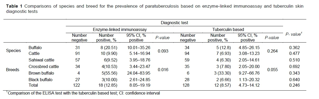
Of the animals tested for MAP, 12.85% tested positive based on ELISA, 8.57% were positive with TST, and the PCR technique (Figure 1) identified 13.57% positive animals. Buffaloes were twice as likely to be positive for MAP compared with cattle (P =0.093) (Table 1). Brown buffaloes were five times more likely to be positive for MAP than black buffaloes, although the numbers of observations were small.
The findings of the current study contradict those of studies in which Rehman et al. (2017b) found prevalence of MAP was 5.66%, and Haji Hajikolaei et al. (2006) observed a 2% incidence. The results of an abattoir-based study conducted at Faisalabad showed the prevalence rate to be higher in cattle compared with buffaloes (Rehman et al., 2017a). Recently an ELISA-based study revealed 14.2% positive animals for MAP antibodies in Tai'an city of China (Cheng et al., 2020).
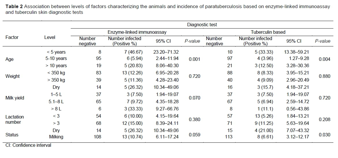
The prevalence rate was comparatively higher in young animals, those with low bodyweight and high lactating animals (Table 2). It seems the chances of finding the infection in cattle and buffalo with low bodyweight might be attributable to the severe diarrhoea that debilitates the body condition of affected animals (Kudahl et al., 2009). The results are in line with those of Borujeni et al. (2021), who reported high disease prevalence in low bodyweight animals. The prevalence of MAP in animals that weighed less than 350 kg was numerically greater based on the ELISA test than on the TST. The salient assumed risk factors had P-values indicating significant variability (Table 2). Milking status (dry vs lactating animals) had a significant effect (P =0.03) with MAP prevalence based on the TST, whereas its effect only approached significance (P =0.059) when MAP was diagnosed with the ELISA test. The difference in P-values for milk yield potential was more significant (P =0.07) when ELISA was used whereas it was non-significant (P =0.72) for TST (Table 2). A higher percentage of milking animals testing positive for MAP was noted with the ELISA test, and TST showed higher prevalence of MAP in animals that were not lactating. In addition, cows with fewer than three lactations were more prone to MAP being identified by ELISA than TST (Table 2).
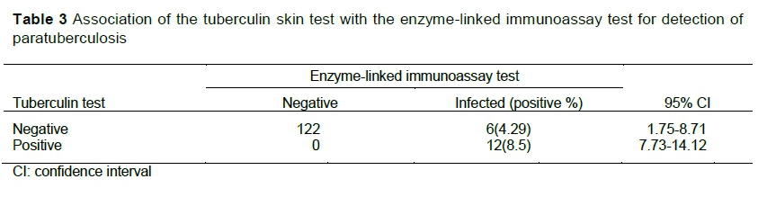
The technique-based variation with PCR throughout the trial was most sensitive followed by ELISA and TST (Table 3). Association of ELISA and TST by replacing each other as a fixed diagnostic tool showed shifting sensitivity and specificity (Table 3). Considering TST as a standard tool, the ELISA specificity and sensitivity were 95.31% and 100% in the current study. On the other hands keeping ELISA as fixed diagnostic tool, the specificity and sensitivity of TST were observed 100% and 66.67% (Table 3). Higher sensitivity results of ELISA for MAP identification were also noted by Cihan et al. (2012), who did studied the efficiency of diverse diagnostic approaches paratuberculosis. The TST-based studies showed this test is a routine diagnostic tool but cross-reactivity with other mycobacteria hampered the accuracy of results (Marassi et al., 2005). Varges et al. (2009) also reported tuberculin skin testing affecting immune system mechanism of animal's body resulting in false positive outcomes. A serological survey on 19627 cattle from Italy reported MAP identification through ELISA an easy, reliable, economical, and useful in dairy herds (Lillini et al., 2005). On the other hand, as the disease progresses, the humoral response of animals can vary resulting in discrepancies in the outcomes of serological diagnostic tests (Collins et al., 2005).
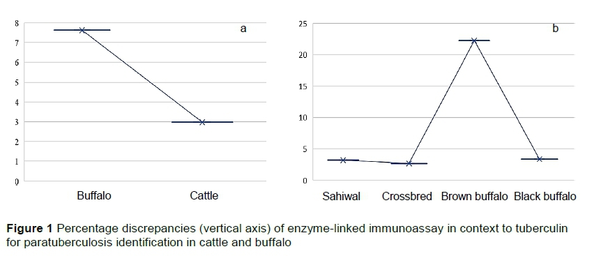
The apparent discrepancies between the ELISA test and TST showed the percentage changes in the identification of paratuberculosis for various species and breeds. In comparing ELISA to TST for buffaloes 7.62% of the results were disparate, whereas for cattle the numbers of cases in which the tests produced conflicting results were only 2.97% of the total number of animals evaluated (Figure 1a). The highest rate of discrepancies between the tests was noted for brown buffalo (Figure 1b). However, the total number of brown buffalo that were sampled was small, so even a discrepancy of results for a single animal produced a very large percentage change.
Percentage discrepancies = (Elisa positive-tuberculin positive/total tested) x 100
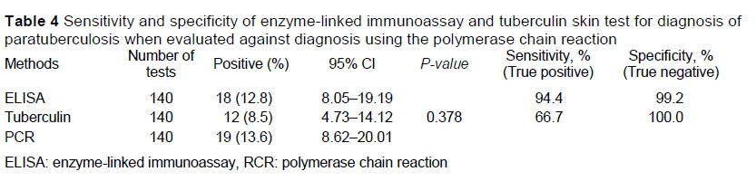
Considering PCR as fixed diagnostic/gold standard test, the current study found variable sensitivity and specificity of ELISA and TST (Table 4). The specificity of TST was similar to that of ELISA indicating that these tests were similarly and highly precise in screening negative samples. However, the sensitivity of TST was low compared with that of ELISA, indicating that TST was not reliable in identifying true positive MAP in bovine (Table 4).
Conclusion
A higher than expected prevalence of MAP was found in bovine maintained at an experimental station where reasonable healthcare and management were provided. There was considerable variation in the detected prevalence of MAP based on diagnostic techniques, and a significant association of risk factors with MAP also showed discrepancies between the test results. Significant variation in sensitivity and specificity of TST and that of ELISA were observed. Moreover, a significant association of assumed risk factors varied with diagnostic tests. Thus, immediate measures are needed to identify a suitable diagnostic test to facilitate the prevention of MAP.
Acknowledgment
The authors acknowledged the farm workers and their management that allowed the study to be conducted with their animals.
Authors contributions
AR (https://orcid.org/0000-0003-4665-781Xcc) and MTJ conceived the idea of research; AR collected the samples and was involved in sample testing and lab activity; MFQ drafted the skeleton; AIA analysed the data; IA, AS (https://orcid.org/0000-0001-7966-772) and MR prepared the draft of the manuscript; MK, LA and MN drafted the details of the manuscript and helped in data editing, data analysis and construction of tables; MKR, TH and MFAK contributed to proofreading, added references and checked reference styling, and helped in revising the draft and editing. All authors read and interpreted the data critically and approved the final version.
Declaration of conflicting interests
The authors received no financial support for the research, authorship, and publication of this article; and they declare there are no potential conflicts of interest.
References
Agrawal, A., Varshney, R., Kirthika, P., Gupta, R., Sulabh, S., Varshney, R., Chakravarti, S., & Thankappan, S., 2021. Global scenario of paratuberculosis: A threat to livestock sector. Biological Rhythm Research 52, 6, 957-972. DOI: 10.1080/09291016.2019.1610858 [ Links ]
Alexopoulou, K., Foka, A., Petinaki, E., Jelastopulu, E., Dimitracopoulos, G. & Spiliopoulou, I., 2006. Comparison of two commercial methods with PCR restriction fragment length polymorphism of the tuf gene in the identification of coagulase-negative staphylococci. Letters in Applied Microbiology 43(4), 450-454. https://doi.org/10.1111/j.1472-765X.2006.01964.x [ Links ]
Andersen, P., Munk, M.E., Pollock, J.M. & Doherty, T.M., 2000. Specific immune-based diagnosis of tuberculosis. The Lancet 356(9235), 1099-1104. https://doi.org/10.1016/S0140-6736(00)02742-2 [ Links ]
Bartos, M., Hlozek, P., Svastova, P., Dvorska, L., Bull, T., Matlova, L., Parmova, I., Kuhn, I., Stubbs, J., Moravkova, M. & Kintr, J., 2006. Identification of members of Mycobacterium avium species by Accu-Probes, serotyping, and single IS900, IS901, IS1245 and IS901-flanking region PCR with internal standards. Journal of Microbiological Methods 64(3), 333-345. https://doi.org/10.1016/j.mimet.2005.05.009 [ Links ]
Borujeni, M.P., Haji, Hajikolaei, M.R.H., Ghorbanpoor, M., Sahar, H.E., Bagheri, S. & Roveyshedzadeh, S., 2021. Comparison of Mycobacterium avium subsp. paratuberculosis infection in cattle, sheep and goats in the Khuzestan Province of Iran: Results of a preliminary survey. Vet. Med. Sci. 7(5), 1970-1979. https://doi.org/10.1002/vms3.559 [ Links ]
Cheng, Z., Liu, M., Wang, P., Liu, P., Chen, M., Zhang, J., Liu, S. & Wang, F., 2020. Characteristics and epidemiological investigation of paratuberculosis in dairy cattle in Tai'an, China. BioMed Research International. https://doi.org/10.1155/2020/3896754 [ Links ]
Chiodini, R.J., Chamberlin, W.M., Sarosiek, J. & McCallum, R.W., 2012. Crohn's disease and the Mycobacterioses: A quarter century later. Causation or simple association? Critical Reviews In Microbiology 38(1), 52-93. https://doi.org/10.3109/1040841X.2011.638273 [ Links ]
Cihan, H., Mecitoglu, Z., Demir, G., Temizel, E.M. & Çentürk, S., 2012. Evaluation of faecal shedding of acid-fast Mycobacterium avium subsp. paratuberculosis (MAP) in both intradermal Johnin test and serologically (ELISA) Map-positive cattle. J. Biol. Environ. Sci. 6(18), 281-283. https://uludag.edu.tr/dosyalar/jbes/18/mak10.pdf [ Links ]
Collins, M.T., Wells, S.J., Petrini, K.R., Collins, J.E., Schultz, R.D. & Whitlock, R.H., 2005. Evaluation of five antibody detection tests for diagnosis of bovine paratuberculosis. Clinical and Vaccine Immunology 12(6), 685-692. https://doi.org/10.1128/CDLI.12.6.685-692.2005 [ Links ]
Fox, N.J., Smith, L.A., Stevenson, K., Davidson, R.S., Marion, G. & Hutchings, M.R., 2020. Infection of non-ruminant w by Mycobacterium avium subsp. paratuberculosis. In: M. Behr, K. Stevenson, V. Kapur (eds). Paratuberculosis organism, disease, control. 2nd edition. CABI, Wallingford, UK. Pp. 200-210. https://www.cabi.org/vetmedresource/ebook/20103091587 [ Links ]
Gupta, A., Rani, S.M., Agrawal, P. & Gupta, P.K., 2012. Sero-prevalence of paratuberculosis (Johne's disease) in cattle population of south-western Bangalore using ELISA kit. http://www.scirp.org/journal/PaperInformation.aspx?PaperID=25395 [ Links ]
Haji Hajikolaei, M.R., Ghorbanpoor, M. & Solaymani, M., 2006. The prevalence of Mycobacterium paratuberculosis infection in ileocecal valve of cattle slaughtered in Ahvaz abattoir, southern Iran. Iranian Journal of Veterinary Research 7(2), 77-80. https://ijvr.shirazu.ac.ir/article_2667.html [ Links ]
Harris, N.B. & Barletta, R.G., 2001. Mycobacterium avium subsp. paratuberculosis in veterinary medicine. Clinical Microbiology Reviews 14(3), 489-512. https://doi.org/10.1128/CMR.14.3.489-512.2001 [ Links ]
Kudahl, A.B. & Nielsen, S.S., 2009. Effect of paratuberculosis on slaughter weight and slaughter value of dairy cows. Journal of Dairy Science 92(9), 4340-4346. https://doi.org/10.3168/jds.2009-2039 [ Links ]
Lillini, E., Bitonti, G., Gamberale, F. & Cersini, A., 2005, August. Prevalence of bovine paratuberculosis in the Latium region (Italy). Proceedings of the 8th International Colloquium on Paratuberculosis, August 14-18, Copenhagen, Denmark. Pages 638-644. [ Links ]
Marassi, C.D., Fráguas, S., Gonzaga, J.S., Ristow, P., Ferreira, R., Oelemann, W.M.R., Fonseca, L.S. & Lilenbaum, W., 2005, August. Interference of anti-M. bovis antibodies in serological tests for paratuberculosis. Proceedings of the 8th International Colloquium of Paratuberculosis, August 14-18, Cophagan, Denmark. Pages 511-515. [ Links ]
McNees A.L., Markesich D., Zayyani N.R. & Graham D.Y., 2015. Mycobacterium paratuberculosis as a cause of Crohn's disease. Expert Review of Gastroenterology & Hepatology. 9(12), 1523-1534. DOI: 10.1586/17474124.2015.1093931. [ Links ]
Raizman, E.A., Fetrow, J., Wells, S.J., Godden, S.M., Oakes, M.J. & Vazquez, G., 2007. The association between Mycobacterium avium subsp. paratuberculosis fecal shedding or clinical Johne's disease and lactation performance on two Minnesota, USA dairy farms. Preventive Veterinary Medicine 78(3-4), 179-195. https://doi.org/10.1016/j.prevetmed.2006.10.006 [ Links ]
Rasmussen, P., Barkema, H.W., Mason. S., Beaulieu, E. & Hall, D.C., 2021, Economic losses due to Johne's disease (paratuberculosis) in dairy cattle. J. Dairy Sci. 104(3), 3123-3143. DOI: 10.3168/jds.2020-19381 [ Links ]
Rehman A., Javed MT., Rizvi, F. & Khan, M.N., 2017a., Prevalence and pathology of paratuberculosis in cattle and buffaloes at Faisalabad Abattoir. Pak. J. Agri. Sci. 54(1), 189-194. https://doi.org/10.21162/PAKJAS/17.5989 [ Links ]
Rehman A., Javed M.T., Rizvi, F. & Khan, M.N., 2017b. Prevalence of paratuberculosis in cattle and buffaloes in Faisalabad and associated risk factors. Journal of Animal & Plant Sciences 27(6), 1867-1872. [ Links ]
Rehman, A., Javed, M.T., Aslam, M.S., Khan, M.N., Hussain, S.M., Ashfaq, K. & Rafique, A., 2018. Prevalence of paratuberculosis in water buffaloes on public livestock farms of Punjab, Pakistan. Veterinaria Italiana 54(4), 287-292. https://doi.org/10.12834/10.12834/VetIt.852.4241.1 [ Links ]
Ruiz, H., Ferrer, L.M., Ramos, J.J., Baselga, C., Alzugure, O., Tejedor, M.T., Miguel, R. & Lacasta, D., 2020. The relevance of Caseous lymphadenitis as a cause of culling in adult sheep. Animals 10:1962. DOI: 10.3390/ani10111962 [ Links ]
Salgado, M., Kruze, J. & Collins, M.T., 2007. Diagnosis of paratuberculosis by fecal culture and ELISA on milk and serum samples in two types of Chilean dairy goat herds. Journal of Veterinary Diagnostic Investigation 19(1), 99-102. https://doi.org/10.1177%2F104063870701900117 [ Links ]
Schiller, I., Vordermeier, H.M., Waters, W.R., Whelan, A.O., Coad, M., Gormley, E., Buddle, B.M., Palmer, M., Thacker, T., McNair, J. & Welsh, M., 2010. Bovine tuberculosis: effect of the tuberculin skin test on in vitro interferon gamma responses. Veterinary Immunology and Immunopathology 136(1-2), 1-11. https://doi.org/10.1016/j.vetimm.2010.02.007 [ Links ]
Sergeant, E.S.G., Marshall, D.J., Eamens, G.J., Kearns, C. & Whittington, R.J., 2003. Evaluation of an absorbed ELISA and an agar-gel immuno-diffusion test for ovine paratuberculosis in sheep in Australia. Preventive Veterinary Medicine 61(4), 235-248. https://doi.org/10.1016/j.prevetmed.2003.08.010. [ Links ]
Sikandar, A., Cheema, A.H., Younus, M., Aslam, A., Zaman, M.A. & Rehman, T., 2012. Histopathological and serological studies on paratuberculosis in cattle and buffaloes. Pak. Vet. J. 32(4), 547-551. [ Links ]
Singh, S.V., Yadav, R.K., Gupta, V.K., Gupta, S., Chaubey, K.K., Agarwal, N.D. & Kumar, N., 2014. Co-incidence of bovine Johne's disease and bovine brucellosis in young bulls of Murrah breed in their native tract (Rohtak, Haryana, India). Adv. Anim. Vet. Sci, 2(15), 23-25. http://dx.doi.org/10.14737/journal.aavs/2014/2.1s.23.25 [ Links ]
Stanley, E.C., Mole, R.J., Smith, R.J., Glenn, S.M., Barer, M.R., McGowan, M. & Rees, C.E., 2007. Development of a new, combined rapid method using phage and PCR for detection and identification of viable Mycobacterium paratuberculosis bacteria within 48 hours. Applied and Environmental Microbiology 73(6), 1851-1857. https://doi.org/10.1128/AEM.01722-06 [ Links ]
Varges, R., Marassi, C.D., Oelemann, W. & Lilenbaum, W., 2009. Interference of intradermal tuberculin tests on the serodiagnosis of paratuberculosis in cattle. Research in Veterinary Science 86(3), 371-372. https://doi.org/10.1016/j.rvsc.2008.08.006 [ Links ]
Whittington, R.J. & Sergeant, E.S.G., 2001. Progress towards understanding the spread, detection and control of Mycobacterium avium subsp paratuberculosis in animal populations. Australian Veterinary Journal 79(4), 267-278. https://doi.org/10.1111/j.1751-0813.2001.tb11980.x [ Links ]
Submitted 24 August 2021
Accepted 5 March 2022
Published 24 April 2022
# Corresponding author: aziz.rehman@uvas.edu.pk

