


Services on Demand
Article
 English (pdf)
 Article in xml format
 Article references
Indicators

Related links
-
 Cited by Google
 -
 Similars in Google
Share

 Permalink
South African Journal of Animal Science
On-line version ISSN 2221-4062
Print version ISSN 0375-1589
S. Afr. j. anim. sci. vol.52 n.1 Pretoria 2022
http://dx.doi.org/10.4314/sajas.v52i1.12
Bone, microbiological and intestinal characteristics of piglets fed diets containing Lithothamnium calcareum
J. L. GenovaI, #; N. T. E. OliveiraI; C. P. SchererI; P. L. O. CarvalhoI; A. B. S. CostaI; S. M. B. ArtoniII; A. C. G. JúniorIII; L. B. A. SantosI; S. T. CarvalhoI; J. BortoluzziIV
IAnimal Science Department, State University of Western Paraná, Marechal Cândido Rondon, PR 85960-000, Brazil
IIAnimal Science Department, Paulista State University, Jaboticabal, SP 14884-900, Brazil
IIIAgricultural Science Center - Agronomy, State University of Western Paraná, Marechal Cândido Rondon, PR 85960000, Brazil
IVAgricultural Science Center - Agronomy, Faculdade de Ensino Superior de São Miguel do Iguaçu Ltda/UNIGUAÇU, São Miguel do Iguaçu, PR 85877-000, Brazil
ABSTRACT
The aim of this study was to assess the effect of calcitic seaweed (CS), calcitic limestone (CL) and monodicalcium phosphate (DP) fed to piglets in various combinations on their gastrointestinal tract and bones. A total of 128 piglets (21 days old, 5.50 ± 0.49 kg bodyweight (BW) were assigned to treatments in a randomized complete block design and eight replications to one of four diets: D1: with CL + DP, D2: with CL, CS + DP, D3, similar to D2, but with 30% less calcium from CS and DP, and D4: with CS + DP. Treatments affected the metatarsal bones, mineral concentration in the heart and liver, and pH in the cecum and colon. Piglets fed D4 tended to have a greater Enterobacteriaceae count in the jejunum and ileum compared with D1 (4.82 vs 4.79 CFU/g). Piglets fed D1 and D3 had a greater Enterobacteriaceae count in their cecum than D4 (4.79 and 4.80 vs 4.76). The D2 and D1 treatments produced greater crypt depth (CD) in the duodenum and ileum compared with D3 and D4, respectively. Feeding D3 resulted in a greater villus height (VH) to CD ratio in the duodenum compared with D2 and showed a 24.5% increase in heart weight compared with fed D1. In conclusion, CS could be an alternative source of calcium source for piglets. The inconsistent findings of the present study suggest the need for further studies to better understand the interplay of effects of Ca + source and level on its metabolism.
Keywords: bone density, calcitic limestone, intestinal microbiota, seaweed, weanling swine
Introduction
Diets composed of grains are deficient in Ca2+, which is essential to animal nutrition. In addition, the Ca2+ in grains is insoluble because the formation of antinutrient complexes, which reduce its availability and absorption (Cowieson et al., 2011). These circumstances make its supplementation necessary in the diet. Calcium is commonly supplemented in diets in the form of calcitic or dolomitic limestone and monocalcium or dicalcium phosphate, because these sources are low in cost. However, supplementation can lead to imbalances with other minerals, resulting in secondary deficiencies (González-Vega & Stein, 2016).
The use of piglets in studies of mineral nutrition has aroused interest because they can serve as a model organism for preclinical investigations with relevance to human nutrition (Knight & Dilger, 2018) In addition to their metabolic effects, minerals have a modulating effect on the intestinal microbiota (Blavi et al., 2018). To reduce the use of inorganic Ca2+ and phosphorus (P) in diets for piglets, studies involving organic Ca2+ sources were performed with seaweed Šimkus et al., 2013; Adeleye & Blomfield, 2014; Gatrell et al., 2014).
Lithothamnium calcareum is a limestone alga of the Corallinacea family, belonging to the red algae group (Almeida et al., 2012). It is an organic source of Ca2+ and magnesium (Zhu et al., 2014) and contains more than 20 trace elements that are bioavailable (Costa Neto et al., 2010; Carlos et al., 2011). Studies evaluating the use of calcitic seaweed include reports on Ca2+ digestibility similar to Ca2+ carbonate (González-Vega et al., 2015) and similar effects on Ca2+ and P metabolism when compared with Ca2+ carbonate (Schlegel & Gutzwiller, 2017). However, it is not known whether L. caícareum influences the basic indicators of bone and gastrointestinal tract parameters of piglets in the nursery phase.
Therefore, the aim of this study was to assess the effect of CS, CL, and DP in various combinations on the relative weight and physicochemical composition of organs, bone structure, pH of contents of the gastrointestinal tract, microbial population, and intestinal morphometry of piglets in the nursery phase.
Materials and methods
The experiment was conducted in the Swine Sector of the Experimental Farm Professor Antonio Carlos dos Santos Pessoa of State University of Western Paraná (UNIOESTE), in Marechal Cândido Rondon, Brazil. All experimental procedures were approved by the UNIOESTE Research Ethics Committee (No. 14/16 - CEUA).
A total of 128 crossbred piglets, male entire of commercial lineage (Landrace x Large White, Agroceresd and DanBred?) weaned at 21 days old with an average initial bodyweight (IBW) of 5.50 ± 0.49 kg were allocated to a randomized complete block design with four treatments, eight replications and four pigs per experimental unit (EU), totaling 32 EU (pens). The blocks were based on the IBW of the piglets.
At the beginning of the experiment, the animals were weighed and identified with numbered ear tags and housed in a nursery facility consisting of suspended pens (1.54 m2), with polyethylene plastic flooring, provided with nipple-type drinking and gutter-type feeders, arranged in two rows, divided by a central aisle. The experiment lasted 37 days.
The ambient temperature and relative humidity were recorded with a digital dial data logger (UNI-T UT 330B digital USB; Pequim, China). The minimum recorded temperature of the indoor environment was 22.00 ± 4.2 °C and the maximum was 25.00 ± 5.1 °C. The nursery facility was ventilated with fans, exhaust fan and tilting-type windows. The heating of the pens was controlled with individual infrared incandescent lamps per EU.
The diets were formulated to meet the piglets' requirements for the pre-starter I (6.01 to 9.42 kg), II (9.43 to 15.47 kg) and starter (15.48 to 22.88 kg) phases, following the nutritional requirements proposed by Rostagno et al. (2011), except for the treatment with 30% reduction in Ca2+ (Sakomura & Rostagno, 2007). The treatments were composed of four experimental diets: D1: with Ca2+ from CL + DP, D2: Ca2+ from CL + CS + DP, D3: 30% reduction in the Ca2+ supply from CS + DP (-30% Ca2+ diet), and D4: with Ca2+ from CS + DP (Table 1). Water and diets were provided ad libitum during the experiment. The animals did not receive antibiotic intervention.
At the end of the nursery experiment, eight animals from each treatment were slaughtered (after 6 hours solid-feed deprivation) following humane slaughter methods (electronarcosis followed by exsanguination). The selected animal was the one whose bodyweight was nearest to the average weight of the animals in its pen.
Immediately after slaughter, the pH of the stomach, jejunum, ileum, cecum, and colon contents was measured with a digital pH meter (model HI 99163, Hanna Instruments Inc., Rhodes Island, USAUSA) by inserting a unipolar electrode, adopting the methods described by Manzanilla et al. (2004) and Guo et al. (2001).
After pH measurement, the digestive organs (empty stomach, liver, and gallbladder, small intestine and pancreas, cecum, and colon) and non-digestive organs (spleen, heart, and kidneys) were removed, washed with saline solution (0.9% sodium chloride) and weighed (stainless steel digital scale, model UL50i; Pequim, China). The relative organ weights were then calculated, considering the animals' weight at slaughter.
The heart, liver and kidneys were pre-dried in a forced ventilation oven (Tecnal, SF-325 NM; Piracicaba, SP, Brazil) at 55 °C for 72 hours, then ground in a closed-chamber ball mill and stored in labelled flasks so that portions could be used to perform dry matter (DM) and mineral matter (MM) analyses. The mineral solutions (Ca2+ and P) were obtained by digestion with nitroperchloric (4:1) and Ca + and P concentrations of the organs were determined by reading inflame atomic absorption spectrophotometry (Tecnal brand, GBC 932 AA dual beam model; Dandenong, VIC, Australia) at the Agricultural Chemistry Laboratory and Environmental (UNIOESTE). The physicochemical analyses of the feeds and tissues were performed according to the methodologies described by AOAC (1990).
After slaughter, content samples from the jejunum, ileum, cecum and colon were collected and destined for the counts of Enterobacteriaceae (Levine EMB) (Kasvi agar) and lactic acid bacteria (LAB) (MRS) (Acumedia agar) populations. The samples were stored in sterile plastic tubes, identified and transported under refrigeration for laboratory analysis according to Weedman et al. (2011).
To assess the structures of the intestinal epithelium, immediately after organ removal, segments approximately 3 cm length from the jejunum (extracted 150 cm from the ileocecal junction) and ileum (extracted 15 cm from the ileocecal junction) were harvested (Guo et al., 2001). The slides were stained with hematoxylin and eosin for histological description (Gao et al., 2000). Fifteen villi measurements and their crypts were analysed by sample.
The hind limbs collected during slaughter were cleaned with a scalpel and surgical scissors to remove the musculature and the third and fourth metatarsals without affecting bone tissue. Afterwards, the clean fresh bones were taken to the UNIOESTE's Animal Nutrition Laboratory for weighing (fresh weight) using an analytical scale and length measurement with a digital caliper (Lorben) with a measurement from 0.01 to 150 mm to calculate the Seedor index (Seedor et al., 1991).
Dry matter was determined in a 105 °C oven (Tecnal TE 393/2, Piracicaba, SP, Brazil) and later the material was calcined in muffle furnace (Fornitec F2 DM single-phase, São Paulo, SP, Brazil) at 600 °C to obtain the MM. The mineral solutions (Ca2+ and P) obtained by digestion with nitroperchloric (3:1) were subjected to reading with atomic absorption equipment (Tecnal GBC 932 AA dual beam, Dandenong, VIC, Australia) at UNIOESTE to obtain the total mineral concentration.
Densitometry analysis of the metatarsal bones was performed in Hologic Discovery Wi® equipment, calibrated with a phantom. The bones were positioned in dorsoventral view for about three minutes. The reading was performed with the passage of two types of x-rays on the bone surface, with results on the bone mineral content (BMC) (g), bone area (cm2), and bone mineral density (BMD) (g/cm2).
For bone resistance to breaking, a three-point mechanical flexion test was performed with a universal mechanical testing machine (EMIC DL 10,000, with an EMIC load-cell of 200 kgf), following the standard (ANSI/ASAE S459 MAR 98) for the three-point flexion test with speed of 5 mm/min, 500 N preload with 30 seconds accommodation time and a distance between points of 60 mm.
The normality of experimental errors and the variance homogeneity between treatments for the characteristics had been evaluated with Shapiro-Wilk and Levine tests. Covariance analysis (ANCOVA) was performed to verify the effects of treatment class and IBW. When the IBW effect (P <0.05) was detected, the statistical model was expressed by:

where: Yyk= average observation of the dependent variable in each plot, measured in the i-th class of feed,
the j-th block and the k-th replication,
m = effect of the overall average,
Ti = effect of treatment classes, for i = (1, 2, 3, and 4),
bj = effect of blocks, for j = (1, 2, 3, and 4),
β = regression coefficient of Y over X,
Xijk= average observation of the covariate (IBW of the animal sampled in the pen) in each plot, measured in the i-th treatment class, the j-th block and the k-th replication,
= overall average for the covariate X, and
= random error of the plot associated with each Yük observation.
Subsequently, for the characteristics in which the effect of IBW was not detected (P >0.05), variance analysis (ANOVA) was performed to verify the effects of the treatments (adopting the statistical model above) without the use of a covariate.
Because post-hoc power-of-the-test was less than 70% for Enterobacteriaceae counts and HV:DC ratio, a 10% level of type I error was used as the critical value in assessing these variables. Comparisons of treatment means for the other variables were performed using t-tests with a 5% level of probability. Statistical analyses were performed using SAS university edition (SAS Inst. Inc., Cary, North Carolina, USA).
Results and Discussion
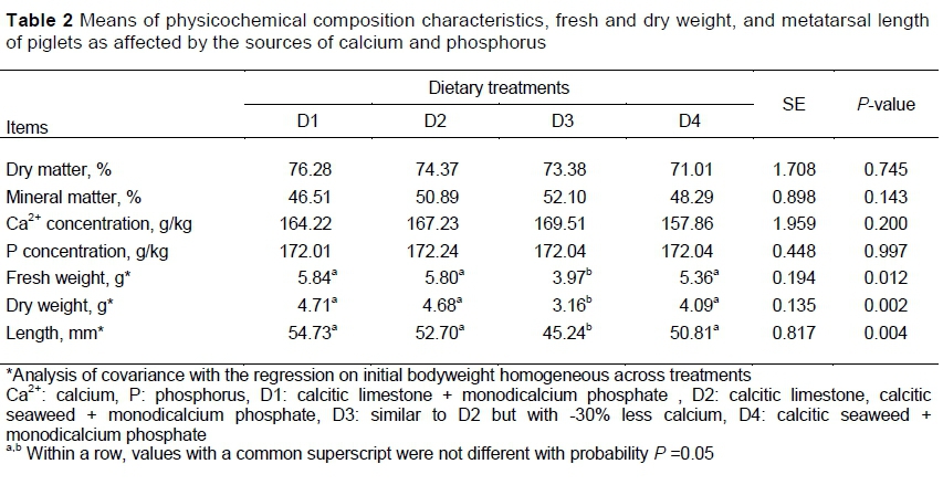
There was no effect of treatments on DM, MM, and concentrations of Ca2+ and P of the metatarsal bone of the piglets. However, there was an effect on fresh weight, dry weight and length of this bone (Table 2).
There was no difference among treatments on BMD. However, the treatments affected the area, BMC, Seedor index and maximum force applied (MFA). D3 had lower area and Seedor index compared with the other treatments (Table 3). D1 and D2 had greater BMC compared with D3. D4 had intermediate BMC between D2 and D3, with the BMC not different from either of these treatments. However, MFA was greater for D2 and D4 than for the D1 and D3 (Table 3).
All the piglets remained healthy. The results showed that Ca2+ sources were similarly effective in maintaining the MM content and the concentrations of Ca2+ and P. Despite this, CS is an organic Ca2+ source with greater bioavailability compared with CL (Avelar et al., 2009). This may explain why D3 piglets maintained their MM and Ca2+ concentrations. Moreover, bones are the largest reserve of Ca2+ in the body, which ensures the maintenance of Ca2+ and P homeostasis in addition to functioning in the structure and support of the body (Gerlinger et al., 2019).
Possibly the minerals in CS were not used for other organic functions of piglet metabolism, resulting in greater concentration in bone tissue and reduction in other tissues (liver, heart, and kidney), altering MM (Avelar et al., 2009). The current findings for MM were similar to those of González-Vega et al. (2016), who evaluated six diets with various calcium levels and reported values of the average ash content in the metacarpal (52.6%) and femur (56.75%), with concentrations of Ca2+ (37.68%) and P (17.90%) in the piglet femur.
The weight and length of the bone are related to the amount of mineral deposited, which may explain the results found in D3, showing that the reduced Ca2+ supply affected the its deposition in bone. Thus, unbalanced diets or those that have lower levels of minerals that are important for bone health result in impaired bone formation and reduced growth (Santana et al., 2017), which promotes change in the size, shape, and cells of the bone matrix (Barcellos et al., 2007).
The literature is limited in studies of CS and its effects on bone parameters of starter phase pigs. Saraiva et al. (2009) tested various levels of total P and Ca2+ (0.68%) and reported average values for the ash content (50.37%) and Ca2+ (163.4 g/kg) in the metatarsal analogous with the current findings. However, Santana et al. (2017) tested different sources of Ca2+ in piglet diets and observed slightly higher values than the present study in metatarsal length, and its DM content and fresh weight.
Changes in BMC promote measurable changes in BMD, although the BMD values were not significant among the treatments. This observation may be related to the proliferation of osteoblasts stimulated by the chemical factor released by osteocytes that function as mechanical sensors and thus respond to dietary insufficiency to promote bone increment (Andreoli et al., 2001). According to Cadore et al. (2005), BMD is a dynamic process of bone formation and remodeling and its maintenance is important for the prevention of bone malformation. However, resorption of bone tissue causes tissue deterioration, which was not observed for BMD in the present study.
Measurement of structural integrity of bone involves the application of mechanical tests. Bone ash content is directly related to mineralization, but the relationship with structural integrity is generally lower. Crenshaw et al. (2011) reported the potential appearance of bone damage because of malnutrition and that results obtained from a sample of one bone could be used to characterize the entire skeleton. Bollen and Bai (2005) observed reduced skeletal measurements of femur and skull in male rats fed diets containing low Ca2+ levels and controlled intake of diet.
Low Ca2+ intake impaired SI, MFA and BMC. These changes can cause negative changes in bone resistance to breaking, as reported by Barcellos et al. (2007). According to Suttle (2010), the swine metatarsus is a fast-growing bone that can be used as an indicator of resistance to breaking to reflect differences in the formation and growth of bone from variations in the deposition of minerals such as Ca2+.
Rufino et al. (2017) reduced the amount of P and concomitantly of Ca2+ in piglet diets and observed lower bone resistance in the metacarpal of pigs. The reason that D3 and D4 showed lower MFA and their bones had less resistance to breaking involved factors such as bone length, diameter, weight and plasma Ca2+ concentration (Santana et al., 2017). Besides being affected by bioavailability, Ca + and P can interact in bone metabolism (Schlegel & Gutzwiller, 2017).
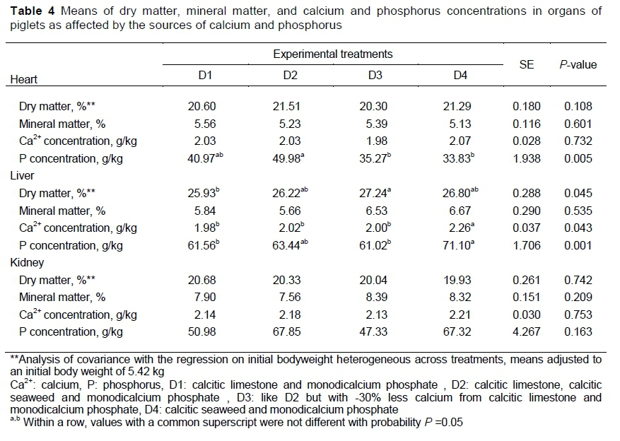
No effect of treatments was observed in DM, MM, Ca2+ concentration in the heart, in MM in the liver, or in DM, MM, Ca2+ and P concentrations in the kidneys of the piglets. However, treatments affected the P concentration in the heart, and DM and concentrations of Ca2+ and P in the liver (Table 4). The results indicated that D2 had greater P concentration in the heart than D3 and D4. The D3 treatment produced more DM in the liver than D1. The Ca2+ concentration in the liver was higher for D4 compared with the other diets. The P concentration in the liver was higher in D4 compared with D1 and D3.
There is no evidence that piglets fed diets that supplied less Ca2+ had greater liver DM compared with those that met the Ca2+ requirement (Table 4). Increased protein, fat and carbohydrates in the liver may decrease its moisture content (Tajik et al., 2012). Diets that restricted the amount of Ca2+ supplied to rats promoted greater fat mass (Bollen & Bai, 2005). More detailed studies are needed to assess Ca2+ kinetics and effects of Ca2+ sources on the physical composition of piglet body tissues because of a possible response of MM content to different sources of P (Queiroz et al., 2008).
Previously, MM in the internal organs of piglets has been related to the dietary source of Ca2+, its bioavailability and the level at which it was provided (Santana et al., 2017). The percentage of MM in organs is related to the use of the mineral by the tissue. Absorbed minerals may not have been fully used by the metabolism, which increases the concentration in the kidneys and liver, concomitantly in the MM content. Although the content of MM is influenced by age, the Ca2+ content remains relatively constant, regulated by physiological processes (Field, 2000).
The results suggested that Ca2+ concentration is established in organs and tissues because the physiological response to Ca2+ is regulated by several factors (Gerlinger et al., 2019). Calcium is found mainly in the endoplasmic reticulum and mitochondria (Bygrave & Benedetti, 1996), bones (99%) and serum (1%) (Beto, 2015). However, the amount of Ca2+ is in a range of 1,200 g in bone tissue (99% or 29.94 mol), 7 g in teeth (0.6% or 174.66 mmol), 7 g in soft tissues (0.6% or 174.66 mmol), 0.7 g in extracellular fluid (0.06% or 17.47 mmol), and 0.35 g in plasma (0.03% or 8.73 mmol) (Nordin, 1997). The present findings corroborate those reported by Moreira et al. (2004), who evaluated Ca2+ and P concentrations in tissues and obtained higher values for bone (83.02 and 75.35 mg/kg DM, respectively) compared with the Ca2+ concentration in the heart (0.32 mg/kg DM), and P concentration in kidney (8.91 mg/kg DM). Georgievskii et al. (1982) reported a concentration of Ca2+ (per 100 g of tissue) within a range of 8 to 25 mg in the heart, 10 to 30 mg in the liver and 6 to 20 mg in the kidney.
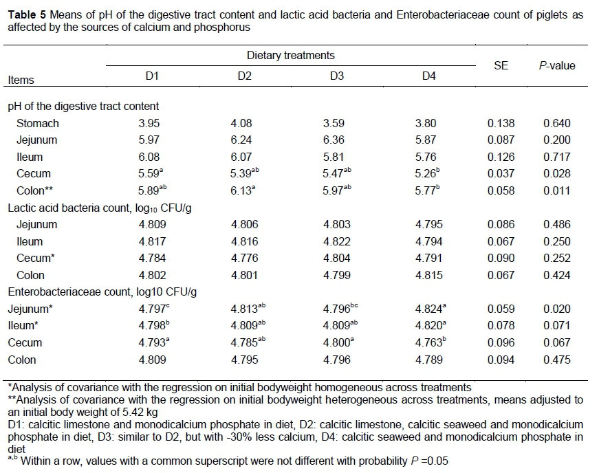
The treatments in this study did not affect (P >0.05) the pH of the digestive tract contents, except in the cecum and colon. D1 showed a higher pH of the cecal contents compared with D4. D2 had colon contents with a higher pH than D4 (Table 5). There was no effect (P >0.05) of treatments on the lactic acid bacteria count of the digesta. However, D4 showed a higher Enterobacteriaceae count in the jejunum compared with D1 and D3. A similar difference between D1 and D4 was observed for the pH of the digesta in the ileum. D3 had a greater Enterobacteriaceae count in the cecum compared with D1 (Table 5).
The results suggested a minor role for the pH of the digestive tract in the modulation of the LAB microbiota. Because pigs fed diets containing CL + DP or CL + CS + DP showed greater pH of the digestive tract content may be a result of the Ca2+ and P absorption. The results were inconsistent with the study by González-Vega et al. (2014), who reported higher pH values in piglets fed diets containing CS than in piglets fed Ca2+ carbonate. These authors thought this was because Ca2+ and P absorption decreased from CS. However, Brun et al. (2014) stated that the Ca2+ absorption was reduced at low pH.
The pH values in the current study are in agreement with Rufino et al. (2017), who found no difference between treatments and intestinal segments for diets that met the supply of Ca2+ and P and those with reduction of these minerals. For Almeida et al. (2012), in a study with rats, L. calcareum (dose of 480 mg/kg) demonstrated a protective effect on low-intensity gastric lesions and increased the gastric pH. However, the pH of the digestive tract contents might vary depending on the diet/feed intake (extrinsic) and animal physiology (intrinsic).
Reduced pH inhibits the growth of pathogenic bacteria such as Escherichia coli and Salmonella sp., which are then unable to harm the gastrointestinal tract (Tran et al., 2016). However, the authors did not find a plausible answer to justify the increase in Enterobacteriaceae in piglets receiving CS + DP owing to lower pH in the jejunum (5.87), cecum (5.26), and colon (5.77), where a higher pH (7.2 to 7.8) is speculated to provide an ideal environment for colonization of E. coli in intestinal villi (Gonzales et al., 2013). Various studies have demonstrated changes in the microbial community with Ca2+ supplementation (Blavi et al., 2018). However, the Enterobacteriaceae growth can be verified over a wide pH range (4.5 - 9.0) (Gonzales et al., 2013).
The reduced pH of the digestive tract content may provide an appropriate environment for the development of beneficial bacteria and inhibit growth of pathogenic bacteria. The authors were not able to identify alterations in the intestinal microbiota for LAB. Despite the absence of significant differences between the treatments, except for the Enterobacteriaceae in the jejunum, reports in the literature indicate that the amount of LAB was significantly higher than the pathogenic bacteria. However, one of the direct effects of Ca2+ supplementation was on the LAB that inhabit the hindgut (Blavi et al., 2018). This was related to Ca2+ levels, sources, and the site of the gastrointestinal tract (Mann et al., 2014).
However, the composition of the intestinal microbiota may present variations along the intestine because the population, quantity and microbial distribution are constantly changing their natural balance due to minimal variations in the physiological condition of the animal such as pH, bile and enzymatic secretion and microbial interactions (Oetting et al., 2006).
There was no effect (P >0.05) of treatments on VH. However, D2 and DI produced greater CD in the duodenum (P =0.004) and ileum (P =0.011) compared with those that consumed D3 and D4 (Table 6). Feeding D3 caused a higher (P =0.091) VH:CD ratio in the duodenum compared with D2 (Table 6).
Piglets fed D4 had slightly smaller villi. This was supported by the greater Enterobacteriaceae count, especially enterotoxigenic E. coli, in which fimbriae F5, F6, and F41 colonize mainly in the distal jejunum and ileum, whereas F4 can colonize the entire jejunum and ileum (Sun & Kim, 2017), compromising intestinal architecture because the size, number, integrity and protection of the villi, in association, determine the intestinal architecture (Maiorka et al., 2002).
Reduced villus size has been associated with post-weaning growth retardation in piglets (Anjos et al., 2019). As a consequence, intestinal architecture had fewer secretory cells and reduced absorption capacity in the small intestine, and a larger amount of unabsorbed dietary material, which could act as a substrate for pathogenic bacteria and increase their growth (Gao et al., 2013).
The variation in villi growth may be a result of greater cell proliferation, which is related to the higher CD to ensure an adequate epithelial renewal rate (Kisielinski et al., 2002), which explains the results for D1 and D2. However, villus growth occurs when the mitosis rate is greater than that of extrusion or when extrusion is not occurring (Maiorka et al., 2002). On the other hand, the capacity to absorb nutrients with lower energy losses owing to cell turnover is improved by greater VH and lower CD, that is, higher VH:CD ratio.
The VH:CD ratio values indicated a greater presence of mature and functional enterocytes (Tucci et al., 2011). Because D3 showed higher VH:CD compared with those D2 suggested the need for further investigation on interactions between organic and inorganic sources, and calcium reduction in diets for piglets. Supposedly, because D3 had a higher VH:CD ratio in an attempt to assimilate the reduced Ca2+ concentrations of unknown physiological mechanisms, Ca2+ metabolism or Ca2+ source.
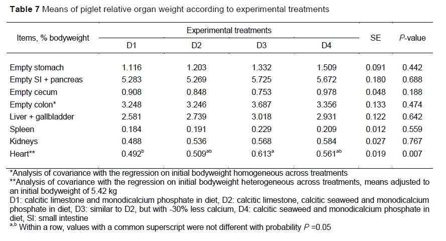
To determine whether CS affected ROW, digestive and non-digestive organs were evaluated. However, there was no effect (P >0.05) of treatments on ROW (bodyweight percentage), but D3 showed greater (P =0.007) heart weight compared with those that consumed CL + DP (Table 7).
There seem to be no reports in the literature about relative organ weights from piglets fed different sources of calcium and L. caícareum. In general, piglets maintained similar organ weights, which demonstrated an apparently normal state of development. The weight of internal organs is associated with the growth, health and general condition of animal metabolism. In Knight and Dilger (2018), the effect of iron in diets for pigs was evaluated and a compensatory growth mechanism was observed after a period of feed restriction. This observation agreed with the present findings, because piglets that received D3 showed greater heart weight. In addition, different sources of P in pig diets affected the relative weight of the kidneys compared with a diet without inorganic P (Teixeira et al., 2013).
The role of Ca2+ and P in animal metabolism and gene expression, involving absorption, resorption and bone remodeling influences the growth of body tissues (González-Vega et al., 2016). The weight of the small intestine has been associated with feed intake and with development of intestinal villi. However, it was reported that Ca2+ propionate promoted the development of internal organs and the gastrointestinal tract (Zhang et al., 2017).
Conclusions
These inconsistent findings suggest a need for further studies to better understand the interplay of effects of Ca2+ source and level on its metabolism.
Acknowledgements
The National Council for Scientific and Technological Development (CNPq, São Paulo, SP, Brazil), the Coordenação de Aperfeiçoamento de Pessoal de Nível Superior - Brazil (CAPES), the company Oceana Minerals (research funding), the COPAGRIL (ingredients and animals supply), the company COPISCES (ingredient supply) and the State University of the West of Paraná (Marechal Cândido Rondon, Paraná, Brazil).
Authors' contributions
All the authors contributed equally and commented on the early and final version of the manuscript.
Conflict of Interest Declaration
There are no conflicts of interest.
References
Adeleye, O.B.M. & Blomfield, D., 2014. The effect of algal biomass supplementation in maternal diets on piglet survival in two housing systems. Livestock Sci. 162, 193-200. DOI: 10.1016/j.livsci.2013.12.030 [ Links ]
Almeida, F., Schiavo, L.V., Vieira, A.D., Araújo, G.L., Queiroz-Junior, C.M., Teixeira, M.M. & Tagliati, C.A., 2012. Gastroprotective and toxicological evaluation of the Lithothamnion calcareum algae. Food Chem. Toxicol. 50, 1399-1404. DOI: 10.1016/j.fct.2012.02.028 [ Links ]
Andreoli, A., Monteleone, M., Van Loan, M., Promenzio, L., Tarantino, U. & De Lorenzo, A., 2001. Effects of different sports on bone density and muscle mass in highly trained athletes. Med. Sci. Sports Exerc. 33, 507-511. DOI: 10.1097/00005768-200104000-00001 [ Links ]
Anjos, C.M.D., Gois, F.D., Anjos, C.M.D., Rocha, V.D.S., Castro, D.E.D.S., Allaman, I.B., Silva, F.L., Carvalho, P.L.O., Meneghetti, C. & Costa, L.B., 2019. Effects of dietary beta-glucans, glucomannans and mannan oligosaccharides or chlorohydroxyquinoline on the performance, diarrhea, hematological parameters, organ weight and intestinal health of weanling pigs. Livestock Sci. 223, 39-46. DOI: 10.1016/j.livsci.2019.02.018 [ Links ]
AOAC (Association of Official Analytical Chemists), 1990. Official methods of analysis of Association of Official Analytical Chemists. 15th edition. AOAC, Arlington VA, USA. [ Links ]
Avelar, A.C., Ferreira, W.M., Brito, W. & Menezes, M.A.B.C., 2009. Composição mineral de fosfatos, calcário e farinha de ossos usados na agropecuária brasileira. Arch Zootec. 58, 737-740. [ Links ]
Barcellos, D.E.S.N.D., Lippke, R.T., Borowski, S.M. & Almeida, M.N.D., 2007. O problema da osteocondrose na suinocultura tecnificada. Acta Sci. Vet. 35, S165-S170. [ Links ]
Beto, J.A., 2015. The role of calcium in human aging. Clin. Nutr. Res. 4, 1-8. DOI: 10.7762/cnr.2015.4.1.1 [ Links ]
Blavi, L., Perez, J.F., Villodre, C., López, P., Martín-Orúe, S.M., Motta, V. & Sola-Oriol, D., 2018. Effects of limestone inclusion on growth performance, intestinal microbiota, and the jejunal transcriptomic profile when fed to weaning pigs. Anim. Feed Sci. Technol. 242, 8-20. DOI: 10.1016/j.anifeedsci.2018.05.008 [ Links ]
Bollen, A.M. & Bai, X.Q., 2006.Effects of long-term calcium intake on body weight, body fat and bone in growing rats. Osteoporos Int. 16, 1864-1870. DOI: 10.1007/s00198-005-1952-y [ Links ]
Brun, L.R., Brance, M.L., Lombarte, M., Lupo, M., Di Loreto, V.E. & Rigalli, A., 2014. Regulation of intestinal calcium absorption by luminal calcium content: role of intestinal alkaline phosphatase. Mol. Nutr. Food Res. 58, 1546-1551. DOI: 10.1002/mnfr.201300686 [ Links ]
Bygrave, F.L. & Benedetti, A., 1996. What is the concentration of calcium ions in the endoplasmic reticulum? Cell Calcium 19. 547-551. DOI: 10.1016/S0143-4160(96)90064-0 [ Links ]
Cadore, E.L., Brentano, M.A. & Kruel, L.F.M., 2005. Efeitos da atividade física na densidade mineral óssea e na remodelação do tecido ósseo. Rev. Bras. Med. Esporte 11, 373-379. DOI: 10.1590/S1517-86922005000600013 [ Links ]
Carlos, A.C., Sakomura, N.K., Pinheiro, S.R.F., Toledano, F.M.M., Giacometti, R. & Silva Júnior, J.W.D., 2011. Uso da alga Lithothamnium calcareum como fonte alternativa de cálcio nas rações de frangos de corte. Cienc. Agrotec. 35, 833-839. DOI: 10.1590/S1413-70542011000400025 [ Links ]
Costa Neto, J.M., Teixeira, R.G., Cavalcanti De Sá, M.J., Lima, A.E., Jacinto-Aragão, G.S., Teixeira, M.W. & Azevedo, A.S.D., 2010. Farinha de algas marinhas (Lithothamnium calcareum) como suplemento mineral na cicatrização óssea de auto enxerto cortical em cães. Rev. Bras. Saude Prod. Anim. 11, 217-230. https://repositorio.ufba.br/handle/ri/5380 [ Links ]
Cowieson, A.J., Wilcock, P. & Bedford, M.R., 2011. Super-dosing effects of phytase in poultry and other monogastrics. Worlds Poult. Sci.J. 67, 225-235. DOI: 10.1017/S0043933911000250 [ Links ]
Crenshaw, T.D., Rortvedt, L.A. & Hassen, Z., 2011. Triennial Growth Symposium: A novel pathway for vitamin D- mediated phosphate homeostasis: Implications for skeleton growth and mineralization. J. Anim. Sci. 89, 1957-1964. DOI: 10.2527/jas.2010-3411 [ Links ]
Field, R.A., 2000. Ash and calcium as measures of bone in meat and bone mixtures. Meat Sci. 55, 255-264. DOI: 10.1016/s0309-1740(99)00147-3 [ Links ]
Gao, C., Zhao, J. & Gregersen, H., 2000. Histomorphometry and strain distribution in pig duodenum with reference to zero-stress state. Dig. Dis. Sci. 45, 1500-1508. DOI: 10.1023/A:1005592306587 [ Links ]
Gao, Y., Han, F., Huang, X., Rong, Y., Yi, H. & Wang, Y., 2013.Changes in gut microbial populations, intestinal morphology, expression of tight junction proteins, and cytokine production between two pig breeds after challenge with Escherichia coli K88: A comparative study. J. Anim. Sci. 91, 5614-5625. DOI: 10.2527/jas.2013-6528. [ Links ]
Gatrell, S., Lum, K., Kim, J. & Lei, G., 2014. Potential of defatted microalgae from the biofuel industry as an ingredient to replace corn and soybean meal in swine and poultry diets. J. Anim. Sci. 92, 1306-1314. DOI: 10.2527/jas2013-7250 [ Links ]
Georgievskii, V.I., Annenkov, B.N. & Samokhin, V.T., 1982. Mineral composition of bodies and tissues of animals. In: V.I. Georgievskii, B.N. Annenkov & V.T. Samokhin (eds). Mineral nutrition of animals. Butterworth-Heinemann, London. Pp. 69-78. [ Links ]
Gerlinger, C., Oster, M., Borgelt, L., Reyer, H., Muráni, E., Ponsuksili, S. & Wimmers, K., 2019. Physiological and transcriptional responses in weaned piglets fed diets with varying phosphorus and calcium levels. Nutrients 11, 436. DOI: 10.3390/nu11020436 [ Links ]
Gonzales, L., Al, Z.B., Nygren, E., Wang, Z., Karlsson, S., Zhu, B. & Sjöling, Å., 2013. Alkaline pH is a signal for optimal production and secretion of the heat labile toxin, LT in enterotoxigenic Escherichia coli (ETEC). PLoS One 8, e74069. DOI: 10.1371/journal.pone.0074069 [ Links ]
González-Vega, J.C. & Stein, H.H., 2016.Digestibility of calcium in feed ingredients and requirements of digestible calcium for growing pigs. Anim. Prod. Sci. 56, 1339-1344. DOI: 10.1071/AN15352 [ Links ]
González-Vega, J.C., Walk, C.L. & Stein, H.H., 2015. Effects of microbial phytase on apparent and standardized total tract digestibility of calcium in calcium supplements fed to growing pigs. J. Anim. Sci. 93, 2255-2264. DOI: 10.2527/jas.2014-8215 [ Links ]
González-Vega, J.C., Walk, C.L., Liu, Y. & Stein, H.H., 2014. The site of net absorption of Ca from the intestinal tract of growing pigs and effect of phytic acid, Ca level and Ca source on Ca digestibility. Arch Anim. Nutr. 68, 126-142. DOI: 10.1080/1745039X.2014.892249 [ Links ]
González-Vega, J.C., Liu, Y., McCann, J.C., Walk, C.L., Loor, J.J. & Stein, H.H., 2016. Requirement for digestible calcium by eleven- to twenty-five-kilogram pigs as determined by growth performance, bone ash concentration, calcium and phosphorus balances, and expression of genes involved in transport of calcium in intestinal and kidney cells. J. Anim. Sci. 94, 3321-3334. DOI: 10.2527/jas.2016-0444 [ Links ]
Guo, M., Hayes, J., Cho, K.O., Parwani, A.V., Lucas, L.M. & Saif, L.J., 2001. Comparative pathogenesis of tissue culture-adapted and wild-type Cowden porcine enteric calicivirus (PEC) in gnotobiotic pigs and induction of diarrhea by intravenous inoculation of wild-type PEC. J. Virol. 75, 9239-9251. DOI: 10.1128/JVI.75.19.9239-9251.2001 [ Links ]
Kisielinski, K., Willis, S., Prescher, A., Klosterhalfen, B. & Schumpelick, V., 2002. A simple new method to calculate small intestine absorptive surface in the rat. Clin. Exp. Med. 2, 131-135. DOI: 10.1007/s102380200018 [ Links ]
Knight, L. & Dilger, R., 2018. Longitudinal effects of iron deficiency anemia and subsequent repletion on blood parameters and the rate and composition of growth in pigs. Nutrients 10, 632. DOI: 10.3390/nu10050632 [ Links ]
Maiorka, A., Boleli, I.C. & Macari, M., 2002. Desenvolvimento e reparo da mucosa intestinal. In: M. Macari, R.L. Furlan, E Gonzales (eds). Fisiologia aviária aplicada a frangos de corte. Funep, São Paulo.Pp. 113-124. [ Links ]
Mann, E., Schmitz-Esser, S., Zebeli, Q., Wagner, M., Ritzmann, M. & Metzler-Zebeli, B.U., 2014. Mucosa-associated bacterial microbiome of the gastrointestinal tract of weaned pigs and dynamics linked to dietary calcium-phosphorus. PLoS One 9, e8695. DOI: 10.1371/journal.pone.0086950 [ Links ]
Manzanilla, E.G., Perez, J.F., Martin, M., Kamel, C., Baucells, F. & Gasa, J., 2004. Effect of plant extracts and formic acid on the intestinal equilibrium of early-weaned pigs. J. Anim. Sci. 82, 3210-3218. DOI: 10.2527/2004.82113210x [ Links ]
Moreira, J.A., Vitti, D.M.S.S., Lopes, J.B. & Trindade Neto, M.A., 2004. Cinética do fósforo em tecidos de suínos alimentados com dietas contendo enzima fitase. Arq. Bras. Med. Vet. Zootec. 56, 74-80. DOI: 10.1590/S0102-09352004000100012 [ Links ]
Nordin, B.E.C., 1997. Calcium and osteoporosis. Nutr. 13, 664-686. DOI: 10.1016/S0899-9007(97)83011-0 [ Links ]
Oetting, L.L., Utiyama, C.E., Giani, P.A., Ruiz, U.S. & Miyada, V.S., 2006. Efeitos de extratosvegetais e antimicrobianossobre a digestibilidade aparente, desempenho, morfometria dos órgãos e histologia do epitélio intestinal de leitões recém-desmamados. R. Bras. Zootec. 35, 1389-1397. DOI: 10.1590/S1516-35982006000500019 [ Links ]
Queiroz, L.S.B., Bertechini, A.G. & Rodrigues, P.B., 2008. Utilização de fosfatos comerciais para frangos de corte na fase inicial. Pesqui. Agropecu. Bras. 43, 1421-1427. DOI: 10.1590/S0100-204X2008001000022 [ Links ]
Rostagno, H.S., Albino, L.F.T., Donzele, J.L., Gomes, P.C., Oliveira, R.F.D., Lopes, D.C., Ferreira, A.S., Barreto, S.L.D.T. & Euclides, R.F., 2011. Tabelas brasileiras para aves e suínos: Composição de alimentos e exigências nutricionais. 3rd edition. UFV, Viçosa MG, Brazil. [ Links ]
Rufino, L.M., Nunes, R.C., Stringhin. J.H., Mascarenhas, A.G., Arnhold, E., Coelho, K.O., Rocha, F.R.T. & Kiefer, C., 2017. Available phosphorus reduction in weaned piglets' diets containing phytase combined with butyric and benzoic acids. Semin. Cienc. Agrar. 38, 1429-1440.DOI: 10.5433/1679-0359.2017v38n3p1439 [ Links ]
Sakomura, N.K. & Rostagno, H.S., 2007. Métodos de pesquisa em animais monogástricos. 1st edition. Funep, Jaboticabal SP, Brazil. [ Links ]
Santana, A.L.A., Carvalho, P.L.O., Oliveira, N.E.T., Gonçalves Junior, A.C., Gazola, A.P., Castro, D.E.S., Carvalho, S.T. & Oliveira, A.C., 2017. Different sources of calcium for starter pig diets. Livestock Sci. 206, 175-181. DOI: 10.1016/j.livsci.2017.10.021 [ Links ]
Saraiva, A., Donzele, J.L., Oliveira, R.F.M., Abreu, M.L.T., Silva, F.C.O. & Santos, F.A., 2009. Available phosphorus levels in diets for swine from 15 to 30 kg genetically selected for meat deposition. R. Bras. Zootec. 38, 307-313. DOI: 10.1590/S1516-35982009000200013 [ Links ]
Schlegel, P. & Gutzwiller, A., 2017.Effect of dietary calcium level and source on mineral utilisation by piglets fed diets containing exogenous phytase. J. Anim. Physiol. Anim. Nutr. 101, e165-e174. DOI: 10.1111/jpn.12582. [ Links ]
Seedor, J.G., Quartuccio, H.A. & Thompson, D.D., 1991. The biophosphonate alendronate (MK-217) inhibits bone loss due to ovariectomy in rats. J. Bone Miner. Res. 6, 339-346. DOI: 10.1002/jbmr.5650060405 [ Links ]
Simkus, A., Simkienè, A. & Cernauskienè, J., 2013. The effect of blue algae Spirulina platensis on pig growth performance and carcass and meat quality. Vet. ir Zootech. 61, 70-74. [ Links ]
Sun, Y. & Kim, S.W., 2017. Intestinal challenge with enterotoxigenic Escherichia coli in pigs, and nutritional intervention to prevent postweaning diarrhea. Anim. Nutr. 3, 322-330. DOI: 10.1016/j.aninu.2017.10.001 [ Links ]
Suttle, N.F., 2010. Calcium. In: N.F. Suttle (ed). Mineral nutrition of livestock. CAB, Oxfordshire, UK. Pp. 54-91. [ Links ]
Tajik, H., Ramin, A., Nozad, S., Jelodari, B., Ashtab, G., Eftekhari, Z. & Ramin, S., 2012. Relationship between liver lipid and liver dry matter in slaughtered ruminants. Vet. Res. Forum 3, 275. [ Links ]
Teixeira, A.D.O., Nogueira, E.T., Corassa, A., Ferreira, V.P.D.A., Rocha Júnior, C.M. & Lopes, D.C., 2013. Evaluation of phosphorus sources on performance, organ weight and blood parameters of pigs. Rev. Bras. Saude Prod. Anim. 14, 808-819. DOI: 10.1590/S1519-99402013000400014 [ Links ]
Tran, T.H.T., Everaert, N. & Bindelle, J., 2016. Review on the effects of potential prebiotics on controlling intestinal enteropathogens Salmonella and Escherichia coli in pig production. J. Anim. Physiol. Anim. Nutr. 102, 17-32. DOI: 10.1111/jpn.12666 [ Links ]
Tucci, F.M., Thomaz, M.C., Nakaghi, L.S.O., Hannas, M.I., Scandolera, A.J. & Budino, F.E.L., 2011. Efeito da adição de agentes tróficos na dieta de leitões desmamados sobre a estrutura e ultraestrutura do intestino delgado e sobre o desempenho. Arq. Bras. Med. Vet. Zootec. 63, 931-940. DOI: 10.1590/S0102-09352011000400019. [ Links ]
Weedman, S.M., Rostagno, M.H., Patterson, J.A., Yoon, I., Fitzner, G. & Eicher, S.D., 2011. Yeast culture supplement during nursing and transport affects immunity and intestinal microbial ecology of weanling pigs. J. Anim. Sci. 89, 1908-1921. DOI: 10.2527/jas.2009-2539 [ Links ]
Zhang, X., Wu, X., Chen, W., Zhang, Y., Jiang, Y., Meng, Q. & Zhou, Z., 2017.Growth performance and development of internal organ, and gastrointestinal tract of calf supplementation with calcium propionate at various stages of growth period. PloS One 12, 1-12. DOI: 10.1371/journal.pone.0179940 [ Links ]
Zhu, Y.S., Connolly, A., Guyon, A. & FitzGerald, R.J., 2014.Solubilisation of calcium and magnesium from the marine red algae Lithothamnion calcareum. Int. J. Food Sci.Technol. 49, 1600-1606. DOI: 10.1111/ijfs.12459 [ Links ]
Submitted 2 July 2021
Accepted 4 November 2021
Published 22 February 2022
# Corresponding author: jansllerg@gmail.com

