


Services on Demand
Article
 English (pdf)
 Article in xml format
 Article references
Indicators

Related links
-
 Cited by Google
 -
 Similars in Google
Share

 Permalink
South African Journal of Animal Science
On-line version ISSN 2221-4062
Print version ISSN 0375-1589
S. Afr. j. anim. sci. vol.47 n.1 Pretoria 2017
http://dx.doi.org/10.4314/sajas.v47i1.6
ARTICLES
Electroencephalographic responses to neck cut and exsanguination in minimally anaesthetized goats
A.B. SabowI, II; Y.M. GohIII, IV, #; I. ZulkifliI, III; A.Q. SaziliI, III, V; M.Z.A. Ab KadirVI; U. KakaVII, VIII; N. KhadijahIX; K.D. AdeyemiI, X; M. EbrahimiIV
IDepartment of Animal Science, Faculty of Agriculture, Universiti Putra Malaysia, 43400 UPM Serdang, Selangor, Malaysia
IIInstitute of Tropical Agriculture, Universiti Putra Malaysia, 43400 UPM Serdang, Selangor, Malaysia
IIIDepartment of Veterinary Preclinical Sciences, Faculty of Veterinary Medicine, Universiti Putra Malaysia, 43400 UPM Serdang, Selangor, Malaysia
IVDepartment of Veterinary Clinical Studies, Faculty of Veterinary Medicine, Universiti Putra Malaysia, 43400 UPM Serdang, Selangor, Malaysia
VDepartment of Electrical and Electronic Engineering, Faculty of Engineering, Universiti Putra Malaysia, 43400 UPM Serdang, Selangor, Malaysia
VIHalal Products Research Institute, Universiti Putra Malaysia, 43400 UPM Serdang, Selangor, Malaysia
VIIDepartment of Animal Resource, University of Salahaddin, Erbil, Kurdistan Region, Iraq
VIIIDepartment of Veterinary Surgery and Obstetrics, Faculty of Animal Husbandry and Veterinary Sciences, Sindh Agriculture University Tandojam, Sindh, Pakistan
IXDepartement of Food Science and Nutrition, Islamic University in Uganda, P.O.Box 2555 Mbale, Uganda
XDepartement of Animal Production, University of Ilorin, Ilorin, Nigeria
ABSTRACT
Conscious animals typically experience sensory (nociception) and emotional pain, whereas unconscious animals that were minimally anesthetized would experience minimal emotional pain. To determine whether 'silencing' the emotional component through a minimally anesthetized model would minimize stress response, and thus improve animal welfare, this study aimed at comparing changes in electroencephalographic (EEG) activities associated with possible noxious stimuli following neck-cut slaughter in conscious non-anesthetized versus minimally anaesthetized Boer cross-bred goats. Ten bucks were randomly assigned to two groups of five animals each, and subjected to neck-cut slaughter when fully conscious (HS) or under minimal anaesthesia (AS) and exsanguinated. The anaesthesia was induced with propofol (5 mg/kg) administered to effect by rapid injection into a cephalic vein and maintained with halothane in 100 % oxygen. Changes in the root mean square (RMS) for each of alpha, beta, delta and theta waves, median frequency (F50) and total power of the EEG (Ptot) were compared in each group before and after neck cut and between groups following treatments. Electroencephalographic parameters did not differ between goats that were fully conscious or slaughtered under minimal anaesthesia. These findings showed that the noxious stimuli from neck cut were present in both conscious and minimally anaesthetized goats. Most importantly, the presence of emotional pain and nociception did not affect the extent of electroencephalographic responses significantly compared with animals that were experiencing nociception only.
Keywords: brain activity, propofol-halothane anaesthetic, noxious stimuli, slaughter
Introduction
The way in which animals are handled, restrained and slaughtered can affect their welfare and the quality of the meat (Zulkifli et al., 2014; Agbeniga & Webb, 2012). The basic welfare requirement stipulates that the animal must be stunned (mechanical, electrical or gas) to render it unconscious prior to slaughter (Rodriguez et al., 2012). However, exceptions were given for animals that were slaughtered for religious purposes or emergencies. During halal and shechita slaughter, the animals are restrained and bled through a transverse incision across the neck, severing the skin, muscles, oesophagus, carotid arteries, trachea, major nerves and jugular veins. The bleeding requires time to provoke brain death. In addition, the cuts involve substantial tissue damage in areas that are well supplied with nociceptors (Rodriguez et al., 2012). Therefore, there are welfare concerns about the slaughter of animals without stunning prior to neck slit and exsanguination, comprising potential pain from the incision itself, and pain and distress prior to the onset of insensibility (Grandin, 2013). However, it has been suggested that the use of a superbly sharp knife produces minimal behavioural reactions in animals and, as a result, the neck cut is not perceived as painful (Rosen, 2004).
Gibson et al. (2009a) made an assessment of the electroencephalographic changes at slaughter by neck cut without pre-stunning in halothane-anaesthetized calves and found that there is a period after slaughtering in which the ventral neck cut signifies a noxious stimulus that would probably be perceived as pain in fully conscious animals. The minimally anesthetized model is widely used in farm animals to assess cerebrocortical responses to noxious stimuli during neck cut and surgical operations without jeopardizing animal welfare in cattle and sheep (Gibson et al., 2009a), but has not yet been reported in goats.
Because the slaughter process could induce stress and noxious stimuli, this is a significant physiological change that could be utilized for animal welfare assessment (Zulkifli et al., 2014). However, stress and nociception involve more than physiological input and response. It is postulated that visual and psychological inputs to the autonomic nervous system, which are not typically present in minimally anaesthetized animals, would affect the intensity of stress and nociception, particularly in fully awake animals. As a consequence, there is a need to study and compare electroencephalographic response following the neck cut in minimally anaesthetized animals versus fully conscious ones. This study strives to evaluate the utility of the minimally anesthetized model in animal slaughter research. Further evidence is required to assess the extent of physiological pain (manifested as nociceptive reflexes and measurable via EEG parameters), and emotional pain (associated with fear, and thus compounded stress factors). Awake animals would typically experience both sensory and emotional pain, whereas unconscious animals that were minimally anesthetized would experience minimal emotional pain. Therefore, this study is aimed at comparing the changes in electroencephalographic responses associated with possible noxious stimuli following neck-cut slaughter in conscious non-anesthetized goats (HS) versus minimally anaesthetized goats (AS).
Materials and Methods
This study was conducted in accordance with the animal ethics guidelines of the Research Policy of Universiti Putra Malaysia (Ethical Clearance No. R052/2015).
The work was conducted on ten male Boer cross-bred goats of the same age (approximately seven months) at the Animal Science Departmental Research Abattoir, Faculty of Agriculture, Universiti Putra Malaysia. Five goats weighing between 21 kg and 24.8 kg (22.84 ± 1.66 kg) were allocated to slaughter by neck cut without stunning (fully conscious) (HS) as defined in MS1500: 2009 (Department of Standards Malaysia, 2009). Another group of five goats weighing between 22 kg and 25 kg (23.40 ± 1.29 kg) were allotted to a similar slaughter technique to mimic the action of the neck cut under minimal anaesthesia (AS). The slaughter procedure was performed by a licensed slaughter man. The head of each animal was pulled dorsally to stretch the neck to facilitate exsanguination. The process involved severing the skin, muscle, oesophagus, trachea, carotid arteries, jugular veins and major nerves without decapitating the animal during the process. A transverse section was performed with a sharp knife.
Animals were anesthetized with 5 mg/kg of propofol (Bharat Serums and Vaccines Ltd, Thane Maharashtra, India). After intubation with a 8-mm cuffed endotracheal tube (TaperGuard™ Oral/Nasal Tracheal Tube, Covidien Limited, Dublin, Ireland), anaesthesia was maintained with inhalation of halothane (Piramal Healthcare Limited, Medak District, Telangana, India) in 100% oxygen delivered via a precision vaporizer and a circle breathing system (Cyprane TEC3 halothane anaesthesia vaporizer, UK), slaughtered and subsequently bled (Sabow et al., 2016; Johnson et al., 2009). Briefly, the vaporizer was adjusted to maintain the end tidal halothane (ETHal) between 0.95% and 1.0%. A blood pressure cuff of 40-60% circumference of the antebrachium was connected to a monitor (Datex-Ohmeda, GE healthcare, Finland Oy, Helsinki, Finland) to measure blood pressure noninvasively. Lactated Ringer's solution was administered at 10 ml/kg/h to maintain mean blood pressure above 60 mm Hg throughout the aesthetic period. The temperature was monitored with an oesophageal thermistor probe and was maintained at 37 °C to 38 °C with a heating pad and warm blanket.
For each animal, prior to neck cut, baseline EEG activities (T1) and immediately post neck cut (T2) were recorded telemetrically with Power LabTM Biopotential Recordings Systems (AD Instruments, Sydney, Australia). Animals were instrumented with needle electrodes to record the EEG according to the technique of Kaka et al. (2015). A personal computer installed with software Chart 5.0 recording (PowerlabTM data acquisition system, Sydney, Australia) was used to record the EEG. The EEG data were analysed offline after the completing the experiments according to the methods described by Zulkifli et al. (2014). The root mean square (RMS) for each of the alpha, beta, delta and theta waveform, EEG median frequency (F50) and total power (Ptot) were calculated continuously for non-overlapping five-second epochs, using Chart 5.0TM software. The data of EEG from 60-second blocks prior to neck cut to 90-second blocks post neck cut were taken for statistical analysis based on the time to loss of pupillary reflex. The average time lapse from the point of slaughter to the loss of pupillary reflex was 2.24 min, with no significant differences (P >0.05) between slaughter methods.
The experiment was a completely randomized design. All data obtained were analysed with the general linear model (GLM) procedure of the Statistical Analysis System package (SAS), in which the EEG parameters were fitted as dependent variables. Slaughter methods, sampling times and interaction between slaughter methods and sampling times were fitted as fixed effects in a repeated measure analysis of variance. Because the interactions between slaughter methods and sampling times were not statistically different, these results were not presented. Duncan's multiple range test was used to separate means at a significance level of P <0.05.
Results
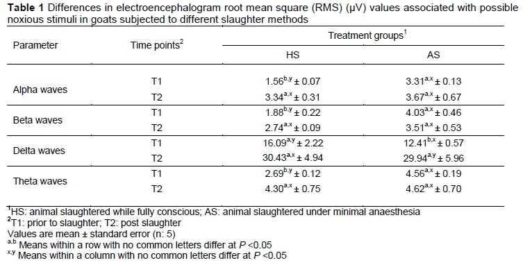
EEG activities for the two slaughter methods are presented in Table 1 and 2. At the beginning of the EEG recording before the neck cut, differences in EEG activities were observed (P <0.05) between slaughter methods with the lower RMS value in the alpha, beta, delta and theta frequency bands for HS. This may partially be caused by movement of the animal. The RMS value for alpha, beta, delta and theta waves among HS animals increased significantly (P <0.05) by almost twofold within 90 seconds of the initial throat cut, although the RMS value for both groups (HS and AS) did not show statistical significance in brain activity within 90 seconds post slaughter (Table 1).
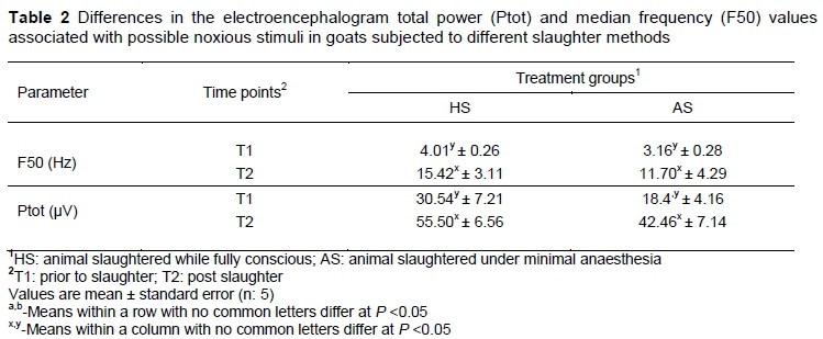
Observed differences in F50 values were only numerical (P >0.05) across treatment groups before and after slaughter (Table 2). The HS goats exhibited higher values of Ptot than the AS ones at T2, although the values were insignificant. The magnitude of F50 and Ptot changes in the present study was somewhat imprecise because of the small number of animals involved in the study. Regardless of the treatments, the overall mean F50 at T2 increased significantly for goats subjected to HS and those assigned to AS compared with T1. Moreover, after slaughter, the average Ptot recorded in the HS and AS goats was greater (P <0.05) compared with those observed prior to slaughter.
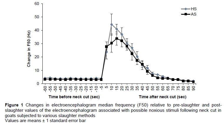
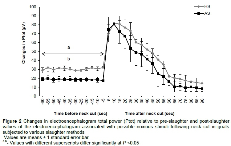
The results for the effectiveness of slaughter methods on changes in F50 and Ptot at various time intervals are shown in Figures 1 and 2. The results revealed no significant difference in F50 between HS and AS at various time intervals. However, in the initial 10 to 15 seconds following neck cut, the increase in mean F50 values were different (P <0.05) from the pre-slaughter values. It remained stable for five seconds and then decreased sharply for both slaughter methods (Figure 1). There were no significant differences in Ptot values among treatments at various time intervals. However, there was a significant increase in total power immediately after slaughter compared with baseline before slaughter. This increase had a mean duration of 35 seconds (Figure 2).
Discussion
An EEG was previously used as a tool to measure pain (Otto & Gerich, 2001; Rodriguez et al., 2012). Recent developments related to quantitative analysis of the EEG have allowed the experience of pain to be assessed more directly than was possible in the past (Johnson et al., 2012). In the current experiment, the results of electroencephalogram recordings indicate that the act of slaughter in non- anaesthetized goats is related to noxious stimulation, which could be painful, as pointed out by Johnson et al. (2012). These authors (Johnson et al., 2012) summarized the outcomes of a number of studies in which the minimal anaesthesia model was used to determine the effect of slaughter of calves without stunning. The results demonstrated that the act of slaughter by ventral neck incision without stunning is associated with pain and distress in the period between slaughter and subsequent loss of consciousness. Changes in the EEG attributable to noxiousness in the current study are within the reported window of possible sensibility in the majority of studies following ventral neck incision and exsanguination. The current observation is in tandem with those of Zulkifli et al. (2014), who found higher levels of alpha and beta wave activities post slaughter in non-stunned cattle compared with prior to slaughter. Their results demonstrated that the RMS for alpha waves increased by almost three times, while the RMS value for beta waves increased two times within 30 seconds of the initial slit of the throat. Animals that were anaesthetized pre-slaughter presented similar alpha, beta, and theta wave values prior to and post slaughter (T1 and T2). On the other hand, there was a notable increase (P <0.05) in delta RMS values by almost 2.2-fold post slaughter in the anaesthetized animals. This result indicates noxious stimuli are present at the point of slaughter in anaesthetized animals, which is consistent with the findings of a number of studies that were summarized by Murrell & Johnson (2006).
The similarity in F50 values indicates that slaughtering goats by rendering them unconscious is not a significant contributor to compromising the welfare of the animals. The high value of Ptot in HS could be related to the high movement and periodic activity of the animal before slaughter. After slaughter, the F50 and Ptot recorded in the HS and AS goats were higher than those observed prior slaughter. In line with the present results, Gibson et al. (2009b) observed that the F50 and Ptot increased significantly (P <0.05) during slaughter without stunning following anaesthesia in calves. The decrease in F50 values by 15 seconds after slaughter in both AS and HS could be because of the large quantity of blood gushing from the body of the animals. Gibson et al. (2009a) reported that the F50 values in calves during the 30 seconds following slaughter changed significantly (P <0.05) compared with the pre-slaughter values. This finding is consistent with those of Gibson et al. (2009b), who showed a significant decrease in Ptot after the initial increase following neck cut. However, this finding is in agreement with the findings of Gibson et al. (2009a), who reported initial increase in total EEG power after ventral neck incision increase. Johnson et al. (2005) reported an initial increase in total EEG power in lambs of different ages undergoing rubber-ring castration. In these cases, the increase in total EEG power could be attributed to the presence of noxious stimuli.
In this study, the decrease in F50 and Ptot 35 seconds after the neck cut in the HS and AS groups represented a reduction in and eventual loss of cerebral cortical electrical activity because of ischaemia. In their study of electrophysiological responses in veal calves, Lambooij et al. (2012) reported that after neck cutting without stunning, percentage power of EEG beta wave fraction decreased slowly to lower values resulting in an induction of unconsciousness lasting on average 80 seconds. In slaughter without stunning, animals become unconscious and consequently die from loss of blood. However, in cattle, brain activity can be maintained for up to two minutes after slaughter for two reasons: i) cattle have an alternative pathway; the vertebral arteries, which arise from the brachiocephalic trunk before the carotid arteries, can supply enough blood to the brain to maintain some level of brain function, even if the carotid arteries have been severed; and ii) the carotid arteries in cattle are prone to spasms at the site of the cut, particularly when a blunt knife is used, which restricts the outflow of blood and maintenance of systemic blood pressure (ballooning) (Gregory et al., 2012).
Conclusion
The results of the current study demonstrated that the noxiousness of the stimuli from the neck cut is evident in both conscious and minimally anaesthetized goats. This confirmed that both groups of animals experienced nociception. Electroencephalographic parameters surveyed in this study were not different between anesthetized and anaesthetized animals. Thus, this study affirms that the presence of emotional pain and nociception did not seem to affect the extent of electroencephalographic and hormonal responses significantly, compared with when the animals experienced nociception only. Minimal anaesthesia is a good research model with which to study goats subjected to neck cut slaughter.
Acknowledgments
The authors are very grateful to the Ministry of Education Malaysia for the research fund provided through the Universiti Putra Malaysia Grant (Project No. GP-IBT/2013/9409300).
Authors' Contributions
ABS, YMG, UK, NK, KDA and ME designed and carried out the studies, interpreted the results and drafted the manuscript. YMG, IZ, AQS and MZAAK supervised the study and interpreted the results for inclusion in the manuscript. ABS, YMG, AQS, UK, NK, and KDA participated in the animal experiment and processed the initial electroencephalography data. UK and YMG carried out the veterinary medical procedures in the experiment. ABS, ME and YMG performed the final electroencephalography and statistical analysis. All authors read and approved the final manuscript.
Conflict of Interest Declaration
Authors declare that there is no conflict of interest for this study.
References
Agbeniga, B., & Webb, E.C., 2012. Effect of slaughter technique on bleed-out, blood in the trachea and blood splash in the lungs of cattle. S. Afr. J. Anim. Sci 42, 524-529. [ Links ]
Department of Standards Malaysia (2009). MS1500: 2009 (1st revision). Halal food-production, preparation, handling and storage-general guideline. Department of Standards Malaysia, Cyberjaya, Selangor, 1-13. [ Links ]
Gibson, T., Johnson, C., Murrell, J., Chambers, J., Stafford, K. & Mellor, D., 2009a. Components of electroencephalographic responses to slaughter in halothane-anaesthetised calves: Effects of cutting neck tissues compared with major blood vessels. N. Z. Vet. J. 57, 84-89. [ Links ]
Gibson, T, Johnson, C., Murrell, J., Hulls, C., Mitchinson, S., Stafford, K., Johnstone, A. & Mellor, D., 2009b. Electroencephalographic responses of halothane-anaesthetised calves to slaughter by ventral-neck incision without prior stunning. N. Z. Vet. J. 57, 77-83. [ Links ]
Grandin. T., 2013. Elctric stunning of pigs and sheep. Colorado State University, USA. Available at: http://wwwgrandincom/humane/elecstunhtml. [ Links ]
Gregory, N.G., Schuster, P., Mirabito, L., Kolesar, R. & McManus, T., 2012. Arrested blood flow during false aneurysm formation in the carotid arteries of cattle slaughtered with and without stunning. Meat Sci. 90(2), 368-372. [ Links ]
Johnson, C., Gibson, T., Stafford, K. & Mellor, D., 2012. Pain perception at slaughter. Anim. Welf. 21, 113-122. [ Links ]
Johnson, C., Sylvester, S.P., Stafford, K.J., Mitchinson, S.L., Ward, R.N. & Mellor, D.J., 2009. Effects of age on the electroencephalographic response to castration in lambs anaesthetized with halothane in oxygen from birth to 6 weeks old. Vet. Anaesth. Analg. 36, 273-279. [ Links ]
Johnson, C., Stafford, K., Sylvester, S., Ward, R., Mitchinson, S. & Mellor, D., 2005 Effects of age on the electroencephalographic response to castration in lambs anaesthetised using halothane in oxygen. N. Z. Vet. J. 53, 433-437. [ Links ]
Kaka, U., Hui Cheng, C., Meng, G.Y., Fakurazi, S., Kaka, A., Behan, A.A. & Ebrahimi, M., 2015. Electroencephalographic changes associated with antinociceptive actions of lidocaine, ketamine, meloxicam, and morphine administration in minimally anaesthetized dogs. BioMed. Res. Int. doi: 10.1155/2015/305367. Available at: http://www.hindawi.com/journals/bmri/2015/305367/. [ Links ]
Lambooij, E., van der Werf, J., Reimert, H. & Hindle, V., 2012. Restraining and neck cutting or stunning and neck cutting of veal calves. Meat Sci., 91, 22-28. [ Links ]
Murrell J. & Johnson C., 2006. Neurophysiological techniques to assess pain in animals. J. Vet. Pharmacol. Ther. 29, 325-335. [ Links ]
Otto, K., & Gerich, T., 2001. Comparison of simultaneous changes in electroencephalographic and haemodynamic variables in sheep anaesthetised with halothane. Vet. Rec. 149, 80-84. [ Links ]
Rodriguez, P., Velarde, A., Dalmau, A. & Llonch, P., 2012. Assessment of unconsciousness during slaughter without stunning in lambs. Anim. Welf. 21, 75-80. [ Links ]
Rosen, S., 2004. Physiological insights into shechita. Vet. Rec., 154(24), 759-765. [ Links ]
Sabow, A., Goh, Y., Zulkifli, I., Sazili, A., Kaka, U., Ab Kadir, M., Ebrahimi, M., Nakyinsige, K. & Adeyemi, K., 2016. Blood parameters and electroencephalographic responses of goats to slaughter without stunning. Meat Sci. 121, 148155. [ Links ]
Zulkifli, I., Goh, Y., Norbaiyah, B., Sazili, A., Lotfi, M., Soleimani, A. & Small, A., 2014. Changes in blood parameters and electroencephalogram of cattle as affected by different stunning and slaughter methods in cattle. Anim. Prod. Sci. 54,187-193. [ Links ]
Received 24 November 2015
Accepted 24 October 2016
First published online 9 December 2016
# Corresponding author: ymgoh@upm.edu.my or gohyongmeng@gmail.com 77

