


Services on Demand
Article
 English (pdf)
 Article in xml format
 Article references
Indicators

Related links
-
 Cited by Google
 -
 Similars in Google
Share

 Permalink
South African Journal of Animal Science
On-line version ISSN 2221-4062
Print version ISSN 0375-1589
S. Afr. j. anim. sci. vol.42 n.5 Pretoria Jan. 2012
Evaluation of silver nanoparticles as a possible coccidiostat in broiler production
N. Chauke; F.K. Siebrits#
Department of Animal Sciences, Tshwane University of Technology, Private Bag X680, Pretoria, 0001, South Africa
ABSTRACT
The effect of administering low (15 mg/L) levels of silver nanoparticles in the drinking water of broilers (n = 40) was investigated as a potential replacement for antibiotic coccidiostats (Salinomycin® or Baycox®) in two trials. Four treatments were used: (1) challenged (with 3.3 x 10⁵ Eimeria tenella oocysts via oral gavage in the first trial and 1.6 x 10⁴ in the second trial) and medicated with silver nanoparticles in drinking water; (2) challenged and medicated with a registered ionophore coccidiostat (Salinomycin® in Trial 1 and Baycox® in Trial 2); (3) challenged and unmedicated and (4) unchallenged and unmedicated (control). Caecal lesions were scored on a scale from 0 - 4, while liver, caecal and kidney samples were taken to determine silver content. Growth performance was subjected to ANOVA using Statistica®. In Trial 1, neither the challenge (587 g vs. 561 g) nor the use of silver nanoparticles (587 vs. 555 g) had a significant effect on the weight gain of chicks from 13 to 27 days of age. The coccidiostat treatment group had a significantly lower weight gain than the unmedicated control (219 vs. 560.5 g) but had the lowest lesion score of 2.3. The silver nanoparticles group had, numerically, a slightly better score than the untreated group. The unchallenged control group had scores of 0. In the second trial there were no significant differences in growth performance between the treatments and there were no lesions, but both the silver nanoparticles group and the coccidiostat group had 50% less oocysts in the faecal samples compared to the control group. The silver content of the livers of the silver nanoparticle group was 0.083 mg/kg compared to 0.001 mg/kg in the control group. The results of this study on the use of silver nanoparticles as a coccidiostat were therefore not conclusive, but holds promise so that further investigation is warranted.
Keywords: Ag, protozoa, oocysts, silver retention, nano technology
Introduction
Coccidiosis in poultry is principally caused by protozoa such as Eimeria spp. which have been the cause of poor performance in broiler chickens for many years. It is controlled by the prophylactic inclusion of ionophores in diets (Chapman et al., 2010). There are signs of resistance development, similar to resistance in bacteria to current antibiotics, leading to a strong incentive to develop new alternatives of control (Kyriacou et al, 2004; Panacek et al, 2006). In the 1950s, silver nanoparticles were used as additive in poultry feeds, but the price of silver nanoparticles could not compete with that of antibiotics (Jeong et al., 2005). Coating of catheters, dental resin composites, burn wounds and homeopathic medicine, with a minimal risk of toxicity in humans is done with the help of silver compounds that are used as antimicrobial agents in a variety of applications (Lansdown, 2006). Silver is a non-toxic, safe, inorganic antibacterial agent used for many years for its capability of killing about 650 types of pathogens (Jeong et al., 2005). According to Singh et al. (2008) elemental silver occurs naturally and it is considered to be non-toxic, non-allergic and not cumulative in body tissue. The small size of nanoparticles of metallic silver (below 200 nm) in solid or colloidal state allows for a higher microbiological effect than silver salts (Atiyeh et al, 2007). Several studies have shown that silver nanoparticles can be successfully used as an antimicrobial agent against various organisms such as Chlamydomonas reinhardtii (Navarro et al, 2008) and Campylobacter jejuni (Spruill, 2006). In this study the efficiency of silver nanoparticles against Eimeria tenella as the predominant organism involved in coccidiosis in broiler chickens, was determined.
Materials and Methods
Two experiments were conducted, each using 40 day-old male chicks raised in a clean environment in wire cages at the Veterinary Faculty at Onderstepoort, South Africa. The birds were fed a starter diet with no medication from day 1 up to 13 days of age. On day 13, chicks were weighed individually and divided into four groups of 10 each. Groups were randomly allocated to treatments. In each trial, three groups were challenged on day 21 by oral gavage with 3.3 x 10⁵ coccidial oocysts (Trial 1) or with 1.6 x 10⁴ oocysts (Trial 2).
Treatments were: (1) Challenged by dosing with coccidial larvae and medicated with silver nanoparticles in drinking water at a dose of 15 mg/L, (2) challenged and medicated with a registered ionophore coccidiostat, (3) challenged and unmedicated and (4) unchallenged and unmedicated (control). In Trial 1, Salinomycin® was used as the coccidiostat, administered daily from day 14 to 27 days of age in the feed. Baycox® was used as the coccidiostat in Trial 2, administered from day 14 to 34 days of age in the water.
In Trial 1 feed wastage was such that feed intakes could not be measured while feed intakes and live weights of chicks were recorded once a week in Trial 2. Pens were checked daily for chick mortality. Faecal material was collected on plastic sheets from under the cages on days 6, 7 and 8 after the birds were challenged. Oocyst counts were done using the "Faecalizer" flotation method and the "McMaster Chamber" method of Holdsworth et al. (2004). Lesion scoring of the gastrointestinal tract (GIT) was done during post mortem examination on three chicks from each group. The lumen of the GIT at specific sites (upper and mid small- intestine and caecae) was examined macroscopically. Lesion scores (Johnson & Reid, 1970) were ascribed from 0 to 4 and recorded.
Liver, caecal and kidney samples were frozen and then ground. Samples were digested in a CEM Mars® microwave digester and their silver content determined using inductively coupled plasma optical emission spectrometry (ICP-OES). Data were statistically analysed by means of analysis of variance (Stata, 2009) while means were separated by means of Least Significant Difference (Steel et al., 1997).
Results and Discussion
Trial 1
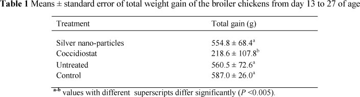
The results of the weight gains of the chicks are presented in Table 1. No mortalities of chicks were recorded during the trial.
The total weight gain was lower (P <0.005) in the birds receiving Salinomycin® in comparison to all the other treatments, including the unmedicated group. There were no differences (P >0.05) between the other three treatments implicating that nano silver had no effect on growth rate. In contrast Ahmadi & Rahimi (2011) found decreased growth of chickens on nano silver treatments.
The oocyst numbers in the excreta of the chickens six to eight days after the challenge are presented in Table 2. During the first six days of the challenge there were no oocysts produced (first generation of the oocysts) and on day seven, when the oocysts entered the second generation, they started to produce vigorously, as expected, while in the control group there were no counts.
By the 7 day after the challenge the groups receiving the nano silver and the coccidiostat had oocyst counts of about 356 000 and 968 000, respectively, while the unmedicated group had counts of 172 000, which was contrary to expectation and called for a repetition of the experiment. The gut lesion scores, on a scale of zero to four, were not statistically analysed and suggest that the lesions were less severe when a coccidiostat was used. The nano silver group and the untreated group were very similar as they all had scores of about 4 which indicated that the caecal walls were distended (severely damaged) with blood cores and normal faecal debris lacking or included in the core. The silver nano-particles group had scores of 2, 3 and 4 (average 3.2), whereby 2 indicated that the caecal contents are somewhat normal but contained streaks of blood and 3 indicated that the caecal walls were gently thickened and caeca had a little bit of faecal contents or blood and white caseous material present, therefore this could have affected the growth of the birds. The average score of the coccidiostat group was 2.4. The control group had a score of 0.
Trial 2
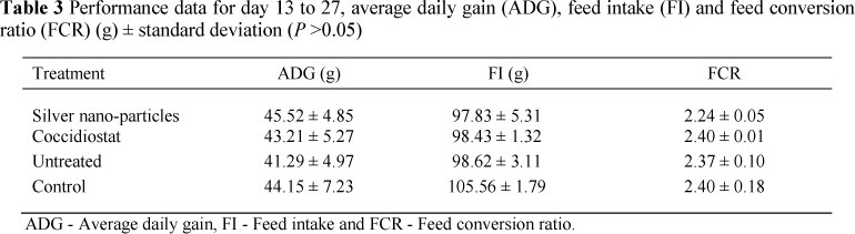
The performance data of the second trial are presented in Table 3 and showed no significant differences between any of the treatment groups (P >0.05), which again confirms that nano-silver is not a growth promoter and neither is the coccidiostat (Baycox®). The data also suggest that the challenge with the coccidia did not affect growth (P >0.05), possibly because there were no caecal lesions caused.
The coccidia oocyst counts in the excreta of the chicks at day 7 after the challenge were 408 000 for the Nano-silver group, 364 000 for the coccidiostat group and 788 000 for the unmedicated group, which suggest that the silver treatment also reduced the oocyst count by about 50% compared to the untreated group. Unfortunately, no substantial lesions were found in the second trial so the results are still inconclusive in terms of lesions.
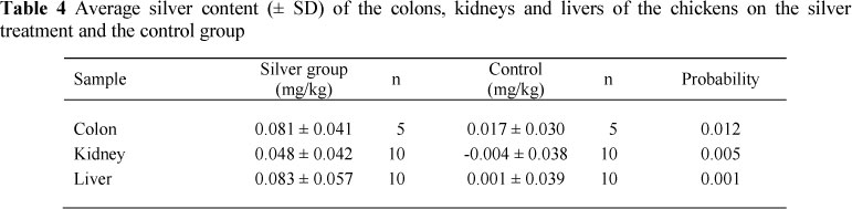
The residual silver contents of the tissues analysed are presented in Table 4. Significantly higher levels of silver were found in all the tissues analysed viz. colon kidney and liver.
Ahmadi and Rahimi (2011) found silver levels in the edible parts of broilers such as breast, femur and liver to be in the region of 0.1 mg/kg. This is not a toxic level for humans, but there is evidence that, as nano particles it may be toxic (Park et al, 2011). It is not known whether the silver in the tissues is in the ionic form or as silver-nano particles.
Conclusion
The results demonstrate that administering silver-nano particles to the drinking water of broilers did not affect their growth performance, though it may kill coccidia in broiler intestines in terms of oocysts excreted. The results suggest that silver-nano particles may be used as an alternative for antibiotic (ionophor) coccidiostats. Further work with a larger numbers of experimental animals is, however, needed.
Acknowledgements
NRF, Biosil, S. Bischopp and A. van Wyk, Poultry Research Unit, Onderstepoort, South Africa. [ Links ]
Ahmadi, F. & Rahimi, F., 2011. The effect of different levels of nanosilver on the performance and retention of silver in edible tissues of broilers. Wrld Appl. Sci. J. 12, 01-04. http://idosi.org/wasj/wasj 12(1)11/1.pdf [ Links ]
Atiyeh, B.S., Costagliola, M., Hayek, S.N. & Dibo, S.A., 2007. Effect of silver on burn wound infection control and healing: review of the literature. Burns 33, 139-148. [ Links ]
Chapman, H.D., Jeffers, T.K. & Williams, R.B., 2010. Forty years of monensin for the control of coccidiosis in poultry. Poult. Sci. 89, 1788-1801. [ Links ]
Holdsworth, P.A., Conway, D.P., Mckenzie, A.D., Dayton, A.D., Chapman, H.D., Mathis, G.F., Skinner, J.T., Mundt, H.C. & Williams, R.B., 2004. World Association for the Advancement of Veterinary Parasitology (WAAVP) guidelines for evaluating the efficacy of anticoccidial drugs in chickens and turkeys. Vet. Parasitol. 121, 189-212. [ Links ]
Jeong S.H., Yeo S.Y & Yi, S.C., 2005. The effect of filler particle size of antibacterial properties of compounded polymer/silver fibres. J. Mater. Sci. 40, 5407-5411. [ Links ]
Johnson, J. & Reid, W.M., 1970. Anticoccidial drugs: Lesion scoring techniques in battery and floor-pen experiments with chickens. Exp. Parasitol. 28, 30-36. [ Links ]
Kyriacou, S.V., Brownlow, W.J. & Xu X.H.N., 2004. Enhancement of antibacterial properties of Ag nanorods by electric field. Biochem. 43, 140-147. [ Links ]
Lansdown, A.B., 2006. Silver in health care: antimicrobial effects and safety in use. Curr. Probl. Dermatol. 33, 17-34. [ Links ]
Navarro, E., Piccapietra, F., Wagner, B., Marconi, F., Kaegi, R., Odzak, N., Sigg, L. & Behra, R., 2008. Toxicity of silver nanoparticles to Chlamydomonas reinhardtii. Environ. Sci. Technol. 42, 8959-8964. [ Links ]
Panacek, A., Kvitek, L., Prucek R., Kolar, M., Vecerova, R., Pizurova, N., Sharma, V. K., Nevecna, T. & Zboril, R., 2006. Silver colloid nanoparticles: synthesis, characterization, and their antibacterial activity. J. Phys. Chem. B. 110, 16248-16253. [ Links ]
Park, M.V.D.Z., Neigh, A.M., Vermeulen, J.P., de la Fonteyne, L.J.J., Verharen, H.W., Briedé, J.J., Van Loveren, H. & de Jong, W.H., 2011. The effect of particle size on the cytotoxicity, inflammation, developmental toxicity and genotoxicity of silver nanoparticles. Biomaterials 32, 9810-9817. [ Links ]
Singh, M., Singh, S., Prasad, S. & Gambhir, I.S., 2008. Nanotechnology in medicine and antibacterial effect of silver nanoparticles. Dig. J. Nanomater. Bios. 3, 115-122. [ Links ]
Spruill, J., 2006. Novel pre-harvest approaches to control enteric food-borne bacteria in poultry. Master of Science thesis. North Carolina State University. Raleigh, N.C., USA. [ Links ]
STATA CORP 2009. Stata: release 11. Statistical Software. College station. TX. USA. [ Links ]
Steel, R.G.D., Torrie, J.H. & Dickey, D.A., 1997. Principles and Procedures of Statistics. A Biochemical Approach (3rd ed.), McGraw Hill Book Co. Inc., New York, USA. [ Links ]
Copyright resides with the authors in terms of the Creative Commons Attribution 2.5 South African Licence. See: http://creativecommons.org/licenses/by/2.5/za/ Condition of use: The user may copy, distribute, transmit and adapt the work, but must recognise the authors and the South African Journal of Animal Science
# Corresponding author: siebritsfk@tut.ac.za

